|
Chorioamnionitis
Chorioamnionitis, also known as intra-amniotic infection (IAI), is inflammation of the fetal membranes ( amnion and chorion), usually due to bacterial infection. In 2015, a National Institute of Child Health and Human Development Workshop expert panel recommended use of the term "triple I" to address the heterogeneity of this disorder. The term triple I refers to intrauterine infection or inflammation or both and is defined by strict diagnostic criteria, but this terminology has not been commonly adopted although the criteria are used. Chorioamnionitis results from an infection caused by bacteria ascending from the vagina into the uterus and is associated with premature or prolonged labor. It triggers an inflammatory response to release various inflammatory signaling molecules, leading to increased prostaglandin and metalloproteinase release. These substances promote uterine contractions and cervical ripening, causations of premature birth. The risk of developing chorioamnioniti ... [...More Info...] [...Related Items...] OR: [Wikipedia] [Google] [Baidu] |
Chorioamnionitis - Intermed Mag
Chorioamnionitis, also known as intra-amniotic infection (IAI), is inflammation of the fetal membranes (amnion and chorion), usually due to bacterial infection. In 2015, a National Institute of Child Health and Human Development Workshop expert panel recommended use of the term "triple I" to address the heterogeneity of this disorder. The term triple I refers to intrauterine infection or inflammation or both and is defined by strict diagnostic criteria, but this terminology has not been commonly adopted although the criteria are used. Chorioamnionitis results from an infection caused by bacteria ascending from the vagina into the uterus and is associated with premature or prolonged labor. It triggers an inflammatory response to release various inflammatory signaling molecules, leading to increased prostaglandin and metalloproteinase release. These substances promote uterine contractions and cervical ripening, causations of premature birth. The risk of developing chorioamnionitis i ... [...More Info...] [...Related Items...] OR: [Wikipedia] [Google] [Baidu] |
Premature Birth
Preterm birth, also known as premature birth, is the birth of a baby at fewer than 37 weeks gestational age, as opposed to full-term delivery at approximately 40 weeks. Extreme preterm is less than 28 weeks, very early preterm birth is between 28 and 32 weeks, early preterm birth occurs between 32 and 36 weeks, late preterm birth is between 34 and 36 weeks' gestation. These babies are also known as premature babies or colloquially preemies (American English) or premmies (Australian English). Symptoms of preterm labor include uterine contractions which occur more often than every ten minutes and/or the leaking of fluid from the vagina before 37 weeks. Premature infants are at greater risk for cerebral palsy, delays in development, hearing problems and problems with their vision. The earlier a baby is born, the greater these risks will be. The cause of spontaneous preterm birth is often not known. Risk factors include diabetes, high blood pressure, multiple gestation (being pregna ... [...More Info...] [...Related Items...] OR: [Wikipedia] [Google] [Baidu] |
Fetal Membrane
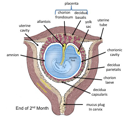
The fetal membranes are the four extraembryonic membranes, associated with the developing embryo, and fetus in humans and other mammals.. They are the amnion, chorion, allantois, and yolk sac. The amnion and the chorion are the chorioamniotic membranes that make up the amniotic sac which surrounds and protects the embryo. The fetal membranes are four of six accessory organs developed by the conceptus that are not part of the embryo itself, the other two are the placenta, and the umbilical cord. Structure The fetal membranes surround the developing embryo and form the fetal-maternal interface. The fetal membranes are derived from the trophoblast layer (outer layer of cells) of the implanting blastocyst. The trophoblast layer differentiates into amnion and the chorion, which then comprise the fetal membranes. The amnion is the innermost layer and, therefore, contacts the amniotic fluid, the fetus and the umbilical cord. The internal pressure of the amniotic fluid causes th ... [...More Info...] [...Related Items...] OR: [Wikipedia] [Google] [Baidu] |
Gonorrhea
Gonorrhea, colloquially known as the clap, is a sexually transmitted infection (STI) caused by the bacterium '' Neisseria gonorrhoeae''. Infection may involve the genitals, mouth, or rectum. Infected men may experience pain or burning with urination, discharge from the penis, or testicular pain. Infected women may experience burning with urination, vaginal discharge, vaginal bleeding between periods, or pelvic pain. Complications in women include pelvic inflammatory disease and in men include inflammation of the epididymis. Many of those infected, however, have no symptoms. If untreated, gonorrhea can spread to joints or heart valves. Gonorrhea is spread through sexual contact with an infected person. This includes oral, anal, and vaginal sex. It can also spread from a mother to a child during birth. Diagnosis is by testing the urine, urethra in males, or cervix in females. Testing all women who are sexually active and less than 25 years of age each year as well as thos ... [...More Info...] [...Related Items...] OR: [Wikipedia] [Google] [Baidu] |
Micrograph
A micrograph or photomicrograph is a photograph or digital image taken through a microscope or similar device to show a magnified image of an object. This is opposed to a macrograph or photomacrograph, an image which is also taken on a microscope but is only slightly magnified, usually less than 10 times. Micrography is the practice or art of using microscopes to make photographs. A micrograph contains extensive details of microstructure. A wealth of information can be obtained from a simple micrograph like behavior of the material under different conditions, the phases found in the system, failure analysis, grain size estimation, elemental analysis and so on. Micrographs are widely used in all fields of microscopy. Types Photomicrograph A light micrograph or photomicrograph is a micrograph prepared using an optical microscope, a process referred to as ''photomicroscopy''. At a basic level, photomicroscopy may be performed simply by connecting a camera to a microscope, th ... [...More Info...] [...Related Items...] OR: [Wikipedia] [Google] [Baidu] |
Ureaplasma
''Ureaplasma'' is a genus of bacteria belonging to the family Mycoplasmataceae. As the name imples, ''Ureaplasma'' is urease positive. Phylogeny The currently accepted taxonomy is based on the List of Prokaryotic names with Standing in Nomenclature (LPSN) and National Center for Biotechnology Information (NCBI) See also * List of bacterial orders * List of bacteria genera This article lists the genera of the bacteria. The currently accepted taxonomy is based on the List of Prokaryotic names with Standing in Nomenclature (LPSN) and National Center for Biotechnology Information (NCBI). However many taxonomic names are ... References External linksUreaplasma Infection: eMedicine Infectious Diseases Bacteria genera {{Bacteria-stub ... [...More Info...] [...Related Items...] OR: [Wikipedia] [Google] [Baidu] |
Fetus
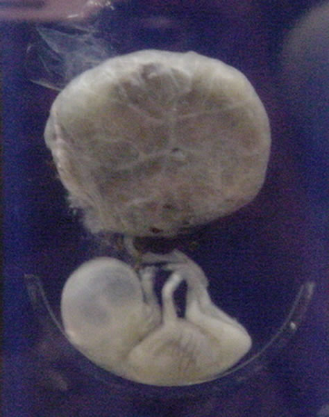
A fetus or foetus (; plural fetuses, feti, foetuses, or foeti) is the unborn offspring that develops from an animal embryo. Following embryonic development the fetal stage of development takes place. In human prenatal development, fetal development begins from the ninth week after fertilization (or eleventh week gestational age) and continues until birth. Prenatal development is a continuum, with no clear defining feature distinguishing an embryo from a fetus. However, a fetus is characterized by the presence of all the major body organs, though they will not yet be fully developed and functional and some not yet situated in their final anatomical location. Etymology The word ''fetus'' (plural ''fetuses'' or '' feti'') is related to the Latin '' fētus'' ("offspring", "bringing forth", "hatching of young") and the Greek "φυτώ" to plant. The word "fetus" was used by Ovid in Metamorphoses, book 1, line 104. The predominant British, Irish, and Commonwealth spelling is '' ... [...More Info...] [...Related Items...] OR: [Wikipedia] [Google] [Baidu] |
Amniotic Fluid
The amniotic fluid is the protective liquid contained by the amniotic sac of a gravid amniote. This fluid serves as a cushion for the growing fetus, but also serves to facilitate the exchange of nutrients, water, and biochemical products between mother and fetus. For humans, the amniotic fluid is commonly called water or waters (Latin liquor amnii). Development Amniotic fluid is present from the formation of the gestational sac. Amniotic fluid is in the amniotic sac. It is generated from maternal plasma, and passes through the fetal membranes by osmotic and hydrostatic forces. When fetal kidneys begin to function around week 16, fetal urine also contributes to the fluid. In earlier times, it was believed that the amniotic fluid was composed entirely of fetal urine. The fluid is absorbed through the fetal tissue and skin. After 22 to 25 week of pregnancy, keratinization of an embryo's skin occurs. When this process completes around the 25th week, the fluid is primarily absor ... [...More Info...] [...Related Items...] OR: [Wikipedia] [Google] [Baidu] |
Meconium
Meconium is the earliest stool of a mammalian infant resulting from defecation. Unlike later feces, meconium is composed of materials ingested during the time the infant spends in the uterus: intestinal epithelial cells, lanugo, mucus, amniotic fluid, bile, and water. Meconium, unlike later feces, is viscous and sticky like tar – its color usually being a very dark olive green and it is almost odorless. When diluted in amniotic fluid, it may appear in various shades of green, brown, or yellow. It should be completely passed by the end of the first few days after birth, with the stools progressing toward yellow (digested milk). Clinical significance Meconium in amniotic fluid Meconium is normally retained in the infant's bowel until after birth, but sometimes it is expelled into the amniotic fluid (also called "amniotic liquor") prior to birth or during labor and delivery. The stained amniotic fluid (called "meconium liquor" or "meconium-stained liquor") is recognized by m ... [...More Info...] [...Related Items...] OR: [Wikipedia] [Google] [Baidu] |
Amniotic Sac
The amniotic sac, also called the bag of waters or the membranes, is the sac in which the embryo and later fetus develops in amniotes. It is a thin but tough transparent pair of membranes that hold a developing embryo (and later fetus) until shortly before birth. The inner of these membranes, the amnion, encloses the amniotic cavity, containing the amniotic fluid and the embryo. The outer membrane, the chorion, contains the amnion and is part of the placenta. On the outer side, the amniotic sac is connected to the yolk sac, the allantois, and via the umbilical cord, the placenta. The yolk sac, amnion, chorion, and allantois are the four extraembryonic membranes that lie outside of the embryo and are involved in providing nutrients and protection to the developing embryo. They form from the inner cell mass; the first to form is the yolk sac followed by the amnion which grows over the developing embryo. The amnion remains an important extraembryonic membrane throughout prenatal dev ... [...More Info...] [...Related Items...] OR: [Wikipedia] [Google] [Baidu] |
Chlamydia
Chlamydia, or more specifically a chlamydia infection, is a sexually transmitted infection caused by the bacterium ''Chlamydia trachomatis''. Most people who are infected have no symptoms. When symptoms do appear they may occur only several weeks after infection; the incubation period between exposure and being able to infect others is thought to be on the order of two to six weeks. Symptoms in women may include vaginal discharge or burning with urination. Symptoms in men may include discharge from the penis, burning with urination, or pain and swelling of one or both testicles. The infection can spread to the upper genital tract in women, causing pelvic inflammatory disease, which may result in future infertility or ectopic pregnancy. Chlamydia infections can occur in other areas besides the genitals, including the anus, eyes, throat, and lymph nodes. Repeated chlamydia infections of the eyes that go without treatment can result in trachoma, a common cause of blindness in th ... [...More Info...] [...Related Items...] OR: [Wikipedia] [Google] [Baidu] |


