|
Cholesteatoma
Cholesteatoma is a destructive and expanding growth consisting of keratinizing squamous epithelium in the middle ear and/or mastoid process. Cholesteatomas are not cancerous as the name may suggest, but can cause significant problems because of their erosive and expansile properties. This can result in the destruction of the bones of the middle ear (ossicles), as well as growth through the base of the skull into the brain. They often become infected and can result in chronically draining ears. Treatment almost always consists of surgical removal. Signs and symptoms Other more common conditions (e.g. otitis externa) may also present with these symptoms, but cholesteatoma is much more serious and should not be overlooked. If a patient presents to a doctor with ear discharge and hearing loss, the doctor should consider cholesteatoma until the disease is definitely excluded. Other less common symptoms (all less than 15%) of cholesteatoma may include pain, balance disruption, tinnitu ... [...More Info...] [...Related Items...] OR: [Wikipedia] [Google] [Baidu] |
Mastoidectomy
A mastoidectomy is a procedure performed to remove the mastoid air cells, air bubbles in the skull, near the inner ears. This can be done as part of treatment for mastoiditis, chronic suppurative otitis media or cholesteatoma. In addition, it is sometimes performed as part of other procedures (cochlear implant) or for access to the middle ear. There are classically 5 different types of mastoidectomy: ;* Radical : Removal of posterior and superior canal wall, meatoplasty and exteriorisation of middle ear. ;* Canal wall down : Removal of posterior and superior canal wall, meatoplasty. Tympanic membrane left in place. ;* Canal wall up : Posterior and superior canal wall are kept intact. A facial recess approach is taken. ;* Cortical : (Also known as schwartze procedure) - Removal of Mastoid air cells The mastoid cells (also called air cells of Lenoir or mastoid cells of Lenoir) are air-filled cavities within the mastoid process of the temporal bone of the cranium. The mastoid cells ... [...More Info...] [...Related Items...] OR: [Wikipedia] [Google] [Baidu] |
Ear Drum
In the anatomy of humans and various other tetrapods, the eardrum, also called the tympanic membrane or myringa, is a thin, cone-shaped membrane that separates the external ear from the middle ear. Its function is to transmit sound from the air to the ossicles inside the middle ear, and then to the oval window in the fluid-filled cochlea. Hence, it ultimately converts and amplifies vibration in the air to vibration in cochlear fluid. The malleus bone bridges the gap between the eardrum and the other ossicles. Rupture or perforation of the eardrum can lead to conductive hearing loss. Collapse or retraction of the eardrum can cause conductive hearing loss or cholesteatoma. Structure Orientation and relations The tympanic membrane is oriented obliquely in the anteroposterior, mediolateral, and superoinferior planes. Consequently, its superoposterior end lies lateral to its anteroinferior end. Anatomically, it relates superiorly to the middle cranial fossa, posteriorly to the os ... [...More Info...] [...Related Items...] OR: [Wikipedia] [Google] [Baidu] |
Eardrum
In the anatomy of humans and various other tetrapods, the eardrum, also called the tympanic membrane or myringa, is a thin, cone-shaped membrane that separates the external ear from the middle ear. Its function is to transmit sound from the air to the ossicles inside the middle ear, and then to the oval window in the fluid-filled cochlea. Hence, it ultimately converts and amplifies vibration in the air to vibration in cochlear fluid. The malleus bone bridges the gap between the eardrum and the other ossicles. Rupture or perforation of the eardrum can lead to conductive hearing loss. Collapse or retraction of the eardrum can cause conductive hearing loss or cholesteatoma. Structure Orientation and relations The tympanic membrane is oriented obliquely in the anteroposterior, mediolateral, and superoinferior planes. Consequently, its superoposterior end lies lateral to its anteroinferior end. Anatomically, it relates superiorly to the middle cranial fossa, posteriorly t ... [...More Info...] [...Related Items...] OR: [Wikipedia] [Google] [Baidu] |
Tympanic Membrane
In the anatomy of humans and various other tetrapods, the eardrum, also called the tympanic membrane or myringa, is a thin, cone-shaped membrane that separates the external ear from the middle ear. Its function is to transmit sound from the air to the ossicles inside the middle ear, and then to the oval window in the fluid-filled cochlea. Hence, it ultimately converts and amplifies vibration in the air to vibration in cochlear fluid. The malleus bone bridges the gap between the eardrum and the other ossicles. Rupture or perforation of the eardrum can lead to conductive hearing loss. Collapse or retraction of the eardrum can cause conductive hearing loss or cholesteatoma. Structure Orientation and relations The tympanic membrane is oriented obliquely in the anteroposterior, mediolateral, and superoinferior planes. Consequently, its superoposterior end lies lateral to its anteroinferior end. Anatomically, it relates superiorly to the middle cranial fossa, posteriorly to ... [...More Info...] [...Related Items...] OR: [Wikipedia] [Google] [Baidu] |
Ear Ache
Ear pain, also known as earache or otalgia, is pain in the ear. Primary ear pain is pain that originates from the ear. Secondary ear pain is a type of referred pain, meaning that the source of the pain differs from the location where the pain is felt. Most causes of ear pain are non-life-threatening. Primary ear pain is more common than secondary ear pain, and it is often due to infection or injury. The conditions that cause secondary (referred) ear pain are broad and range from temporomandibular joint syndrome to inflammation of the throat. In general, the reason for ear pain can be discovered by taking a thorough history of all symptoms and performing a physical examination, without need for imaging tools like a CT scan. However, further testing may be needed if red flags are present like hearing loss, dizziness, ringing in the ear or unexpected weight loss. Management of ear pain depends on the cause. If there is a bacterial infection, antibiotics are sometimes recommended ... [...More Info...] [...Related Items...] OR: [Wikipedia] [Google] [Baidu] |
Squamous Epithelium
Epithelium or epithelial tissue is one of the four basic types of animal tissue, along with connective tissue, muscle tissue and nervous tissue. It is a thin, continuous, protective layer of compactly packed cells with a little intercellular matrix. Epithelial tissues line the outer surfaces of organs and blood vessels throughout the body, as well as the inner surfaces of cavities in many internal organs. An example is the epidermis, the outermost layer of the skin. There are three principal shapes of epithelial cell: squamous (scaly), columnar, and cuboidal. These can be arranged in a singular layer of cells as simple epithelium, either squamous, columnar, or cuboidal, or in layers of two or more cells deep as stratified (layered), or ''compound'', either squamous, columnar or cuboidal. In some tissues, a layer of columnar cells may appear to be stratified due to the placement of the nuclei. This sort of tissue is called pseudostratified. All glands are made up of epithel ... [...More Info...] [...Related Items...] OR: [Wikipedia] [Google] [Baidu] |
Brain Abscess
Brain abscess (or cerebral abscess) is an abscess caused by inflammation and collection of infected material, coming from local (ear infection, dental abscess, infection of paranasal sinuses, infection of the mastoid air cells of the temporal bone, epidural abscess) or remote ( lung, heart, kidney etc.) infectious sources, within the brain tissue. The infection may also be introduced through a skull fracture following a head trauma or surgical procedures. Brain abscess is usually associated with congenital heart disease in young children. It may occur at any age but is most frequent in the third decade of life. Signs and symptoms Fever, headache, and neurological problems, while classic, only occur in 20% of people with brain abscess. The famous triad of fever, headache and focal neurologic findings are highly suggestive of brain abscess. These symptoms are caused by a combination of increased intracranial pressure due to a space-occupying lesion (headache, vomiting, confusion ... [...More Info...] [...Related Items...] OR: [Wikipedia] [Google] [Baidu] |
Sepsis
Sepsis, formerly known as septicemia (septicaemia in British English) or blood poisoning, is a life-threatening condition that arises when the body's response to infection causes injury to its own tissues and organs. This initial stage is followed by suppression of the immune system. Common signs and symptoms include fever, increased heart rate, increased breathing rate, and confusion. There may also be symptoms related to a specific infection, such as a cough with pneumonia, or painful urination with a kidney infection. The very young, old, and people with a weakened immune system may have no symptoms of a specific infection, and the body temperature may be low or normal instead of having a fever. Severe sepsis causes poor organ function or blood flow. The presence of low blood pressure, high blood lactate, or low urine output may suggest poor blood flow. Septic shock is low blood pressure due to sepsis that does not improve after fluid replacement. Sepsis is caused ... [...More Info...] [...Related Items...] OR: [Wikipedia] [Google] [Baidu] |
Mastoid Portion Of The Temporal Bone
The mastoid part of the temporal bone is the posterior (back) part of the temporal bone, one of the bones of the skull. Its rough surface gives attachment to various muscles (via tendons) and it has openings for blood vessels. From its borders, the mastoid part articulates with two other bones. Etymology The word "mastoid" is derived from the Greek word for "breast", a reference to the shape of this bone. Surfaces Outer surface Its outer surface is rough and gives attachment to the occipitalis and posterior auricular muscles. It is perforated by numerous foramina (holes); for example, the mastoid foramen is situated near the posterior border and transmits a vein to the transverse sinus and a small branch of the occipital artery to the dura mater. The position and size of this foramen are very variable; it is not always present; sometimes it is situated in the occipital bone, or in the suture between the temporal and the occipital. Mastoid process The mastoid process is ... [...More Info...] [...Related Items...] OR: [Wikipedia] [Google] [Baidu] |
Epidermoid Cyst
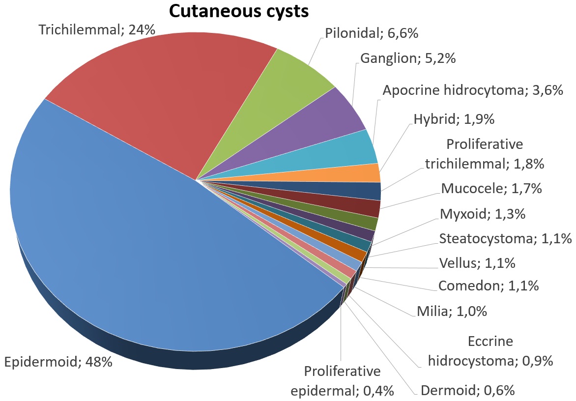
An epidermoid cyst or epidermal inclusion cyst is a benign cyst usually found on the skin. The cyst develops out of ectodermal tissue. Histologically, it is made of a thin layer of squamous epithelium. Signs and symptoms The epidermoid cyst may have no symptoms, or it may be painful when touched. It can release macerated keratin. In contrast to pilar cysts, epidermoid cysts are usually present on parts of the body with relatively little hair. An epidermoid cyst is one type of vaginal cysts. Although they are not malignant, there are rare cases of malignant tumors arising from an epidermoid cyst. Epidermal inclusion cysts account for approximately 85–95% of all excised cysts, malignant transformation is exceedingly rare. The incidence of squamous cell carcinoma developing from an epidermal inclusion cyst has been estimated to range from 0.011 to 0.045%. Diagnosis Epidermoid cysts are usually diagnosed when a person notices a bump on their skin and seeks medical attention. Th ... [...More Info...] [...Related Items...] OR: [Wikipedia] [Google] [Baidu] |
Middle Ear
The middle ear is the portion of the ear medial to the eardrum, and distal to the oval window of the cochlea (of the inner ear). The mammalian middle ear contains three ossicles, which transfer the vibrations of the eardrum into waves in the fluid and membranes of the inner ear. The hollow space of the middle ear is also known as the tympanic cavity and is surrounded by the tympanic part of the temporal bone. The auditory tube (also known as the Eustachian tube or the pharyngotympanic tube) joins the tympanic cavity with the nasal cavity (nasopharynx), allowing pressure to equalize between the middle ear and throat. The primary function of the middle ear is to efficiently transfer acoustic energy from compression waves in air to fluid–membrane waves within the cochlea. Structure Ossicles The middle ear contains three tiny bones known as the ossicles: '' malleus'', '' incus'', and ''stapes''. The ossicles were given their Latin names for their distinctive shapes; they ar ... [...More Info...] [...Related Items...] OR: [Wikipedia] [Google] [Baidu] |
Vertigo (medical)
Vertigo is a condition where a person has the sensation of movement or of surrounding objects moving when they are not. Often it feels like a spinning or swaying movement. This may be associated with nausea, vomiting, sweating, or difficulties walking. It is typically worse when the head is moved. Vertigo is the most common type of dizziness. The most common disorders that result in vertigo are benign paroxysmal positional vertigo (BPPV), Ménière's disease, and labyrinthitis. Less common causes include stroke, brain tumors, brain injury, multiple sclerosis, migraines, trauma, and uneven pressures between the middle ears. Physiologic vertigo may occur following being exposed to motion for a prolonged period such as when on a ship or simply following spinning with the eyes closed. Other causes may include toxin exposures such as to carbon monoxide, alcohol, or aspirin. Vertigo typically indicates a problem in a part of the vestibular system. Other causes of dizziness inclu ... [...More Info...] [...Related Items...] OR: [Wikipedia] [Google] [Baidu] |


