|
Cardiorespiratory Fitness
Cardiorespiratory fitness (CRF) refers to the ability of the circulatory and respiratory systems to supply oxygen to skeletal muscles during sustained physical activity. Scientists and researchers use CRF to assess the functional capacity of the respiratory and cardiovascular systems. These functions include ventilation, perfusion, gas exchange, vasodilation, and delivery of oxygen to the body's tissues. As these body's functions are vital to an individual's health, CRF allows observers to quantify an individual's morbidity and mortality risk as a function of cardiorespiratory health. There are a multitude of ways scientists have developed to measure and estimate an individual's cardiovascular and respiratory fitness. One such way is using an exercise stress test, either treadmill or cycling, that entails using a graded-intensity aerobic stress to assess whether or not an individual can maintain physical exertion up to a heart rate of 85% of their age-predicted maximum. Another metho ... [...More Info...] [...Related Items...] OR: [Wikipedia] [Google] [Baidu] |
Circulatory System
The blood circulatory system is a system of organs that includes the heart, blood vessels, and blood which is circulated throughout the entire body of a human or other vertebrate. It includes the cardiovascular system, or vascular system, that consists of the heart and blood vessels (from Greek ''kardia'' meaning ''heart'', and from Latin ''vascula'' meaning ''vessels''). The circulatory system has two divisions, a systemic circulation or circuit, and a pulmonary circulation or circuit. Some sources use the terms ''cardiovascular system'' and ''vascular system'' interchangeably with the ''circulatory system''. The network of blood vessels are the great vessels of the heart including large elastic arteries, and large veins; other arteries, smaller arterioles, capillaries that join with venules (small veins), and other veins. The circulatory system is closed in vertebrates, which means that the blood never leaves the network of blood vessels. Some invertebrates such as a ... [...More Info...] [...Related Items...] OR: [Wikipedia] [Google] [Baidu] |
Blood Pressure
Blood pressure (BP) is the pressure of circulating blood against the walls of blood vessels. Most of this pressure results from the heart pumping blood through the circulatory system. When used without qualification, the term "blood pressure" refers to the pressure in the large arteries. Blood pressure is usually expressed in terms of the systolic pressure (maximum pressure during one heartbeat) over diastolic pressure (minimum pressure between two heartbeats) in the cardiac cycle. It is measured in millimeters of mercury (mmHg) above the surrounding atmospheric pressure. Blood pressure is one of the vital signs—together with respiratory rate, heart rate, oxygen saturation, and body temperature—that healthcare professionals use in evaluating a patient's health. Normal resting blood pressure, in an adult is approximately systolic over diastolic, denoted as "120/80 mmHg". Globally, the average blood pressure, age standardized, has remained about the same sin ... [...More Info...] [...Related Items...] OR: [Wikipedia] [Google] [Baidu] |
Sarcoplasmic Reticulum
The sarcoplasmic reticulum (SR) is a membrane-bound structure found within muscle cells that is similar to the smooth endoplasmic reticulum in other cells. The main function of the SR is to store calcium ions (Ca2+). Calcium ion levels are kept relatively constant, with the concentration of calcium ions within a cell being 10,000 times smaller than the concentration of calcium ions outside the cell. This means that small increases in calcium ions within the cell are easily detected and can bring about important cellular changes (the calcium is said to be a second messenger). Calcium is used to make calcium carbonate (found in chalk) and calcium phosphate, two compounds that the body uses to make teeth and bones. This means that too much calcium within the cells can lead to hardening (calcification) of certain intracellular structures, including the mitochondria, leading to cell death. Therefore, it is vital that calcium ion levels are controlled tightly, and can be released i ... [...More Info...] [...Related Items...] OR: [Wikipedia] [Google] [Baidu] |
Sympathetic Nervous System
The sympathetic nervous system (SNS) is one of the three divisions of the autonomic nervous system, the others being the parasympathetic nervous system and the enteric nervous system. The enteric nervous system is sometimes considered part of the autonomic nervous system, and sometimes considered an independent system. The autonomic nervous system functions to regulate the body's unconscious actions. The sympathetic nervous system's primary process is to stimulate the body's fight or flight response. It is, however, constantly active at a basic level to maintain homeostasis. The sympathetic nervous system is described as being antagonistic to the parasympathetic nervous system which stimulates the body to "feed and breed" and to (then) "rest-and-digest". Structure There are two kinds of neurons involved in the transmission of any signal through the sympathetic system: pre-ganglionic and post-ganglionic. The shorter preganglionic neurons originate in the thoracolumbar divisi ... [...More Info...] [...Related Items...] OR: [Wikipedia] [Google] [Baidu] |
Parasympathetic Nervous System
The parasympathetic nervous system (PSNS) is one of the three divisions of the autonomic nervous system, the others being the sympathetic nervous system and the enteric nervous system. The enteric nervous system is sometimes considered part of the autonomic nervous system, and sometimes considered an independent system. The autonomic nervous system is responsible for regulating the body's unconscious actions. The parasympathetic system is responsible for stimulation of "rest-and-digest" or "feed and breed" activities that occur when the body is at rest, especially after eating, including sexual arousal, salivation, lacrimation (tears), urination, digestion, and defecation. Its action is described as being complementary to that of the sympathetic nervous system, which is responsible for stimulating activities associated with the fight-or-flight response. Nerve fibres of the parasympathetic nervous system arise from the central nervous system. Specific nerves include sev ... [...More Info...] [...Related Items...] OR: [Wikipedia] [Google] [Baidu] |
Autonomic Nervous System
The autonomic nervous system (ANS), formerly referred to as the vegetative nervous system, is a division of the peripheral nervous system that supplies internal organs, smooth muscle and glands. The autonomic nervous system is a control system that acts largely unconsciously and regulates bodily functions, such as the heart rate, its force of contraction, digestion, respiratory rate, pupillary response, urination, and sexual arousal. This system is the primary mechanism in control of the fight-or-flight response. The autonomic nervous system is regulated by integrated reflexes through the brainstem to the spinal cord and organs. Autonomic functions include control of respiration, cardiac regulation (the cardiac control center), vasomotor activity (the vasomotor center), and certain reflex actions such as coughing, sneezing, swallowing and vomiting. Those are then subdivided into other areas and are also linked to autonomic subsystems and the peripheral nervous system ... [...More Info...] [...Related Items...] OR: [Wikipedia] [Google] [Baidu] |
Myofibril
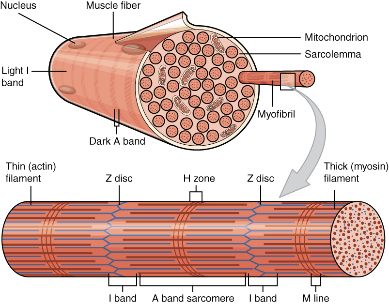
A myofibril (also known as a muscle fibril or sarcostyle) is a basic rod-like organelle of a muscle cell. Skeletal muscles are composed of long, tubular cells known as muscle fibers, and these cells contain many chains of myofibrils. Each myofibril has a diameter of 1–2 micrometres. They are created during embryonic development in a process known as myogenesis. Myofibrils are composed of long proteins including actin, myosin, and titin, and other proteins that hold them together. These proteins are organized into thick, thin, and elastic myofilaments, which repeat along the length of the myofibril in sections or units of contraction called sarcomeres. Muscles contract by sliding the thick myosin, and thin actin myofilaments along each other. Structure Each myofibril has a diameter of between 1 and 2 micrometres (μm). The filaments of myofibrils, myofilaments, consist of three types, thick, thin, and elastic filaments. *Thin filaments consist primarily of the pr ... [...More Info...] [...Related Items...] OR: [Wikipedia] [Google] [Baidu] |
Sarcomere
A sarcomere (Greek σάρξ ''sarx'' "flesh", μέρος ''meros'' "part") is the smallest functional unit of striated muscle tissue. It is the repeating unit between two Z-lines. Skeletal muscles are composed of tubular muscle cells (called muscle fibers or myofibers) which are formed during embryonic myogenesis. Muscle fibers contain numerous tubular myofibrils. Myofibrils are composed of repeating sections of sarcomeres, which appear under the microscope as alternating dark and light bands. Sarcomeres are composed of long, fibrous proteins as filaments that slide past each other when a muscle contracts or relaxes. The costamere is a different component that connects the sarcomere to the sarcolemma. Two of the important proteins are myosin, which forms the thick filament, and actin, which forms the thin filament. Myosin has a long, fibrous tail and a globular head, which binds to actin. The myosin head also binds to ATP, which is the source of energy for muscle movement. ... [...More Info...] [...Related Items...] OR: [Wikipedia] [Google] [Baidu] |
Afterload
Afterload is the pressure that the heart must work against to eject blood during systole (ventricular contraction). Afterload is proportional to the average arterial pressure. As aortic and pulmonary pressures increase, the afterload increases on the left and right ventricles respectively. Afterload changes to adapt to the continually changing demands on an animal's cardiovascular system. Afterload is proportional to mean systolic blood pressure and is measured in millimeters of mercury (mm Hg). Hemodynamics Afterload is a determinant of cardiac output. Cardiac output is the product of stroke volume and heart rate. Afterload is a determinant of stroke volume (in addition to preload, and strength of myocardial contraction). Following Laplace's law, the tension upon the muscle fibers in the heart wall is the pressure within the ventricle multiplied by the volume within the ventricle divided by the wall thickness (this ratio is the other factor in setting the afterload). T ... [...More Info...] [...Related Items...] OR: [Wikipedia] [Google] [Baidu] |
Preload (cardiology)
In cardiac physiology, preload is the amount of sarcomere stretch experienced by cardiac muscle cells, called cardiomyocytes, at the end of ventricular filling during diastole. Preload is directly related to ventricular filling. As the relaxed ventricle fills during diastole, the walls are stretched and the length of sarcomeres increases. Sarcomere length can be approximated by the volume of the ventricle because each shape has a conserved surface-area-to-volume ratio. This is useful clinically because measuring the sarcomere length is destructive to heart tissue. It requires cutting out a piece of cardiac muscle to look at the sarcomeres under a microscope. It is currently not possible to directly measure preload in the beating heart of a living animal. Preload is estimated from end-diastolic ventricular pressure and is measured in millimeters of mercury (mmHg). Estimating preload Though not exactly equivalent to the strict definition of ''preload,'' end-diastolic volu ... [...More Info...] [...Related Items...] OR: [Wikipedia] [Google] [Baidu] |
Myocardial Contractility
Myocardial contractility represents the innate ability of the heart muscle (cardiac muscle or myocardium) to contract. The ability to produce changes in force during contraction result from incremental degrees of binding between different types of tissue, that is, between filaments of myosin (thick) and actin (thin) tissue. The degree of binding depends upon the concentration of calcium ions in the cell. Within an in vivo intact heart, the action/response of the sympathetic nervous system is driven by precisely timed releases of a catecholamine, which is a process that determines the concentration of calcium ions in the cytosol of cardiac muscle cells. The factors causing an increase in contractility work by causing an increase in intracellular calcium ions (Ca++) during contraction. Mechanisms for altering contractility Increasing contractility is done primarily through increasing the influx of calcium or maintaining higher calcium levels in the cytosol of cardiac myocytes during a ... [...More Info...] [...Related Items...] OR: [Wikipedia] [Google] [Baidu] |
Systole
Systole ( ) is the part of the cardiac cycle during which some chambers of the heart contract after refilling with blood. The term originates, via New Latin, from Ancient Greek (''sustolē''), from (''sustéllein'' 'to contract'; from ''sun'' 'together' + ''stéllein'' 'to send'), and is similar to the use of the English term ''to squeeze''. The mammalian heart has four chambers: the left atrium above the left ventricle (lighter pink, see graphic), which two are connected through the mitral (or bicuspid) valve; and the right atrium above the right ventricle (lighter blue), connected through the tricuspid valve. The atria are the receiving blood chambers for the circulation of blood and the ventricles are the discharging chambers. In late ventricular diastole, the atrial chambers contract and send blood to the larger, lower ventricle chambers. This flow fills the ventricles with blood, and the resulting pressure closes the valves to the atria. The ventricles now ... [...More Info...] [...Related Items...] OR: [Wikipedia] [Google] [Baidu] |


