|
Calcium-binding Protein 1
Calcium binding protein 1 is a protein that in humans is encoded by the CABP1 gene. Calcium-binding protein 1 is a calcium-binding protein discovered in 1999. It has two EF hand motifs and is expressed in neuronal cells in such areas as hippocampus, habenular nucleus of the epithalamus, Purkinje cell layer of the cerebellum, and the amacrine cells and cone bipolar cells of the retina. Calcium-binding protein 1 which is a neuron -specific member of the calmodulin (CaM) superfamily which modulates Ca2+-dependent activity of inositol trisphosphate receptors (InsP3RS). L-CaBP1 is also associated with the cytoskeleton structures. But the S-CaBP1 is situated in or near the plasma membrane. In brain, CaBp1 is found in the cerebral cortex and hippocampus and in the protein, Cabp1 is found in cone bipolar and amacrine cells. We can also express that CaBP1 may regulate Ca2+ dependent activity of InSP3Rs by promoting structural contacts between suppressor and core domains but has no effect o ... [...More Info...] [...Related Items...] OR: [Wikipedia] [Google] [Baidu] |
Protein
Proteins are large biomolecules and macromolecules that comprise one or more long chains of amino acid residues. Proteins perform a vast array of functions within organisms, including catalysing metabolic reactions, DNA replication, responding to stimuli, providing structure to cells and organisms, and transporting molecules from one location to another. Proteins differ from one another primarily in their sequence of amino acids, which is dictated by the nucleotide sequence of their genes, and which usually results in protein folding into a specific 3D structure that determines its activity. A linear chain of amino acid residues is called a polypeptide. A protein contains at least one long polypeptide. Short polypeptides, containing less than 20–30 residues, are rarely considered to be proteins and are commonly called peptides. The individual amino acid residues are bonded together by peptide bonds and adjacent amino acid residues. The sequence of amino acid residue ... [...More Info...] [...Related Items...] OR: [Wikipedia] [Google] [Baidu] |
CACNA1A
Cav2.1, also called the P/ Q voltage-dependent calcium channel, is a calcium channel found mainly in the brain. Specifically, it is found on the presynaptic terminals of neurons in the brain and cerebellum. Cav2.1 plays an important role in controlling the release of neurotransmitters between neurons. It is composed of multiple subunits, including alpha-1, beta, alpha-2/delta, and gamma subunits. The alpha-1 subunit is the pore-forming subunit, meaning that the calcium ions flow through it. Different kinds of calcium channels have different isoforms (versions) of the alpha-1 subunit. Cav2.1 has the alpha-1A subunit, which is encoded by the ''CACNA1A'' gene. Mutations in ''CACNA1A'' have been associated with various neurologic disorders, including familial hemiplegic migraine, episodic ataxia type 2, and spinocerebellar ataxia type 6. Function "Voltage-dependent calcium channels mediate the entry of calcium ions into excitable cells, and are also involved in a variety of calci ... [...More Info...] [...Related Items...] OR: [Wikipedia] [Google] [Baidu] |
Axon
An axon (from Greek ἄξων ''áxōn'', axis), or nerve fiber (or nerve fibre: see spelling differences), is a long, slender projection of a nerve cell, or neuron, in vertebrates, that typically conducts electrical impulses known as action potentials away from the nerve cell body. The function of the axon is to transmit information to different neurons, muscles, and glands. In certain sensory neurons (pseudounipolar neurons), such as those for touch and warmth, the axons are called afferent nerve fibers and the electrical impulse travels along these from the periphery to the cell body and from the cell body to the spinal cord along another branch of the same axon. Axon dysfunction can be the cause of many inherited and acquired neurological disorders that affect both the peripheral and central neurons. Nerve fibers are classed into three typesgroup A nerve fibers, group B nerve fibers, and group C nerve fibers. Groups A and B are myelinated, and group C are unmyelinated. ... [...More Info...] [...Related Items...] OR: [Wikipedia] [Google] [Baidu] |
Hypothalamus
The hypothalamus () is a part of the brain that contains a number of small nuclei with a variety of functions. One of the most important functions is to link the nervous system to the endocrine system via the pituitary gland. The hypothalamus is located below the thalamus and is part of the limbic system. In the terminology of neuroanatomy, it forms the ventral part of the diencephalon. All vertebrate brains contain a hypothalamus. In humans, it is the size of an almond. The hypothalamus is responsible for regulating certain metabolic processes and other activities of the autonomic nervous system. It synthesizes and secretes certain neurohormones, called releasing hormones or hypothalamic hormones, and these in turn stimulate or inhibit the secretion of hormones from the pituitary gland. The hypothalamus controls body temperature, hunger, important aspects of parenting and maternal attachment behaviours, thirst, fatigue, sleep, and circadian rhythms. Structure T ... [...More Info...] [...Related Items...] OR: [Wikipedia] [Google] [Baidu] |
Somatodendritic
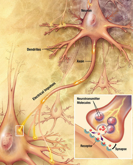
Chemical synapses are biological junctions through which neurons' signals can be sent to each other and to non-neuronal cells such as those in muscles or glands. Chemical synapses allow neurons to form circuits within the central nervous system. They are crucial to the biological computations that underlie perception and thought. They allow the nervous system to connect to and control other systems of the body. At a chemical synapse, one neuron releases neurotransmitter molecules into a small space (the synaptic cleft) that is adjacent to another neuron. The neurotransmitters are contained within small sacs called synaptic vesicles, and are released into the synaptic cleft by exocytosis. These molecules then bind to neurotransmitter receptors on the postsynaptic cell. Finally, the neurotransmitters are cleared from the synapse through one of several potential mechanisms including enzymatic degradation or re-uptake by specific transporters either on the presynaptic cell or on ... [...More Info...] [...Related Items...] OR: [Wikipedia] [Google] [Baidu] |
Caldendrin
Calcium binding protein 1 is a protein that in humans is encoded by the CABP1 gene. Calcium-binding protein 1 is a calcium-binding protein discovered in 1999. It has two EF hand motifs and is expressed in neuronal cells in such areas as hippocampus, habenular nucleus of the epithalamus, Purkinje cell layer of the cerebellum, and the amacrine cells and cone bipolar cells of the retina. Calcium-binding protein 1 which is a neuron -specific member of the calmodulin (CaM) superfamily which modulates Ca2+-dependent activity of inositol trisphosphate receptors (InsP3RS). L-CaBP1 is also associated with the cytoskeleton structures. But the S-CaBP1 is situated in or near the plasma membrane. In brain, CaBp1 is found in the cerebral cortex and hippocampus and in the protein, Cabp1 is found in cone bipolar and amacrine cells. We can also express that CaBP1 may regulate Ca2+ dependent activity of InSP3Rs by promoting structural contacts between suppressor and core domains but has no effect o ... [...More Info...] [...Related Items...] OR: [Wikipedia] [Google] [Baidu] |
HeLa Cells
HeLa (; also Hela or hela) is an immortalized cell line used in scientific research. It is the oldest and most commonly used human cell line. The line is derived from cervical cancer cells taken on February 8, 1951, named after Henrietta Lacks, a 31-year-old African-American mother of five, who died of cancer on October 4, 1951. The cell line was found to be remarkably durable and prolific, which allows it to be used extensively in scientific study. The cells from Lacks's cancerous cervical tumor were taken without her knowledge or consent, which was common practice in the United States at the time. Cell biologist George Otto Gey found that they could be kept alive, and developed a cell line. Previously, cells cultured from other human cells would only survive for a few days. Cells from Lacks's tumor behaved differently. History Origin In 1951, a patient named Henrietta Lacks was admitted to the Johns Hopkins Hospital with symptoms of irregular vaginal bleeding, and was s ... [...More Info...] [...Related Items...] OR: [Wikipedia] [Google] [Baidu] |
PC12 Cells
PC12 is a cell line derived from a pheochromocytoma of the rat adrenal medulla, that have an embryonic origin from the neural crest that has a mixture of neuroblastic cells and eosinophilic cells. Background This cell line was first cultured by Greene and Tischler in 1976. It was developed in parallel to the adrenal chromaffin cell model because of its extreme versatility for pharmacological manipulation, ease of culture, and the large amount of information on their proliferation and differentiation. These qualities provide advantage even though they have smaller vesicles and quantal size, holding only an average of 1.9x10⁻¹⁹ moles of neurotransmitter released. The vesicles hold catecholamines, mostly dopamine, but also limited amount of norepinephrine, and release of these neurotransmitters give rise to spikes due to changes in current similar to chromaffin cells. PC12 cell line use has given much information to the function of proteins underlying vesicle fusion. This ... [...More Info...] [...Related Items...] OR: [Wikipedia] [Google] [Baidu] |
IQ Calmodulin-binding Motif
The IQ calmodulin-binding motif is an amino acid sequence motif containing the following sequence: * ILVxxx Kxxx Kx ILVWY The term "IQ" refers to the first two amino acids of the motif: isoleucine (commonly) and glutamine (invariably). Function Calmodulin (CaM) is recognized as a major calcium (Ca2+) sensor and orchestrator of regulatory events through its interaction with a diverse group of cellular proteins. Three classes of recognition motifs exist for many of the known CaM binding proteins; the IQ motif as a consensus for Ca2+-independent binding and two related motifs for Ca2+-dependent binding, termed 1-14 and 1-5-10 based on the position of conserved hydrophobic residues. Example The regulatory domain of scallop myosin is a three-chain protein complex that switches on this motor in response to Ca2+ binding. Side-chain interactions link the two light chains in tandem to adjacent segments of the heavy chain bearing the IQ-sequence motif. The Ca2+-binding site is a ... [...More Info...] [...Related Items...] OR: [Wikipedia] [Google] [Baidu] |
TRPC5
Short transient receptor potential channel 5 (TrpC5) also known as transient receptor protein 5 (TRP-5) is a protein that in humans is encoded by the ''TRPC5'' gene. TrpC5 is subtype of the TRPC family of mammalian transient receptor potential ion channels. Function TrpC5 is one of the seven mammalian TRPC (transient receptor potential canonical) proteins. TrpC5 is a multi-pass membrane protein and is thought to form a receptor-activated non-selective calcium permeant cation channel. The protein is active alone or as a heteromultimeric assembly with TRPC1, TRPC3, and TRPC4. It also interacts with multiple proteins including calmodulin, CABP1, enkurin, Na+–H+ exchange regulatory factor (NHERF), interferon-induced GTP-binding protein ( MX1), ring finger protein 24 (RNF24), and SEC14 domain and spectrin repeat-containing protein 1 (SESTD1). TRPC4 and TRPC5 have been implicated in the mechanism of mercury toxicity and neurological behavior. It was established in 2021 that TRPC ... [...More Info...] [...Related Items...] OR: [Wikipedia] [Google] [Baidu] |
CACNA1D
Calcium channel, voltage-dependent, L type, alpha 1D subunit (also known as Cav1.3) is a protein that in humans is encoded by the ''CACNA1D'' gene. Cav1.3 channels belong to the Cav1 family, which form L-type calcium currents and are sensitive to selective inhibition by dihydropyridines (DHP). Structure and function Voltage-dependent calcium channels (VDCC) are selectively permeable to calcium ions, mediating the movement of these ions in and out of excitable cells. At resting potential, these channels are closed, but when the membrane potential is depolarised these channels open. The influx of calcium ions into the cell can initiate a myriad of calcium-dependent processes including muscle contraction, gene expression, and secretion. Calcium-dependent processes can be halted by lowering intracellular calcium levels, which, for example, can be accomplished by calcium pumps. Voltage-dependent calcium channels are multi-proteins composed of α1, β, α2δ and γ subunits. The ma ... [...More Info...] [...Related Items...] OR: [Wikipedia] [Google] [Baidu] |
Nasal Embryonic LHRH Factor
Nasal embryonic luteinizing hormone-releasing hormone factor is a protein that in humans is encoded by the ''NELF'' gene In biology, the word gene (from , ; "...Wilhelm Johannsen coined the word gene to describe the Mendelian units of heredity..." meaning ''generation'' or ''birth'' or ''gender'') can have several different meanings. The Mendelian gene is a ba .... References Further reading * * * {{protein-stub ... [...More Info...] [...Related Items...] OR: [Wikipedia] [Google] [Baidu] |


