|
CDON
Cell adhesion molecule-related/down-regulated by oncogenes is a protein that in humans is encoded by the ''CDON'' gene. CDON and BOC are cell surface receptors of the immunoglobulin (Ig)/fibronectin type III ( FNIII) repeat family involved in myogenic differentiation. CDON and BOC are coexpressed during development, form complexes with each other in a cis fashion, and are related to each other in their ectodomains, but each has a unique long cytoplasmic tail. Structure and function Cell adhesion molecule-related/down-regulated by oncogenes (CDON) is a conserved transmembrane glycoprotein belonging to a subgroup of the immunoglobulin superfamily of cell adhesion molecules. It is highly expressed in both the somites and dorsal lips of the neural tube of embryonic day 8.5 mice. It is expressed in proliferating and differentiating myoblast cell lines, there is evidence showing its role in mediating the effects of cell–cell interactions between muscle precursors that are critical ... [...More Info...] [...Related Items...] OR: [Wikipedia] [Google] [Baidu] |
BOC (gene)
Brother of CDO is a protein that in humans is encoded by the ''BOC'' gene. CDON (MI608707 and BOC are cell surface receptors of the immunoglobulin (Ig)/fibronectin type III (FNIII; see MI135600 repeat family involved in myogenic differentiation. CDON and BOC are coexpressed during development, form complexes with each other in a cis fashion, and are related to each other in their ectodomains, but each has a unique long cytoplasmic tail. upplied by OMIMref name="entrez" /> Interactions BOC (gene) has been shown to interact with CDON Cell adhesion molecule-related/down-regulated by oncogenes is a protein that in humans is encoded by the ''CDON'' gene. CDON and BOC are cell surface receptors of the immunoglobulin (Ig)/fibronectin type III ( FNIII) repeat family involved in my .... References External links * Further reading * * * * * * * * {{gene-3-stub ... [...More Info...] [...Related Items...] OR: [Wikipedia] [Google] [Baidu] |
FNIII
The Fibronectin type III domain is an evolutionarily conserved protein domain that is widely found in animal proteins. The fibronectin protein in which this domain was first identified contains 16 copies of this domain. The domain is about 100 amino acids long and possesses a beta sandwich structure. Of the three fibronectin-type domains, type III is the only one without disulfide bonding present. Fibronectin domains are found in a wide variety of extracellular proteins. They are widely distributed in animal species, but also found sporadically in yeast, plant and bacterial proteins. Human proteins containing this domain ABI3BP; ANKFN1; ASTN2; AXL; BOC; BZRAP1; C20orf75; CDON; CHL1; CMYA5; CNTFR; CNTN1; CNTN2; CNTN3; CNTN4; CNTN5; CNTN6; COL12A1; COL14A1; COL20A1; COL7A1; CRLF1; CRLF3; CSF2RB; CSF3R; DCC; DSCAM; DSCAML1; EBI3; EGFLAM; EPHA1; EPHA10; EPHA ... [...More Info...] [...Related Items...] OR: [Wikipedia] [Google] [Baidu] |
Protein
Proteins are large biomolecules and macromolecules that comprise one or more long chains of amino acid residues. Proteins perform a vast array of functions within organisms, including catalysing metabolic reactions, DNA replication, responding to stimuli, providing structure to cells and organisms, and transporting molecules from one location to another. Proteins differ from one another primarily in their sequence of amino acids, which is dictated by the nucleotide sequence of their genes, and which usually results in protein folding into a specific 3D structure that determines its activity. A linear chain of amino acid residues is called a polypeptide. A protein contains at least one long polypeptide. Short polypeptides, containing less than 20–30 residues, are rarely considered to be proteins and are commonly called peptides. The individual amino acid residues are bonded together by peptide bonds and adjacent amino acid residues. The sequence of amino acid residue ... [...More Info...] [...Related Items...] OR: [Wikipedia] [Google] [Baidu] |
Sonic Hedgehog
Sonic hedgehog protein (SHH) is encoded for by the ''SHH'' gene. The protein is named after the character ''Sonic the Hedgehog''. This signaling molecule is key in regulating embryonic morphogenesis in all animals. SHH controls organogenesis and the organization of the central nervous system, limbs, digits and many other parts of the body. Sonic hedgehog is a morphogen that patterns the developing embryo using a concentration gradient characterized by the French flag model. This model has a non-uniform distribution of SHH molecules which governs different cell fates according to concentration. Mutations in this gene can cause holoprosencephaly, a failure of splitting in the cerebral hemispheres, as demonstrated in an experiment using SHH knock-out mice in which the forebrain midline failed to develop and instead only a single fused telencephalic vesicle resulted. Sonic hedgehog still plays a role in differentiation, proliferation, and maintenance of adult tissues. Abnormal activ ... [...More Info...] [...Related Items...] OR: [Wikipedia] [Google] [Baidu] |
Wnt Signaling Pathway
The Wnt signaling pathways are a group of signal transduction pathways which begin with proteins that pass signals into a cell through cell surface receptors. The name Wnt is a portmanteau created from the names Wingless and Int-1. Wnt signaling pathways use either nearby cell-cell communication (paracrine) or same-cell communication (autocrine). They are highly evolutionarily conserved in animals, which means they are similar across animal species from fruit flies to humans. Three Wnt signaling pathways have been characterized: the canonical Wnt pathway, the noncanonical planar cell polarity pathway, and the noncanonical Wnt/calcium pathway. All three pathways are activated by the binding of a Wnt-protein ligand to a Frizzled family receptor, which passes the biological signal to the Dishevelled protein inside the cell. The canonical Wnt pathway leads to regulation of gene transcription, and is thought to be negatively regulated in part by the SPATS1 gene. The noncanonical plana ... [...More Info...] [...Related Items...] OR: [Wikipedia] [Google] [Baidu] |
Fibrosis
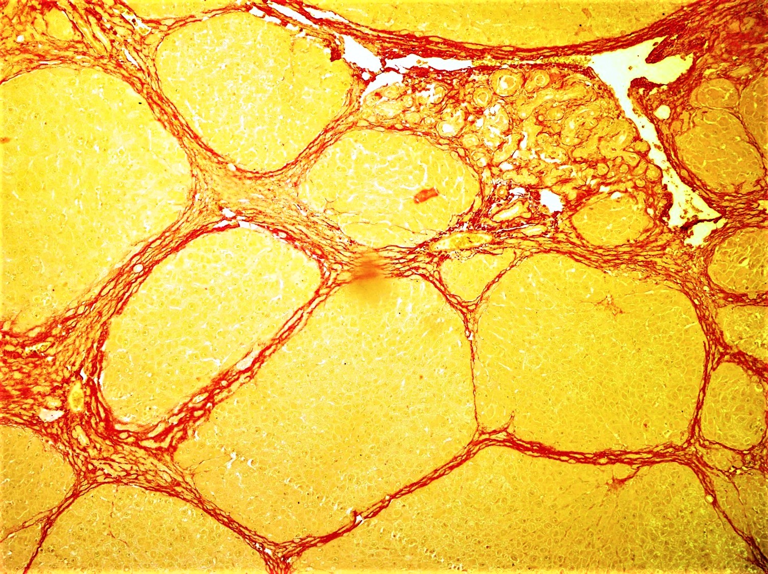
Fibrosis, also known as fibrotic scarring, is a pathological wound healing in which connective tissue replaces normal parenchymal tissue to the extent that it goes unchecked, leading to considerable tissue remodelling and the formation of permanent scar tissue. Repeated injuries, chronic inflammation and repair are susceptible to fibrosis where an accidental excessive accumulation of extracellular matrix components, such as the collagen is produced by fibroblasts, leading to the formation of a permanent fibrotic scar. In response to injury, this is called scarring, and if fibrosis arises from a single cell line, this is called a fibroma. Physiologically, fibrosis acts to deposit connective tissue, which can interfere with or totally inhibit the normal architecture and function of the underlying organ or tissue. Fibrosis can be used to describe the pathological state of excess deposition of fibrous tissue, as well as the process of connective tissue deposition in healing. Define ... [...More Info...] [...Related Items...] OR: [Wikipedia] [Google] [Baidu] |
Septo-optic Dysplasia
Septo-optic dysplasia (SOD), known also as de Morsier syndrome, is a rare congenital malformation syndrome that features a combination of the underdevelopment of the optic nerve, pituitary gland dysfunction, and absence of the septum pellucidum (a midline part of the brain). Two or more of these features need to be present for a clinical diagnosis — only 30% of patients have all three. French-Swiss doctor Georges de Morsier first recognized the relation of a rudimentary or absent septum pellucidum with hypoplasia of the optic nerves and chiasm in 1956. Signs and symptoms The symptoms of SOD can be divided into those related to optic nerve underdevelopment, pituitary hormone abnormalities, or mid-line brain abnormalities. Symptoms may vary greatly in their severity. Optic nerve underdevelopment About one quarter of people with SOD have significant visual impairment in one or both eyes, as a result of optic nerve underdevelopment. Developmental delays are more common in chi ... [...More Info...] [...Related Items...] OR: [Wikipedia] [Google] [Baidu] |
Optic Nerve Hypoplasia
Optic nerve hypoplasia (ONH) is a medical condition arising from the underdevelopment of the optic nerve(s). This condition is the most common congenital optic nerve anomaly. The optic disc appears abnormally small, because not all the optic nerve axons have developed properly.Sadun, Alfredo A., and Michelle Y. Wang. ''Handbook of Clinical Neurology''. p. 37. In press. It is often associated with endocrinopathies (hormone deficiencies), developmental delay, and brain malformations. The optic nerve, which is responsible for transmitting visual signals from the retina to the brain, has approximately 1.2 million nerve fibers in the average person. In those diagnosed with ONH, however, there are noticeably fewer nerves. Symptoms ONH may be found in isolation or in conjunction with myriad functional and anatomic abnormalities of the central nervous system. Nearly 80% of those affected with ONH will experience hypothalamic dysfunction and/or impaired development of the brain, regardless ... [...More Info...] [...Related Items...] OR: [Wikipedia] [Google] [Baidu] |
Morpholino
A Morpholino, also known as a Morpholino oligomer and as a phosphorodiamidate Morpholino oligomer (PMO), is a type of oligomer molecule (colloquially, an oligo) used in molecular biology to modify gene expression. Its molecular structure contains DNA bases attached to a backbone of methylenemorpholine rings linked through phosphorodiamidate groups. Morpholinos block access of other molecules to small (~25 base) specific sequences of the base-pairing surfaces of ribonucleic acid (RNA). Morpholinos are used as research tools for reverse genetics by knocking down gene function. This article discusses only the Morpholino antisense oligomers, which are nucleic acid analogs. The word "Morpholino" can occur in other chemical names, referring to chemicals containing a six-membered morpholine ring. To help avoid confusion with other morpholine-containing molecules, when describing oligos "Morpholino" is often capitalized as a trade name, but this usage is not consistent across scienti ... [...More Info...] [...Related Items...] OR: [Wikipedia] [Google] [Baidu] |
Gene Knockdown
Gene knockdown is an experimental technique by which the expression of one or more of an organism's genes is reduced. The reduction can occur either through genetic modification or by treatment with a reagent such as a short DNA or RNA oligonucleotide that has a sequence complementary to either gene or an mRNA transcript. Versus transient knockdown If a DNA of an organism is genetically modified, the resulting organism is called a "knockdown organism." If the change in gene expression is caused by an oligonucleotide binding to an mRNA or temporarily binding to a gene, this leads to a temporary change in gene expression that does not modify the chromosomal DNA, and the result is referred to as a "transient knockdown". In a transient knockdown, the binding of this oligonucleotide to the active gene or its transcripts causes decreased expression through a variety of processes. Binding can occur either through the blocking of transcription (in the case of gene-binding), the degradati ... [...More Info...] [...Related Items...] OR: [Wikipedia] [Google] [Baidu] |
Holoprosencephaly
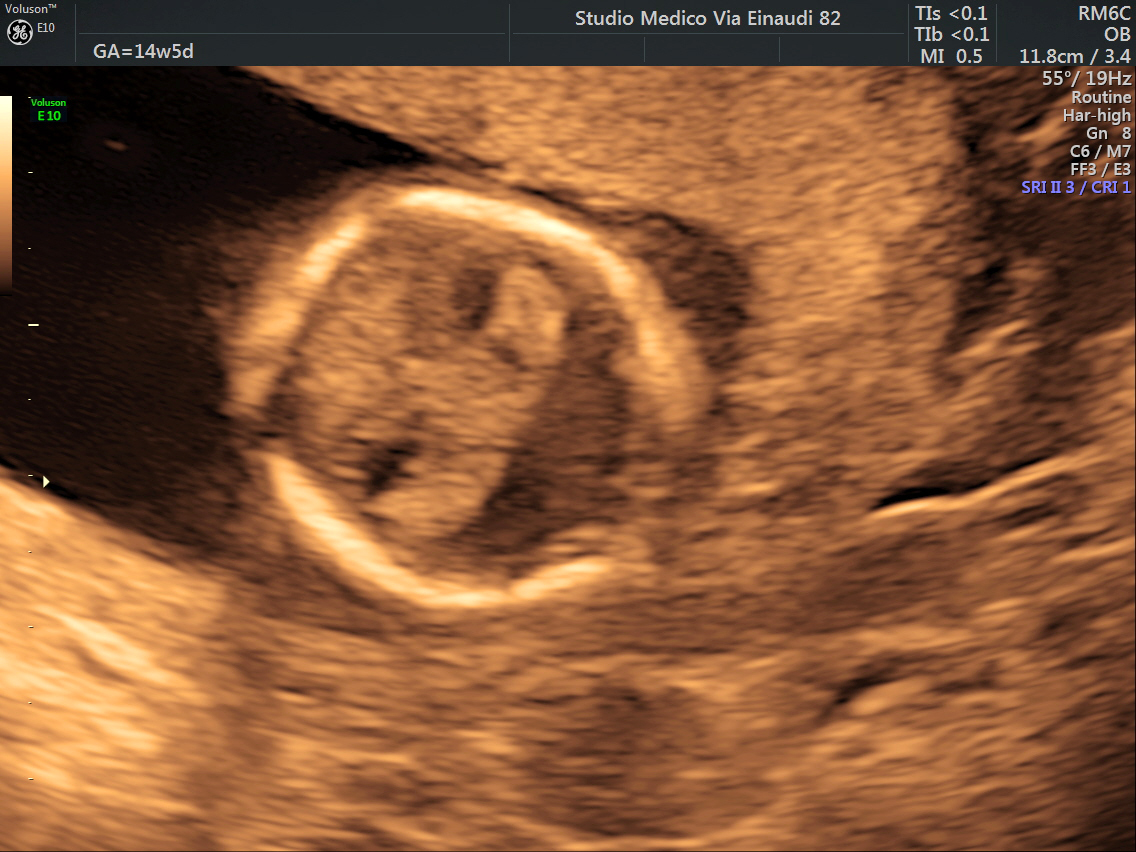
Holoprosencephaly (HPE) is a cephalic disorder in which the prosencephalon (the forebrain of the embryo) fails to develop into two hemispheres, typically occurring between the 18th and 28th day of gestation. Normally, the forebrain is formed and the face begins to develop in the fifth and sixth weeks of human pregnancy. The condition also occurs in other species. Holoprosencephaly is estimated to occur in approximately 1 in every 250 conceptions and most cases are not compatible with life and result in fetal death in utero due to deformities to the skull and brain. However, holoprosencephaly is still estimated to occur in approximately 1 in every 8,000 live births. When the embryo's forebrain does not divide to form bilateral cerebral hemispheres (the left and right halves of the brain), it causes defects in the development of the face and in brain structure and function. The severity of holoprosencephaly is highly variable. In less severe cases, babies are born with normal or ... [...More Info...] [...Related Items...] OR: [Wikipedia] [Google] [Baidu] |


