|
CDA Type III
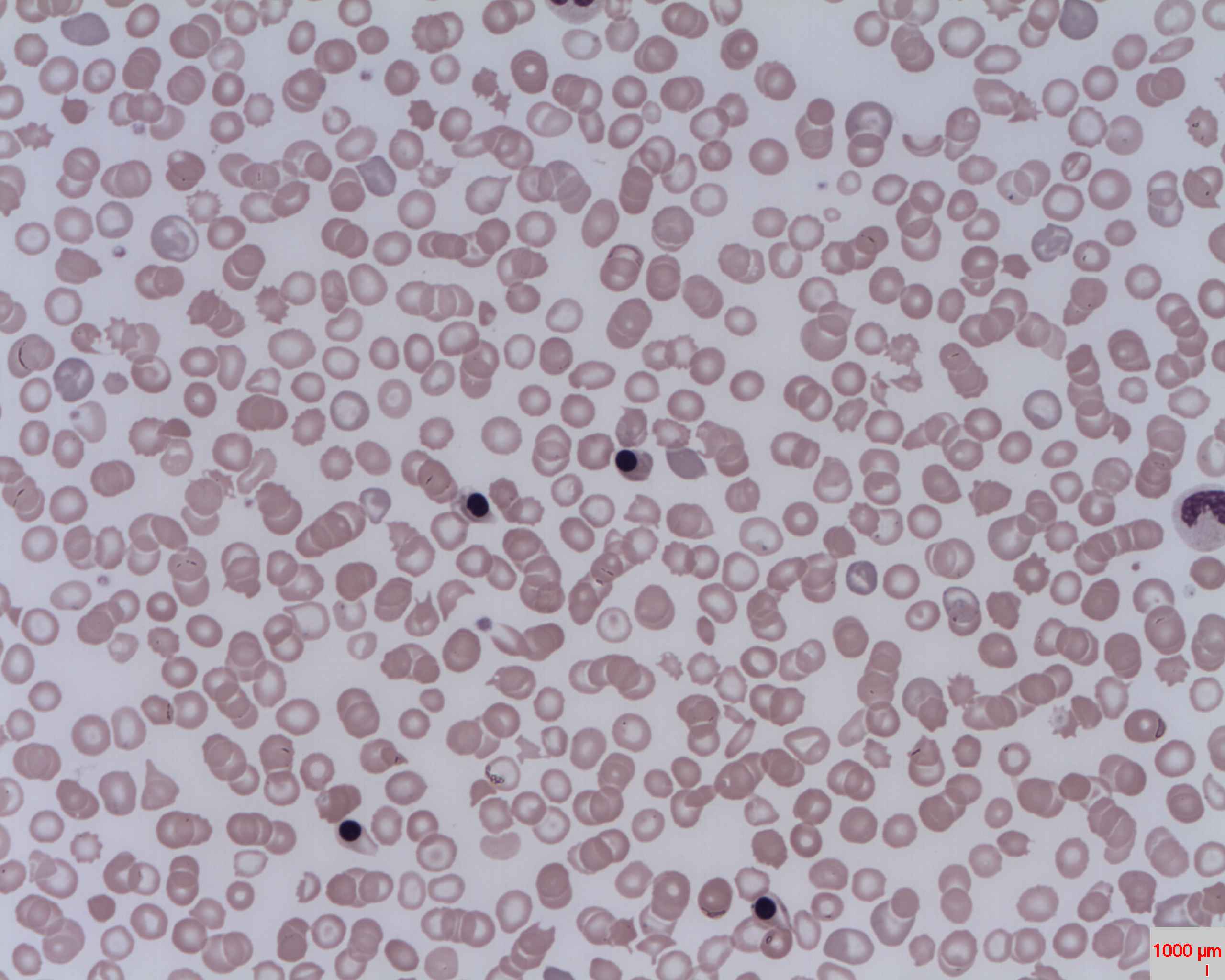
Congenital dyserythropoietic anemia type III (CDA III) is a rare autosomal dominant disorder characterized by macrocytic anemia, bone marrow erythroid hyperplasia and giant multinucleate erythroblasts. New evidence suggests that this may be passed on recessively as well. Presentation The signs and symptoms of CDA type III tend to be milder than those of the other types. Most affected individuals do not have hepatosplenomegaly, and iron does not build up in tissues and organs. In adulthood, abnormalities of a specialized tissue at the back of the eye (the retina) can cause vision impairment. Some people with CDA type III also have a blood disorder known as monoclonal gammopathy, which can lead to a cancer of white blood cells (multiple myeloma). - Genetic Home References ... [...More Info...] [...Related Items...] OR: [Wikipedia] [Google] [Baidu] |
Macrocytic Anemia
The term ''macrocytic'' is from Greek words meaning "large cell". A macrocytic class of anemia is an ''anemia'' (defined as blood with an insufficient concentration of hemoglobin) in which the red blood cells (erythrocytes) are larger than their normal volume. The normal erythrocyte volume in humans is about 80 to 100 femtoliters (fL= 10−15 L). In metric terms the size is given in equivalent cubic micrometers (1 μm3 = 1 fL). The condition of having erythrocytes which (on average) are too large, is called macrocytosis. In contrast, in microcytic anemia, the erythrocytes are smaller than normal. In a macrocytic anemia, the larger red cells are always associated with insufficient ''numbers'' of cells and often also insufficient hemoglobin content per cell. Both of these factors work to the opposite effect of larger cell size, to finally result in a ''total blood hemoglobin concentration'' that is less than normal (i.e., anemia). Macrocytic anemia is not a disease in the sense of h ... [...More Info...] [...Related Items...] OR: [Wikipedia] [Google] [Baidu] |
Hyperplasia
Hyperplasia (from ancient Greek ὑπέρ ''huper'' 'over' + πλάσις ''plasis'' 'formation'), or hypergenesis, is an enlargement of an organ or tissue caused by an increase in the amount of organic tissue that results from cell proliferation. It may lead to the gross enlargement of an organ, and the term is sometimes confused with benign neoplasia or benign tumor. Hyperplasia is a common preneoplastic response to stimulus. Microscopically, cells resemble normal cells but are increased in numbers. Sometimes cells may also be increased in size (hypertrophy). Hyperplasia is different from hypertrophy in that the adaptive cell change in hypertrophy is an increase in the ''size'' of cells, whereas hyperplasia involves an increase in the ''number'' of cells. Causes Hyperplasia may be due to any number of causes, including proliferation of basal layer of epidermis to compensate skin loss, chronic inflammatory response, hormonal dysfunctions, or compensation for damage o ... [...More Info...] [...Related Items...] OR: [Wikipedia] [Google] [Baidu] |
Multinucleate
Multinucleate cells (also known as multinucleated or polynuclear cells) are eukaryotic cells that have more than one nucleus per cell, i.e., multiple nuclei share one common cytoplasm. Mitosis in multinucleate cells can occur either in a coordinated, synchronous manner where all nuclei divide simultaneously or asynchronously where individual nuclei divide independently in time and space. Certain organisms may have a multinuclear stage of their life cycle. For example, slime molds have a vegetative, multinucleate life stage called a plasmodium. Although not normally viewed as a case of multinucleation, plant cells share a common cytoplasm by plasmodesmata, and most cells in animal tissues are in communication with their neighbors via gap junctions. Multinucleate cells, depending on the mechanism by which they are formed, can be divided into "syncytia" (formed by cell fusion) or "coenocytes" (formed by nuclear division not being followed by cytokinesis). A number of dinoflagellat ... [...More Info...] [...Related Items...] OR: [Wikipedia] [Google] [Baidu] |
Erythroblast
A nucleated red blood cell (NRBC), also known by several other names, is a red blood cell that contains a cell nucleus. Almost all vertebrate organisms have hemoglobin-containing cells in their blood, and with the exception of mammals, all of these red blood cells are nucleated. In mammals, NRBCs occur in normal development as precursors to mature red blood cells in erythropoiesis, the process by which the body produces red blood cells. NRBCs are normally found in the bone marrow of humans of all ages and in the blood of fetuses and newborn infants. After infancy, RBCs normally contain a nucleus only during the very early stages of the cell's life, and the nucleus is ejected as a normal part of cellular differentiation before the cell is released into the bloodstream. Thus, if NRBCs are identified on an adult's complete blood count or peripheral blood smear, it suggests that there is a very high demand for the bone marrow to produce RBCs, and immature RBCs are being released i ... [...More Info...] [...Related Items...] OR: [Wikipedia] [Google] [Baidu] |
OMIM
Online Mendelian Inheritance in Man (OMIM) is a continuously updated catalog of human genes and genetic disorders and traits, with a particular focus on the gene-phenotype relationship. , approximately 9,000 of the over 25,000 entries in OMIM represented phenotypes; the rest represented genes, many of which were related to known phenotypes. Versions and history OMIM is the online continuation of Dr. Victor A. McKusick's ''Mendelian Inheritance in Man'' (MIM), which was published in 12 editions between 1966 and 1998.McKusick, V. A. ''Mendelian Inheritance in Man. Catalogs of Autosomal Dominant, Autosomal Recessive and X-Linked Phenotypes.'' Baltimore, MD: Johns Hopkins University Press, 1st ed, 1996; 2nd ed, 1969; 3rd ed, 1971; 4th ed, 1975; 5th ed, 1978; 6th ed, 1983; 7th ed, 1986; 8th ed, 1988; 9th ed, 1990; 10th ed, 1992. Nearly all of the 1,486 entries in the first edition of MIM discussed phenotypes. MIM/OMIM is produced and curated at the Johns Hopkins School of Medicine ... [...More Info...] [...Related Items...] OR: [Wikipedia] [Google] [Baidu] |
KIF23
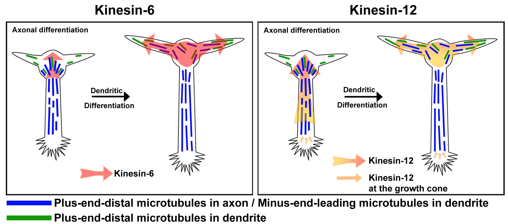
Kinesin-like protein KIF23 is a protein that in humans is encoded by the ''KIF23'' gene. Function In cell division KIF23 (also known as Kinesin-6, CHO1/MKLP1, C. elegans ZEN-4 and Drosophila Pavarotti) is a member of kinesin-like protein family. This family includes microtubule-dependent molecular motors that transport organelles within cells and move chromosomes during cell division. This protein has been shown to cross-bridge antiparallel microtubules and drive microtubule movement in vitro. Alternate splicing of this gene results in two transcript variants encoding two different isoforms, better known as CHO1, the larger isoform and MKLP1, the smaller isoform. KIF23 is a plus-end directed motor protein expressed in mitosis, involved in the formation of the cleavage furrow in late anaphase and in cytokinesis. KIF23 is part of the centralspindlin complex that includes PRC1, Aurora B and 14-3-3 which cluster together at the spindle midzone to enable anaphase in dividing ce ... [...More Info...] [...Related Items...] OR: [Wikipedia] [Google] [Baidu] |
Blood Transfusions
Blood transfusion is the process of transferring blood products into a person's circulation intravenously. Transfusions are used for various medical conditions to replace lost components of the blood. Early transfusions used whole blood, but modern medical practice commonly uses only components of the blood, such as red blood cells, white blood cells, plasma, clotting factors and platelets. Red blood cells (RBC) contain hemoglobin, and supply the cells of the body with oxygen. White blood cells are not commonly used during transfusion, but they are part of the immune system, and also fight infections. Plasma is the "yellowish" liquid part of blood, which acts as a buffer, and contains proteins and important substances needed for the body's overall health. Platelets are involved in blood clotting, preventing the body from bleeding. Before these components were known, doctors believed that blood was homogeneous. Because of this scientific misunderstanding, many patients died becau ... [...More Info...] [...Related Items...] OR: [Wikipedia] [Google] [Baidu] |
Chelation Therapy
Chelation therapy is a medical procedure that involves the administration of Chelation, chelating agents to remove heavy metals from the body. Chelation therapy has a long history of use in clinical toxicology and remains in use for some very specific medical treatments, although it is administered under very careful medical supervision due to various inherent risks, including the mobilization of mercury and other metals through the brain and other parts of the body by the use of weak chelating agents that unbind with metals before elimination, exacerbating existing damage. To avoid mobilization, some practitioners of chelation use strong chelators, such as selenium, taken at low doses over a long period of time. Chelation therapy must be administered with care as it has a number of possible side effects, including death. In response to increasing use of chelation therapy as alternative medicine and in circumstances in which the therapy should not be used in conventional medicine ... [...More Info...] [...Related Items...] OR: [Wikipedia] [Google] [Baidu] |
Bone Marrow Transplantation
Hematopoietic stem-cell transplantation (HSCT) is the transplantation of multipotent hematopoietic stem cells, usually derived from bone marrow, peripheral blood, or umbilical cord blood in order to replicate inside of a patient and to produce additional normal blood cells. It may be autologous (the patient's own stem cells are used), allogeneic (the stem cells come from a donor) or syngeneic (from an identical twin). It is most often performed for patients with certain cancers of the blood or bone marrow, such as multiple myeloma or leukemia. In these cases, the recipient's immune system is usually destroyed with radiation or chemotherapy before the transplantation. Infection and graft-versus-host disease are major complications of allogeneic HSCT. HSCT remains a dangerous procedure with many possible complications; it is reserved for patients with life-threatening diseases. As survival following the procedure has increased, its use has expanded beyond cancer to autoimm ... [...More Info...] [...Related Items...] OR: [Wikipedia] [Google] [Baidu] |
Gene Therapy
Gene therapy is a medical field which focuses on the genetic modification of cells to produce a therapeutic effect or the treatment of disease by repairing or reconstructing defective genetic material. The first attempt at modifying human DNA was performed in 1980, by Martin Cline, but the first successful nuclear gene transfer in humans, approved by the National Institutes of Health, was performed in May 1989. The first therapeutic use of gene transfer as well as the first direct insertion of human DNA into the nuclear genome was performed by French Anderson in a trial starting in September 1990. It is thought to be able to cure many genetic disorders or treat them over time. Between 1989 and December 2018, over 2,900 clinical trials were conducted, with more than half of them in phase I.Gene Therapy Cli ... [...More Info...] [...Related Items...] OR: [Wikipedia] [Google] [Baidu] |
Congenital Dyserythropoietic Anemia
Congenital dyserythropoietic anemia (CDA) is a rare blood disorder, similar to the thalassemias. CDA is one of many types of anemia, characterized by ineffective erythropoiesis, and resulting from a decrease in the number of red blood cells (RBCs) in the body and a less than normal quantity of hemoglobin in the blood. CDA may be transmitted by both parents autosomal recessively or dominantly. Signs and symptoms The symptoms and signs of congenital dyserythropoietic anemia are consistent with: * Tiredness (fatigue) * Weakness * Pale skin Types Congenital dyserythropoietic anemia has four different subtypes, CDA Type I, CDA Type II, CDA Type III, and CDA Type IV. CDA type II (CDA II) is the most frequent type of congenital dyserythropoietic anemias. Diagnosis The diagnosis of congenital dyserythropoietic anemia can be done via sequence analysis of the entire coding region, types I, II, III and IV ( is a relatively new form of CDA that had been found, just 4 cases have been r ... [...More Info...] [...Related Items...] OR: [Wikipedia] [Google] [Baidu] |
Thalassemia
Thalassemias are inherited blood disorders characterized by decreased hemoglobin production. Symptoms depend on the type and can vary from none to severe. Often there is mild to severe anemia (low red blood cells or hemoglobin). Anemia can result in feeling tired and pale skin. There may also be bone problems, an enlarged spleen, yellowish skin, and dark urine. Slow growth may occur in children. Thalassemias are genetic disorders inherited from a person's parents. There are two main types, alpha thalassemia and beta thalassemia. The severity of alpha and beta thalassemia depends on how many of the four genes for alpha globin or two genes for beta globin are missing. Diagnosis is typically by blood tests including a complete blood count, special hemoglobin tests, and genetic tests. Diagnosis may occur before birth through prenatal testing. Treatment depends on the type and severity. Treatment for those with more severe disease often includes regular blood transfusions, iron chel ... [...More Info...] [...Related Items...] OR: [Wikipedia] [Google] [Baidu] |


