|
Patella
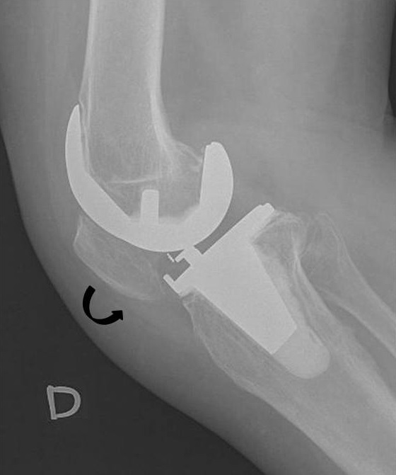
The patella, also known as the kneecap, is a flat, rounded triangular bone which articulates with the femur (thigh bone) and covers and protects the anterior articular surface of the knee joint. The patella is found in many tetrapods, such as mice, cats, birds and dogs, but not in whales, or most reptiles. In humans, the patella is the largest sesamoid bone (i.e., embedded within a tendon or a muscle) in the body. Babies are born with a patella of soft cartilage which begins to ossify into bone at about four years of age. Structure The patella is a sesamoid bone roughly triangular in shape, with the apex of the patella facing downwards. The apex is the most inferior (lowest) part of the patella. It is pointed in shape, and gives attachment to the patellar ligament. The front and back surfaces are joined by a thin margin and towards centre by a thicker margin. The tendon of the quadriceps femoris muscle attaches to the base of the patella., with the vastus intermedius muscle ... [...More Info...] [...Related Items...] OR: [Wikipedia] [Google] [Baidu] |
Patellar Dislocation
A patellar dislocation is a knee injury in which the patella (kneecap) slips out of its normal position. Often the knee is partly bent, painful and swollen. The patella is also often felt and seen out of place. Complications may include a patella fracture or arthritis. A patellar dislocation typically occurs when the knee is straight and the lower leg is bent outwards when twisting. Occasionally it occurs when the knee is bent and the patella is hit. Commonly associated sports include soccer, gymnastics, and ice hockey. Dislocations nearly always occur away from the midline. Diagnosis is typically based on symptoms and supported by X-rays. Reduction is generally done by pushing the patella towards the midline while straightening the knee. After reduction the leg is generally splinted in a straight position for a few weeks. This is then followed by physical therapy. Surgery after a first dislocation is generally of unclear benefit. Surgery may be indicated in those who have bro ... [...More Info...] [...Related Items...] OR: [Wikipedia] [Google] [Baidu] |
Patella Baja
The patella, also known as the kneecap, is a flat, rounded triangular bone which articulates with the femur (thigh bone) and covers and protects the anterior articular surface of the knee joint. The patella is found in many tetrapods, such as mice, cats, birds and dogs, but not in whales, or most reptiles. In humans, the patella is the largest sesamoid bone (i.e., embedded within a tendon or a muscle) in the body. Babies are born with a patella of soft cartilage which begins to ossify into bone at about four years of age. Structure The patella is a sesamoid bone roughly triangular in shape, with the apex of the patella facing downwards. The apex is the most inferior (lowest) part of the patella. It is pointed in shape, and gives attachment to the patellar ligament. The front and back surfaces are joined by a thin margin and towards centre by a thicker margin. The tendon of the quadriceps femoris muscle attaches to the base of the patella., with the vastus intermedius muscle ... [...More Info...] [...Related Items...] OR: [Wikipedia] [Google] [Baidu] |
Bipartite Patella
Bipartite patella is a condition where the patella, or kneecap, is composed of two separate bones. Instead of fusing together as normally occurs in early childhood, the bones of the patella remain separated. The condition occurs in approximately 12% of the population and is no more likely to occur in males than females. It is often asymptomatic and most commonly diagnosed as an incidental finding, with about 2% of cases becoming symptomatic Signs and symptoms are the observed or detectable signs, and experienced symptoms of an illness, injury, or condition. A sign for example may be a higher or lower temperature than normal, raised or lowered blood pressure or an abnormality showi .... Saupe introduced a classification system for Bipartite Patella back in 1921. Type 1: Fragment is located at the bottom of the kneecap (5% of cases) Type 2: Fragment is located on the lateral side of the kneecap (20% of cases) Type 3: Fragment is located on the upper lateral border of the knee ... [...More Info...] [...Related Items...] OR: [Wikipedia] [Google] [Baidu] |
Patella Bipartita
Bipartite patella is a condition where the patella, or kneecap, is composed of two separate bones. Instead of fusing together as normally occurs in early childhood, the bones of the patella remain separated. The condition occurs in approximately 12% of the population and is no more likely to occur in males than females. It is often asymptomatic and most commonly diagnosed as an incidental finding, with about 2% of cases becoming symptomatic Signs and symptoms are the observed or detectable signs, and experienced symptoms of an illness, injury, or condition. A sign for example may be a higher or lower temperature than normal, raised or lowered blood pressure or an abnormality showin .... Saupe introduced a classification system for Bipartite Patella back in 1921. Type 1: Fragment is located at the bottom of the kneecap (5% of cases) Type 2: Fragment is located on the lateral side of the kneecap (20% of cases) Type 3: Fragment is located on the upper lateral border of the kneec ... [...More Info...] [...Related Items...] OR: [Wikipedia] [Google] [Baidu] |
Knee
In humans and other primates, the knee joins the thigh with the leg and consists of two joints: one between the femur and tibia (tibiofemoral joint), and one between the femur and patella (patellofemoral joint). It is the largest joint in the human body. The knee is a modified hinge joint, which permits flexion and extension as well as slight internal and external rotation. The knee is vulnerable to injury and to the development of osteoarthritis. It is often termed a ''compound joint'' having tibiofemoral and patellofemoral components. (The fibular collateral ligament is often considered with tibiofemoral components.) Structure The knee is a modified hinge joint, a type of synovial joint, which is composed of three functional compartments: the patellofemoral articulation, consisting of the patella, or "kneecap", and the patellar groove on the front of the femur through which it slides; and the medial and lateral tibiofemoral articulations linking the femur, or thigh bone ... [...More Info...] [...Related Items...] OR: [Wikipedia] [Google] [Baidu] |
Patellar Ligament
The patellar tendon is the distal portion of the common tendon of the quadriceps femoris, which is continued from the patella to the tibial tuberosity. It is also sometimes called the patellar ligament as it forms a bone to bone connection when the patella is fully ossified. Structure The patellar tendon is a strong, flat ligament, which originates on the apex of the patella distally and adjoining margins of the patella and the rough depression on its posterior surface; below, it inserts on the tuberosity of the tibia; its superficial fibers are continuous over the front of the patella with those of the tendon of the quadriceps femoris. It is about 4.5 cm long in adults (range from 3 to 6 cm). The medial and lateral portions of the quadriceps tendon pass down on either side of the patella to be inserted into the upper extremity of the tibia on either side of the tuberosity; these portions merge into the capsule, as stated above, forming the medial and lateral patellar retina ... [...More Info...] [...Related Items...] OR: [Wikipedia] [Google] [Baidu] |
Quadriceps Tendon
In human anatomy, the quadriceps tendon works with the quadriceps muscle to extend the leg. All four parts of the quadriceps muscle attach to the shin via the patella (knee cap), where the quadriceps tendon becomes the patellar ligament. It attaches the quadriceps to the top of the patella, which in turn is connected to the shin from its bottom by the patellar ligament. A tendon connects muscle to bone, while a ligament connects bone to bone.Saladin, Kenneth S. Anatomy & Physiology: The Unity of Form and Function. 6th ed. New York: McGraw-Hill, 2012. Print. Injuries are common to this tendon, with tears, either partial or complete, being the most common. If the quadriceps tendon is completely torn, surgery will be required to regain function of the knee."Patellar Tendon Tear." OrthoInfo - AAOS. American Academy of Orthopaedic Surgeons, Aug. 2009. Web. 07 Dec. 2014. Without the quadriceps tendon, the knee cannot extend. Often, when the tendon is completely torn, part of the kneec ... [...More Info...] [...Related Items...] OR: [Wikipedia] [Google] [Baidu] |
Vastus Lateralis
The vastus lateralis (), also called the vastus externus, is the largest and most powerful part of the quadriceps femoris, a muscle in the thigh. Together with other muscles of the quadriceps group, it serves to extend the knee joint, moving the lower leg forward. It arises from a series of flat, broad tendons attached to the femur, and attaches to the outer border of the patella. It ultimately joins with the other muscles that make up the quadriceps in the quadriceps tendon, which travels over the knee to connect to the tibia. The vastus lateralis is the recommended site for intramuscular injection in infants less than 7 months old and those unable to walk, with loss of muscular tone.Mann, E. (2016). ''Injection (Intramuscular): Clinician Information.'' The Johanna Briggs Institute. Structure The vastus lateralis muscle arises from several areas of the femur, including the upper part of the intertrochanteric line; the lower, anterior borders of the greater trochanter, to the out ... [...More Info...] [...Related Items...] OR: [Wikipedia] [Google] [Baidu] |
Exostosis
An exostosis, also known as bone spur, is the formation of new bone on the surface of a bone. Exostoses can cause chronic pain ranging from mild to debilitatingly severe, depending on the shape, size, and location of the lesion. It is most commonly found in places like the ribs, where small bone growths form, but sometimes larger growths can grow on places like the ankles, knees, shoulders, elbows and hips. Very rarely are they on the skull. Exostoses are sometimes shaped like spurs, such as calcaneal spurs. Osteomyelitis, a bone infection, may leave the adjacent bone with exostosis formation. Charcot foot, the neuropathic breakdown of the feet seen primarily in diabetics, can also leave bone spurs that may then become symptomatic. They normally form on the bones of joints, and can grow upwards. For example, if an extra bone formed on the ankle, it might grow up to the shin. When used in the phrases "cartilaginous exostosis" or "osteocartilaginous exostosis", the term is consid ... [...More Info...] [...Related Items...] OR: [Wikipedia] [Google] [Baidu] |
Vascular
The blood vessels are the components of the circulatory system that transport blood throughout the human body. These vessels transport blood cells, nutrients, and oxygen to the tissues of the body. They also take waste and carbon dioxide away from the tissues. Blood vessels are needed to sustain life, because all of the body's tissues rely on their functionality. There are five types of blood vessels: the arteries, which carry the blood away from the heart; the arterioles; the capillaries, where the exchange of water and chemicals between the blood and the tissues occurs; the venules; and the veins, which carry blood from the capillaries back towards the heart. The word ''vascular'', meaning relating to the blood vessels, is derived from the Latin ''vas'', meaning vessel. Some structures – such as cartilage, the epithelium, and the lens and cornea of the eye – do not contain blood vessels and are labeled ''avascular''. Etymology * artery: late Middle English; from Latin ' ... [...More Info...] [...Related Items...] OR: [Wikipedia] [Google] [Baidu] |
Canaliculus (bone)
Bone canaliculi are microscopic canals between the lacunae of ossified bone. The radiating processes of the osteocytes (called filopodia) project into these canals. These cytoplasmic processes are joined together by gap junctions. Osteocytes do not entirely fill up the canaliculi. The remaining space is known as the periosteocytic space, which is filled with periosteocytic fluid. This fluid contains substances too large to be transported through the gap junctions that connect the osteocytes. In cartilage, the lacunae and hence, the chondrocytes, are isolated from each other. Materials picked up by osteocytes adjacent to blood vessels are distributed throughout the bone matrix via the canaliculi. Dental canaliculi The dental canaliculi (sometimes called dentinal tubules) are the blood supply of a tooth. Odontoblast process run in the canaliculi that transverse the dentin layer and are referred as dentinal tubules. The number and size of the canaliculi decrease as the tubul ... [...More Info...] [...Related Items...] OR: [Wikipedia] [Google] [Baidu] |
Hypoplasia
Hypoplasia (from Ancient Greek ὑπo- ''hypo-'' 'under' + πλάσις ''plasis'' 'formation'; adjective form ''hypoplastic'') is underdevelopment or incomplete development of a tissue or organ. Dictionary of Cell and Molecular Biology (11 March 2008) Although the term is not always used precisely, it properly refers to an inadequate or below-normal number of cells.Hypoplasia Stedman's Medical Dictionary. lww.com Hypoplasia is similar to |


