|
Cytochrome B5 Reductase
Cytochrome-''b''5 reductase is a NADH-dependent enzyme that converts ferricytochrome from a Fe3+ form to a Fe2+ form. It contains flavin adenine dinucleotide, FAD and catalyzes the reaction: In its b5-reducing capacity, this enzyme is involved in desaturation and elongation of fatty acids, cholesterol biosynthesis, and drug metabolism. This enzyme can also reduce methemoglobin to normal hemoglobin, gaining it the inaccurate synonym methemoglobin reductase. Isoforms expressed in erythrocytes (CYB5R1, CYB5R3) perform this function ''in vivo''. Ferricyanide is another substrate ''in vitro''. The following four human genes encode cytochrome-''b''5 reductases: * CYB5R1 * CYB5R2 * CYB5R3 * CYB5R4 * CYB5RL See also * Cytochrome b5 * Diaphorase * Methemoglobinemia * Reductase * Leghemoglobin reductase References External links * {{Portal bar, Biology, border=no EC 1.6.2 ... [...More Info...] [...Related Items...] OR: [Wikipedia] [Google] [Baidu] |
Ribbon Diagram
Ribbon diagrams, also known as Richardson diagrams, are three-dimensional space, 3D schematic representations of protein structure and are one of the most common methods of protein depiction used today. The ribbon shows the overall path and organization of the protein backbone in 3D, and serves as a visual framework on which to hang details of the full atomic structure, such as the balls for the oxygen atoms bound to the active site of myoglobin in the adjacent image. Ribbon diagrams are generated by interpolating a smooth curve through the polypeptide backbone. Alpha helix, α-helices are shown as coiled ribbons or thick tubes, Beta strand, β-strands as arrows, and non-repetitive coils or loops as lines or thin tubes. The direction of the Peptide, polypeptide chain is shown locally by the arrows, and may be indicated overall by a colour ramp along the length of the ribbon. Ribbon diagrams are simple yet powerful, expressing the visual basics of a molecular structure (twist, fold ... [...More Info...] [...Related Items...] OR: [Wikipedia] [Google] [Baidu] |
CYB5R3
NADH-cytochrome b5 reductase 3 is an enzyme that in humans is encoded by the ''CYB5R3'' gene. Structure The CYB5R3 gene is located on the 22nd chromosome, with its specific location being 22q13.2. The gene contains 12 exons. CYB5R3 encodes a 34.2 kDa protein that is composed of 301 amino acids; 63 peptides have been observed through mass spectrometry data. The entire gene is about 31 kb in length. Exon 2 contains the junction of the membrane-binding domain and the catalytic domain of b5R, which shows that there are two forms of b5R: a soluble form and a membrane-bound form. The 5' portion of this gene does not have typical regulatory transcriptional elements, but has the sequence G-G-G-C-G-G a total of five times. The GC content of this 5' portion of the gene is 86%, much higher than the average GC of the entire gene, which is 55%. There is also an atypical polyadenylation signal in the 3'-untranslated region of the gene. The protein encoded by the CYB5R3 gene is cytochrome b5 re ... [...More Info...] [...Related Items...] OR: [Wikipedia] [Google] [Baidu] |
Reductase
A reductase is an enzyme that catalyzes a reduction reaction. Examples * 5α-Reductase * 5β-Reductase * Dihydrofolate reductase * HMG-CoA reductase * Methemoglobin reductase * Ribonucleotide reductase * Thioredoxin reductase * ''E. coli'' nitroreductase * Methylenetetrahydrofolate reductase See also * Oxidase * Oxidoreductase In biochemistry, an oxidoreductase is an enzyme that catalyzes the transfer of electrons from one molecule, the reductant, also called the electron donor, to another, the oxidant, also called the electron acceptor. This group of enzymes usually u ... References Oxidoreductases {{Enzyme-stub ... [...More Info...] [...Related Items...] OR: [Wikipedia] [Google] [Baidu] |
Methemoglobinemia
Methemoglobinemia, or methaemoglobinaemia, is a condition of elevated methemoglobin in the blood. Symptoms may include headache, dizziness, shortness of breath, nausea, poor muscle coordination, and blue-colored skin (cyanosis). Complications may include seizures and heart arrhythmias. Methemoglobinemia can be due to certain medications, chemicals, or food or it can be inherited from a person's parents. Substances involved may include benzocaine, nitrates, or dapsone. The underlying mechanism involves some of the iron in hemoglobin being converted from the ferrous e2+to the ferric e3+form. The diagnosis is often suspected based on symptoms and a low blood oxygen that does not improve with oxygen therapy. Diagnosis is confirmed by a blood gas. Treatment is generally with oxygen therapy and methylene blue. Other treatments may include vitamin C, exchange transfusion, and hyperbaric oxygen therapy. Outcomes are generally good with treatment. Methemoglobinemia is relatively u ... [...More Info...] [...Related Items...] OR: [Wikipedia] [Google] [Baidu] |
Diaphorase
Diaphorase may refer to: * Cytochrome b5 reductase, an enzyme * NADH dehydrogenase, an enzyme * NADPH dehydrogenase In enzymology, a NADPH dehydrogenase () is an enzyme that catalysis, catalyzes the chemical reaction :NADPH + H+ + acceptor \rightleftharpoons NADP+ + reduced acceptor The 3 substrate (biochemistry), substrates of this enzyme are nicotinamide ade ..., an enzyme {{Short pages monitor ... [...More Info...] [...Related Items...] OR: [Wikipedia] [Google] [Baidu] |
Cytochrome B5
Cytochromes ''b''5 are ubiquitous electron transport hemoproteins found in animals, plants, fungi and purple phototrophic bacteria. The microsomal and mitochondrial variants are membrane-bound, while bacterial and those from erythrocytes and other animal tissues are water-soluble. The family of cytochrome ''b''5-like proteins includes (besides cytochrome ''b''5 itself) hemoprotein domains covalently associated with other redox domains in flavocytochrome cytochrome ''b''2 (L-lactate dehydrogenase; ), sulfite oxidase (), plant and fungal nitrate reductases (, , ), and plant and fungal cytochrome ''b''5/acyl lipid desaturase fusion proteins. Structure 3-D structures of a number of cytochrome ''b''5 and yeast flavocytochrome ''b''2 are known. The fold belongs to the α+β class, with two hydrophobic cores on each side of a β-sheet. The larger hydrophobic core constitutes the heme-binding pocket, closed off on each side by a pair of helices connected by a turn. The smaller hydro ... [...More Info...] [...Related Items...] OR: [Wikipedia] [Google] [Baidu] |
CYB5R4
Cytochrome b5 reductase 4 is an enzyme that in humans is encoded by the ''CYB5R4'' gene. NCB5OR is a flavohemoprotein that contains functional domains found in both cytochrome Cytochromes are redox-active proteins containing a heme, with a central Fe atom at its core, as a cofactor. They are involved in electron transport chain and redox catalysis. They are classified according to the type of heme and its mode of bi ... b5 (CYB5; MIM 250790) and CYB5 reductase (DIA1; MIM 250800). upplied by OMIMref name="entrez"/> References Further reading * * * * * * * * External links * {{gene-6-stub ... [...More Info...] [...Related Items...] OR: [Wikipedia] [Google] [Baidu] |
CYB5R2
Cytochrome b5 reductase 2 is an enzyme that in humans is encoded by the ''CYB5R2'' gene In biology, the word gene (from , ; "... Wilhelm Johannsen coined the word gene to describe the Mendelian units of heredity..." meaning ''generation'' or ''birth'' or ''gender'') can have several different meanings. The Mendelian gene is a b .... References External links * Further reading * * * * * * * {{gene-11-stub ... [...More Info...] [...Related Items...] OR: [Wikipedia] [Google] [Baidu] |
Red Blood Cell
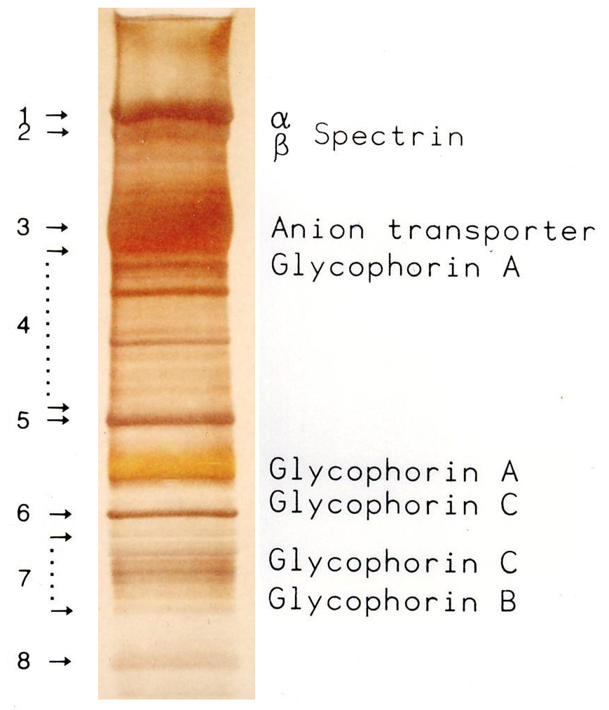
Red blood cells (RBCs), also referred to as red cells, red blood corpuscles (in humans or other animals not having nucleus in red blood cells), haematids, erythroid cells or erythrocytes (from Greek ''erythros'' for "red" and ''kytos'' for "hollow vessel", with ''-cyte'' translated as "cell" in modern usage), are the most common type of blood cell and the vertebrate's principal means of delivering oxygen (O2) to the body tissues—via blood flow through the circulatory system. RBCs take up oxygen in the lungs, or in fish the gills, and release it into tissues while squeezing through the body's capillaries. The cytoplasm of a red blood cell is rich in hemoglobin, an iron-containing biomolecule that can bind oxygen and is responsible for the red color of the cells and the blood. Each human red blood cell contains approximately 270 million hemoglobin molecules. The cell membrane is composed of proteins and lipids, and this structure provides properties essential for physiolo ... [...More Info...] [...Related Items...] OR: [Wikipedia] [Google] [Baidu] |
CYB5R1
NADH-cytochrome b5 reductase 1 is an enzyme that in humans is encoded by the ''CYB5R1'' gene. Structure The CYB5R1 gene is located on the 1st chromosome, with its specific location being 1q32.1. The gene contains 9 exons. CYB5R1 encodes a 34.1 kDa protein that is composed of 305 amino acids; 56 peptides have been observed through mass spectrometry Mass spectrometry (MS) is an analytical technique that is used to measure the mass-to-charge ratio of ions. The results are presented as a ''mass spectrum'', a plot of intensity as a function of the mass-to-charge ratio. Mass spectrometry is use ... data. References External links * Further reading * * * * * Enzymes Genes {{gene-1-stub ... [...More Info...] [...Related Items...] OR: [Wikipedia] [Google] [Baidu] |
Erythrocyte
Red blood cells (RBCs), also referred to as red cells, red blood corpuscles (in humans or other animals not having nucleus in red blood cells), haematids, erythroid cells or erythrocytes (from Greek ''erythros'' for "red" and ''kytos'' for "hollow vessel", with ''-cyte'' translated as "cell" in modern usage), are the most common type of blood cell and the vertebrate's principal means of delivering oxygen (O2) to the body tissues—via blood flow through the circulatory system. RBCs take up oxygen in the lungs, or in fish the gills, and release it into tissues while squeezing through the body's capillaries. The cytoplasm of a red blood cell is rich in hemoglobin, an iron-containing biomolecule that can bind oxygen and is responsible for the red color of the cells and the blood. Each human red blood cell contains approximately 270 million hemoglobin molecules. The cell membrane is composed of proteins and lipids, and this structure provides properties essential for physiologi ... [...More Info...] [...Related Items...] OR: [Wikipedia] [Google] [Baidu] |

2.jpg)

