|
Cyclocryotherapy
Cyclodestruction or cycloablation is a surgical procedure done in management of glaucoma. Cyclodestruction reduce intraocular pressure (IOP) of the eye by decreasing production of aqueous humor by the destruction of ciliary body. Until the development of safer and less destructive techniques like micropulse diode cyclophotocoagulation and endocyclophotocoagulation, cyclodestructive surgeries were mainly done in refractory glaucoma, or advanced glaucomatous eyes with poor visual prognosis. Types Cyclodestruction may be done by using diathermy, penetrating cyclodiathermy, cryotherapy, ultrasound, laser or by surgical excision. Cyclophotocoagulation Cyclophotocoagulation (CPC), the most common cyclodestructive procedure is done using laser beam of different wavelengths. Ruby laser (693 nm wavelength), Nd:YAG laser (1064 nm wavelength) or diode laser (810 nm wavelength) can be used to perform CPC. Commomon cyclophotocoagulation techniques include transscleral cyclopho ... [...More Info...] [...Related Items...] OR: [Wikipedia] [Google] [Baidu] |
Glaucoma
Glaucoma is a group of eye diseases that result in damage to the optic nerve (or retina) and cause vision loss. The most common type is open-angle (wide angle, chronic simple) glaucoma, in which the drainage angle for fluid within the eye remains open, with less common types including closed-angle (narrow angle, acute congestive) glaucoma and normal-tension glaucoma. Open-angle glaucoma develops slowly over time and there is no pain. Peripheral vision may begin to decrease, followed by central vision, resulting in blindness if not treated. Closed-angle glaucoma can present gradually or suddenly. The sudden presentation may involve severe eye pain, blurred vision, mid-dilated pupil, redness of the eye, and nausea. Vision loss from glaucoma, once it has occurred, is permanent. Eyes affected by glaucoma are referred to as being glaucomatous. Risk factors for glaucoma include increasing age, high pressure in the eye, a family history of glaucoma, and use of steroid medication. F ... [...More Info...] [...Related Items...] OR: [Wikipedia] [Google] [Baidu] |
Neovascular Glaucoma
Glaucoma is a group of eye diseases that result in damage to the optic nerve (or retina) and cause vision loss. The most common type is open-angle (wide angle, chronic simple) glaucoma, in which the drainage angle for fluid within the eye remains open, with less common types including closed-angle (narrow angle, acute congestive) glaucoma and normal-tension glaucoma. Open-angle glaucoma develops slowly over time and there is no pain. Peripheral vision may begin to decrease, followed by central vision, resulting in blindness if not treated. Closed-angle glaucoma can present gradually or suddenly. The sudden presentation may involve severe eye pain, blurred vision, mid-dilated pupil, redness of the eye, and nausea. Vision loss from glaucoma, once it has occurred, is permanent. Eyes affected by glaucoma are referred to as being glaucomatous. Risk factors for glaucoma include increasing age, high pressure in the eye, a family history of glaucoma, and use of steroid medication. F ... [...More Info...] [...Related Items...] OR: [Wikipedia] [Google] [Baidu] |
Glaucoma
Glaucoma is a group of eye diseases that result in damage to the optic nerve (or retina) and cause vision loss. The most common type is open-angle (wide angle, chronic simple) glaucoma, in which the drainage angle for fluid within the eye remains open, with less common types including closed-angle (narrow angle, acute congestive) glaucoma and normal-tension glaucoma. Open-angle glaucoma develops slowly over time and there is no pain. Peripheral vision may begin to decrease, followed by central vision, resulting in blindness if not treated. Closed-angle glaucoma can present gradually or suddenly. The sudden presentation may involve severe eye pain, blurred vision, mid-dilated pupil, redness of the eye, and nausea. Vision loss from glaucoma, once it has occurred, is permanent. Eyes affected by glaucoma are referred to as being glaucomatous. Risk factors for glaucoma include increasing age, high pressure in the eye, a family history of glaucoma, and use of steroid medication. F ... [...More Info...] [...Related Items...] OR: [Wikipedia] [Google] [Baidu] |
Ciliary Body
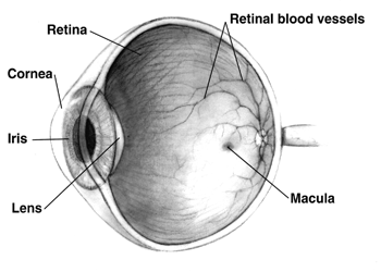
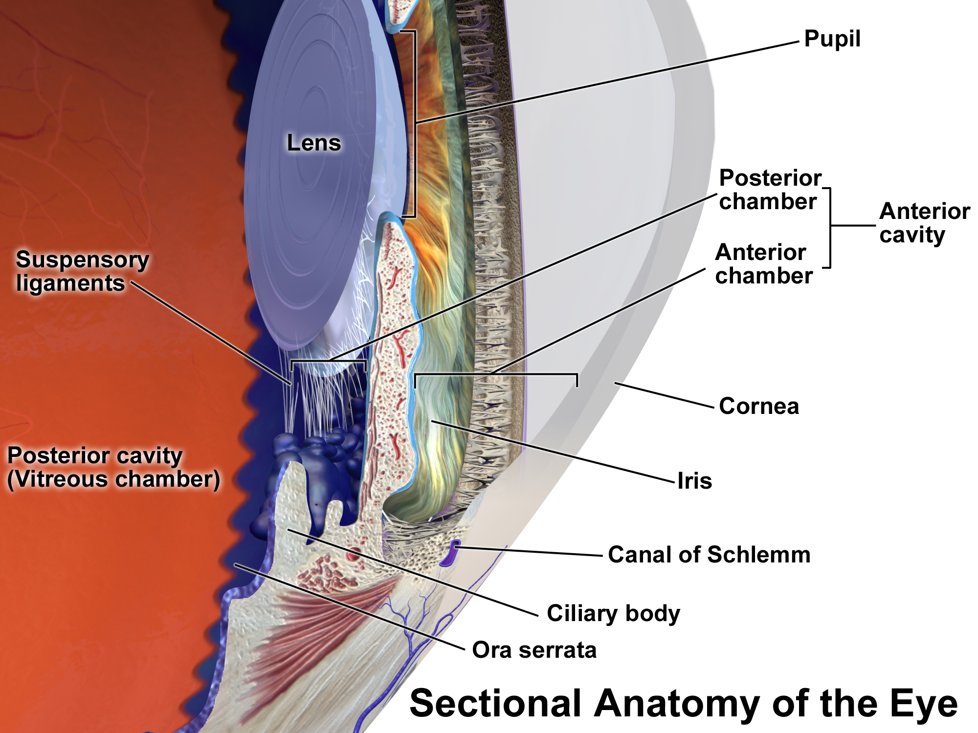
The ciliary body is a part of the eye that includes the ciliary muscle, which controls the shape of the lens, and the ciliary epithelium, which produces the aqueous humor. The aqueous humor is produced in the non-pigmented portion of the ciliary body. The ciliary body is part of the uvea, the layer of tissue that delivers oxygen and nutrients to the eye tissues. The ciliary body joins the ora serrata of the choroid to the root of the iris.Cassin, B. and Solomon, S. ''Dictionary of Eye Terminology''. Gainesville, Florida: Triad Publishing Company, 1990. Structure The ciliary body is a ring-shaped thickening of tissue inside the eye that divides the posterior chamber from the vitreous body. It contains the ciliary muscle, vessels, and fibrous connective tissue. Folds on the inner ciliary epithelium are called ciliary processes, and these secrete aqueous humor into the posterior chamber. The aqueous humor then flows through the pupil into the anterior chamber. The ciliary bo ... [...More Info...] [...Related Items...] OR: [Wikipedia] [Google] [Baidu] |
Anesthesia
Anesthesia is a state of controlled, temporary loss of sensation or awareness that is induced for medical or veterinary purposes. It may include some or all of analgesia (relief from or prevention of pain), paralysis (muscle relaxation), amnesia (loss of memory), and unconsciousness. An individual under the effects of anesthetic drugs is referred to as being anesthetized. Anesthesia enables the painless performance of procedures that would otherwise cause severe or intolerable pain in a non-anesthetized individual, or would otherwise be technically unfeasible. Three broad categories of anesthesia exist: * General anesthesia suppresses central nervous system activity and results in unconsciousness and total lack of sensation, using either injected or inhaled drugs. * Sedation suppresses the central nervous system to a lesser degree, inhibiting both anxiety and creation of long-term memories without resulting in unconsciousness. * Regional and local anesthesia, which blo ... [...More Info...] [...Related Items...] OR: [Wikipedia] [Google] [Baidu] |
Laser Medicine
Laser medicine consists in the use of lasers in medical diagnosis, treatments, or therapies, such as laser photodynamic therapy, photorejuvenation, and laser surgery. Lasers Lasers used in medicine include in principle any type of laser, but especially: * CO2 lasers, used to cut, vaporize, ablate and photo-coagulate soft tissue. * diode lasers * dye lasers * excimer lasers * fiber lasers * gas lasers * free electron lasers * semiconductor diode lasers Applications in medicine Examples of procedures, practices, devices, and specialties where lasers are utilized include: * angioplasty * cancer diagnosis *cancer treatment * Dentistry * cosmetic dermatology such as scar revision, skin resurfacing, laser hair removal, tattoo removal * dermatology, to treat melanoma * frenectomy * lithotripsy *laser mammography * medical imaging * microscopy * ophthalmology (includes Lasik and laser photocoagulation) * optical coherence tomography * optogenetics * prostatectomy * plastic surg ... [...More Info...] [...Related Items...] OR: [Wikipedia] [Google] [Baidu] |
Iridocyclitis
Uveitis () is inflammation of the uvea, the pigmented layer of the eye between the inner retina and the outer fibrous layer composed of the sclera and cornea. The uvea consists of the middle layer of pigmented vascular structures of the eye and includes the iris, ciliary body, and choroid. Uveitis is described anatomically, by the part of the eye affected, as anterior, intermediate or posterior, or panuveitic if all parts are involved. Anterior uveitis ( iridocyclytis) is the most common, with the incidence of uveitis overall affecting approximately 1:4500, most commonly those between the ages of 20-60. Symptoms include eye pain, eye redness, floaters and blurred vision, and ophthalmic examination may show dilated ciliary blood vessels and the presence of cells in the anterior chamber. Uveitis may arise spontaneously, have a genetic component, or be associated with an autoimmune disease or infection. While the eye is a relatively protected environment, its immune mechanisms may ... [...More Info...] [...Related Items...] OR: [Wikipedia] [Google] [Baidu] |
Hyphema
Hyphema is a condition that occurs when blood enters the front (anterior) chamber of the eye between the iris and the cornea. People usually first notice a loss of vision or decrease in vision. The eye may also appear to have a reddish tinge, or it may appear as a small pool of blood at the bottom of the iris or in the cornea. A traumatic hyphema is caused by a hit to the eye from a projected object or a blow to the eye. A hyphema can also occur spontaneously. Presentation A decrease in vision or a loss of vision is often the first sign of a hyphema. People with microhyphema may have slightly blurred or normal vision. A person with a full hyphema may not be able to see at all (complete loss of vision). The person's vision may improve over time as the blood moves by gravity lower in the anterior chamber of the eye, between the iris and the cornea. In many people, the vision will improve, however some people may have other injuries related to trauma to the eye or complications rel ... [...More Info...] [...Related Items...] OR: [Wikipedia] [Google] [Baidu] |
Uveitis
Uveitis () is inflammation of the uvea, the pigmented layer of the eye between the inner retina and the outer fibrous layer composed of the sclera and cornea. The uvea consists of the middle layer of pigmented vascular structures of the eye and includes the iris, ciliary body, and choroid. Uveitis is described anatomically, by the part of the eye affected, as anterior, intermediate or posterior, or panuveitic if all parts are involved. Anterior uveitis ( iridocyclytis) is the most common, with the incidence of uveitis overall affecting approximately 1:4500, most commonly those between the ages of 20-60. Symptoms include eye pain, eye redness, floaters and blurred vision, and ophthalmic examination may show dilated ciliary blood vessels and the presence of cells in the anterior chamber. Uveitis may arise spontaneously, have a genetic component, or be associated with an autoimmune disease or infection. While the eye is a relatively protected environment, its immune mechanisms ... [...More Info...] [...Related Items...] OR: [Wikipedia] [Google] [Baidu] |
Phthisis Bulbi
Phthisis bulbi is a shrunken, non-functional eye. It may result from severe eye disease, inflammation or injury, or it may represent a complication of eye surgery. Treatment options include insertion of a prosthesis, which may be preceded by enucleation of the eye. Symptoms The affected eye is shrunken, and has little to no vision. The intraocular pressure in the affected eye is very low or nonexistent. The layers in the eye may be fused together, thickened, or edematous. The eyelids may be glued shut. The eye may be soft when palpated. Under a microscope there may be deposits of calcium or bone, and the lens is often affected by cataract A cataract is a cloudy area in the lens of the eye that leads to a decrease in vision. Cataracts often develop slowly and can affect one or both eyes. Symptoms may include faded colors, blurry or double vision, halos around light, trouble w ...s. Causes It can be caused by injury, including burns to the eye, or long-term eye diseas ... [...More Info...] [...Related Items...] OR: [Wikipedia] [Google] [Baidu] |
Hypotony
Ocular hypotony, or ocular hypotension, or shortly hypotony, is the medical condition in which intraocular pressure (IOP) of the eye is very low. Description Normal IOP ranges between 10–20 mm Hg. The eye is considered hypotonous if the IOP is ≤5 mm Hg (some sources say IOP less than 6.5 mmHg). Types Ocular hypotony is divided into statistical and clinical types. If intraocular pressure is low (less than 6.5 mm Hg) it is called statistical hypotony, and if the reduced IOP causes a decrease in vision, it is called clinical. Causes Hypotony may occur either due to decreased production of aqueous humor or due to increased outflow. Hypotony has many causes including post-surgical wound leak from the eye, chronic inflammation within the eye including iridocyclitis, hypoperfusion, tractional ciliary body detachment or retinal detachment. Eye inflammation, medications including anti glaucoma drugs, or proliferative vitreoretinopathy causes decreased production. Increased outflow or ... [...More Info...] [...Related Items...] OR: [Wikipedia] [Google] [Baidu] |
Retinal Detachment
Retinal detachment is a disorder of the eye in which the retina peels away from its underlying layer of support tissue. Initial detachment may be localized, but without rapid treatment the entire retina may detach, leading to vision loss and blindness. It is a surgical emergency. The retina is a thin layer of light-sensitive tissue on the back wall of the eye. The optical system of the eye focuses light on the retina much like light is focused on the film in a camera. The retina translates that focused image into neural impulses and sends them to the brain via the optic nerve. Occasionally, posterior vitreous detachment, injury or trauma to the eye or head may cause a small tear in the retina. The tear allows vitreous fluid to seep through it under the retina, and peel it away like a bubble in wallpaper. Diagnosis Symptoms As the retina is responsible for vision, persons experiencing a retinal detachment have vision loss. This can be painful or painless. Imaging Ultraso ... [...More Info...] [...Related Items...] OR: [Wikipedia] [Google] [Baidu] |


