|
Crypts Of Henle
Crypts of Henle are microscopic pockets found in scattered sections of the conjunctiva around the eyeball. They are responsible for secreting mucin, a proteinous substance that makes up the inner layer of tears. It coats the cornea to provide a hydrophilic layer that allows for even distribution of the tear film. The layer of mucin allows tears to glide evenly across the eye's surface. The crypts of Henle are named after German anatomist Anatomy () is the branch of biology concerned with the study of the structure of organisms and their parts. Anatomy is a branch of natural science that deals with the structural organization of living things. It is an old science, having it ... Friedrich Gustav Jakob Henle (1809-1885). Another anatomical structure called the ''glands of Manz'' perform a similar function. They are located in the eyeball's conjunctiva, arranged in a ring around the cornea, near the scleral junction. They also are responsible for secreting mucin into tears. ... [...More Info...] [...Related Items...] OR: [Wikipedia] [Google] [Baidu] |
Conjunctiva
The conjunctiva is a thin mucous membrane that lines the inside of the eyelids and covers the sclera (the white of the eye). It is composed of non-keratinized, stratified squamous epithelium with goblet cells, stratified columnar epithelium and stratified cuboidal epithelium (depending on the zone). The conjunctiva is highly vascularised, with many microvessels easily accessible for imaging studies. Structure The conjunctiva is typically divided into three parts: Blood supply Blood to the bulbar conjunctiva is primarily derived from the ophthalmic artery. The blood supply to the palpebral conjunctiva (the eyelid) is derived from the external carotid artery. However, the circulations of the bulbar conjunctiva and palpebral conjunctiva are linked, so both bulbar conjunctival and palpebral conjunctival vessels are supplied by both the ophthalmic artery and the external carotid artery, to varying extents. Nerve supply Sensory innervation of the conjunctiva is divided into ... [...More Info...] [...Related Items...] OR: [Wikipedia] [Google] [Baidu] |
Human Eyeball
The human eye is a sensory organ, part of the sensory nervous system, that reacts to visible light and allows humans to use visual information for various purposes including seeing things, keeping balance, and maintaining circadian rhythm. The eye can be considered as a living optical device. It is approximately spherical in shape, with its outer layers, such as the outermost, white part of the eye (the sclera) and one of its inner layers (the pigmented choroid) keeping the eye essentially light tight except on the eye's optic axis. In order, along the optic axis, the optical components consist of a first lens (the cornea—the clear part of the eye) that accomplishes most of the focussing of light from the outside world; then an aperture (the pupil) in a diaphragm (the iris—the coloured part of the eye) that controls the amount of light entering the interior of the eye; then another lens (the crystalline lens) that accomplishes the remaining focussing of light into ... [...More Info...] [...Related Items...] OR: [Wikipedia] [Google] [Baidu] |
Mucin
Mucins () are a family of high molecular weight, heavily glycosylated proteins (glycoconjugates) produced by epithelial tissues in most animals. Mucins' key characteristic is their ability to form gels; therefore they are a key component in most gel-like secretions, serving functions from lubrication to cell signalling to forming chemical barriers. They often take an inhibitory role. Some mucins are associated with controlling mineralization, including nacre formation in mollusks, calcification in echinoderms and bone formation in vertebrates. They bind to pathogens as part of the immune system. Overexpression of the mucin proteins, especially MUC1, is associated with many types of cancer. Although some mucins are membrane-bound due to the presence of a hydrophobic membrane-spanning domain that favors retention in the plasma membrane, most mucins are secreted as principal components of mucus by mucous membranes or are secreted to become a component of saliva. Genes Human muci ... [...More Info...] [...Related Items...] OR: [Wikipedia] [Google] [Baidu] |
Tears
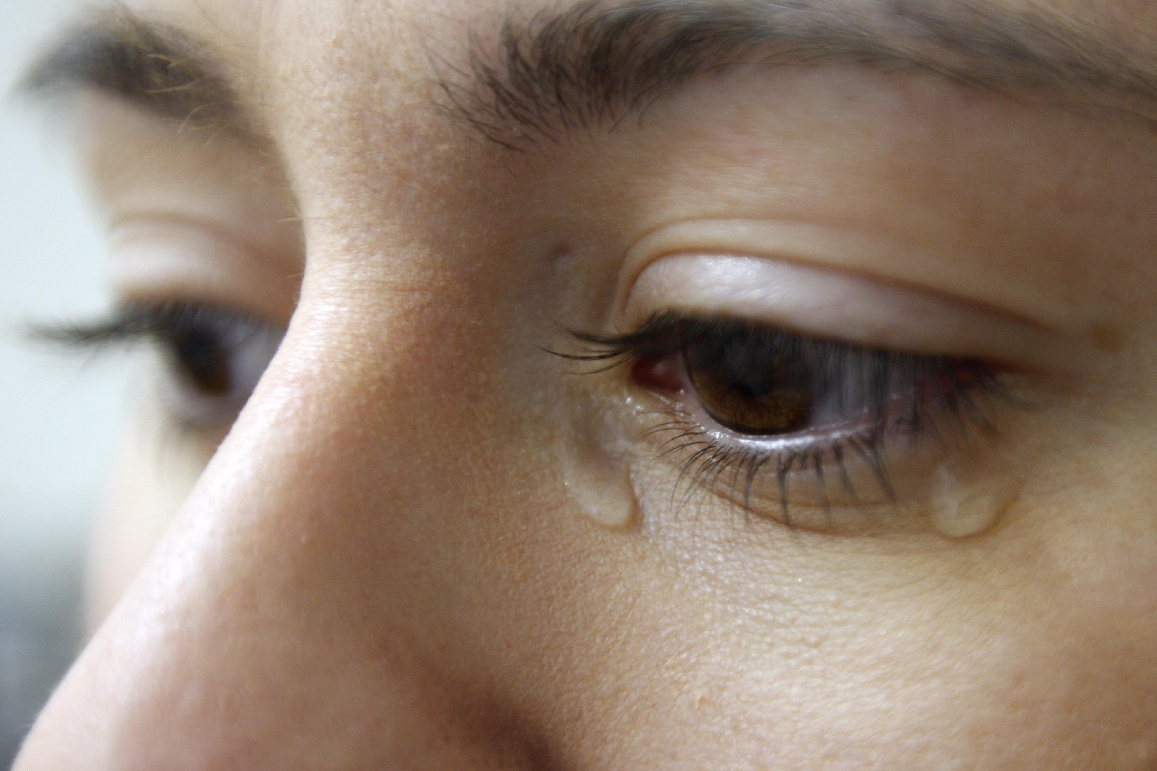
Tears are a clear liquid secreted by the lacrimal glands (tear gland) found in the eyes of all land mammals. Tears are made up of water, electrolytes, proteins, lipids, and mucins that form layers on the surface of eyes. The different types of tears—basal, reflex, and emotional—vary significantly in composition. The functions of tears include lubricating the eyes (basal tears), removing irritants (reflex tears), and also aiding the immune system. Tears also occur as a part of the body's natural pain response. Emotional secretion of tears may serve a biological function by excreting stress-inducing hormones built up through times of emotional distress. Tears have symbolic significance among humans. Physiology Chemical composition Tears are made up of three layers: lipid, aqueous, and mucous. Tears are composed of water, salts, antibodies, and lysozymes (antibacterial enzymes); though composition varies among different tear types. The composition of tears caused by an ... [...More Info...] [...Related Items...] OR: [Wikipedia] [Google] [Baidu] |
Cornea
The cornea is the transparent front part of the eye that covers the iris, pupil, and anterior chamber. Along with the anterior chamber and lens, the cornea refracts light, accounting for approximately two-thirds of the eye's total optical power. In humans, the refractive power of the cornea is approximately 43 dioptres. The cornea can be reshaped by surgical procedures such as LASIK. While the cornea contributes most of the eye's focusing power, its focus is fixed. Accommodation (the refocusing of light to better view near objects) is accomplished by changing the geometry of the lens. Medical terms related to the cornea often start with the prefix "'' kerat-''" from the Greek word κέρας, ''horn''. Structure The cornea has unmyelinated nerve endings sensitive to touch, temperature and chemicals; a touch of the cornea causes an involuntary reflex to close the eyelid. Because transparency is of prime importance, the healthy cornea does not have or need blood vessels with ... [...More Info...] [...Related Items...] OR: [Wikipedia] [Google] [Baidu] |
Hydrophilic
A hydrophile is a molecule or other molecular entity that is attracted to water molecules and tends to be dissolved by water.Liddell, H.G. & Scott, R. (1940). ''A Greek-English Lexicon'' Oxford: Clarendon Press. In contrast, hydrophobes are not attracted to water and may seem to be repelled by it. Hygroscopics ''are'' attracted to water, but are not dissolved by water. Molecules A hydrophilic molecule or portion of a molecule is one whose interactions with water and other polar substances are more thermodynamically favorable than their interactions with oil or other hydrophobic solvents. They are typically charge-polarized and capable of hydrogen bonding. This makes these molecules soluble not only in water but also in other polar solvents. Hydrophilic molecules (and portions of molecules) can be contrasted with hydrophobic molecules (and portions of molecules). In some cases, both hydrophilic and hydrophobic properties occur in a single molecule. An example of these amphiph ... [...More Info...] [...Related Items...] OR: [Wikipedia] [Google] [Baidu] |
Anatomist
Anatomy () is the branch of biology concerned with the study of the structure of organisms and their parts. Anatomy is a branch of natural science that deals with the structural organization of living things. It is an old science, having its beginnings in prehistoric times. Anatomy is inherently tied to developmental biology, embryology, comparative anatomy, evolutionary biology, and phylogeny, as these are the processes by which anatomy is generated, both over immediate and long-term timescales. Anatomy and physiology, which study the structure and function of organisms and their parts respectively, make a natural pair of related disciplines, and are often studied together. Human anatomy is one of the essential basic sciences that are applied in medicine. The discipline of anatomy is divided into macroscopic and microscopic. Macroscopic anatomy, or gross anatomy, is the examination of an animal's body parts using unaided eyesight. Gross anatomy also includes the branch of ... [...More Info...] [...Related Items...] OR: [Wikipedia] [Google] [Baidu] |
Friedrich Gustav Jakob Henle
Friedrich Gustav Jakob Henle (; 9 July 1809 – 13 May 1885) was a German physician, pathologist, and anatomist. He is credited with the discovery of the loop of Henle in the kidney. His essay, "On Miasma and Contagia," was an early argument for the germ theory of disease. He was an important figure in the development of modern medicine. Biography Henle was born in Fürth, Bavaria, to Simon and Rachel Diesbach Henle (Hähnlein). He was Jewish. After studying medicine at Heidelberg and at Bonn, where he took his doctor's degree in 1832, he became prosector in anatomy to Johannes Müller at Berlin. During the six years he spent in that position he published a large amount of work, including three anatomical monographs on new species of animals and papers on the structure of the lymphatic system, the distribution of epithelium in the human body, the structure and development of the hair, and the formation of mucus and pus. In 1840, he accepted the chair of anatomy at Zürich an ... [...More Info...] [...Related Items...] OR: [Wikipedia] [Google] [Baidu] |
Sclera
The sclera, also known as the white of the eye or, in older literature, as the tunica albuginea oculi, is the opaque, fibrous, protective, outer layer of the human eye containing mainly collagen and some crucial elastic fiber. In humans, and some other vertebrates, the whole sclera is white, contrasting with the coloured iris, but in most mammals, the visible part of the sclera matches the colour of the iris, so the white part does not normally show while other vertebrates have distinct colors for both of them. In the development of the embryo, the sclera is derived from the neural crest. In children, it is thinner and shows some of the underlying pigment, appearing slightly blue. In the elderly, fatty deposits on the sclera can make it appear slightly yellow. People with dark skin can have naturally darkened sclerae, the result of melanin pigmentation. The human eye is relatively rare for having a pale sclera (relative to the iris). This makes it easier for one individual to ide ... [...More Info...] [...Related Items...] OR: [Wikipedia] [Google] [Baidu] |


