|
Crutch Paralysis
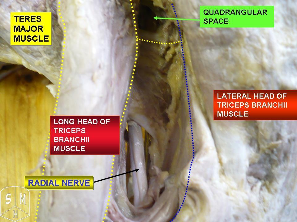
Crutch paralysis is a form of paralysis which can occur when either the radial nerve or part of the brachial plexus, containing various nerves that innervate sense and motor function to the arm and hand, is under constant pressure, such as by the use of a crutch. This can lead to paralysis of the muscles innervated by the compressed nerve. Generally, crutches that are not adjusted to the correct height can cause the radial nerve to be constantly pushed against the humerus. This can cause any muscle that is innervated by the radial nerve to become partially or fully paralyzed. An example of this is wrist drop, in which the fingers, hand, or wrist is chronically in a flexed position because the radial nerve cannot innervate the extensor muscles due to paralysis. This condition, like other injuries from compressed nerves, normally improves quickly through therapy.Anatomy Physiology - The Unity of Form and Function by Saladin 6th Edition See also * Brachial plexus injury A brachial ... [...More Info...] [...Related Items...] OR: [Wikipedia] [Google] [Baidu] |
Paralysis
Paralysis (also known as plegia) is a loss of motor function in one or more muscles. Paralysis can also be accompanied by a loss of feeling (sensory loss) in the affected area if there is sensory damage. In the United States, roughly 1 in 50 people have been diagnosed with some form of permanent or transient paralysis. The word "paralysis" derives from the Greek παράλυσις, meaning "disabling of the nerves" from παρά (''para'') meaning "beside, by" and λύσις (''lysis'') meaning "making loose". A paralysis accompanied by involuntary tremors is usually called "palsy". Causes Paralysis is most often caused by damage in the nervous system, especially the spinal cord. Other major causes are stroke, trauma with nerve injury, poliomyelitis, cerebral palsy, peripheral neuropathy, Parkinson's disease, ALS, botulism, spina bifida, multiple sclerosis, and Guillain–Barré syndrome. Temporary paralysis occurs during REM sleep, and dysregulation of this system can lead ... [...More Info...] [...Related Items...] OR: [Wikipedia] [Google] [Baidu] |
Radial Nerve
The radial nerve is a nerve in the human body that supplies the posterior portion of the upper limb. It innervates the medial and lateral heads of the triceps brachii muscle of the arm, as well as all 12 muscles in the posterior osteofascial compartment of the forearm and the associated joints and overlying skin. It originates from the brachial plexus, carrying fibers from the ventral roots of spinal nerves C5, C6, C7, C8 & T1. The radial nerve and its branches provide motor innervation to the dorsal arm muscles (the triceps brachii and the anconeus) and the extrinsic extensors of the wrists and hands; it also provides cutaneous sensory innervation to most of the back of the hand, except for the back of the little finger and adjacent half of the ring finger (which are innervated by the ulnar nerve). The radial nerve divides into a deep branch, which becomes the posterior interosseous nerve, and a superficial branch, which goes on to innervate the dorsum (back) of the hand. Th ... [...More Info...] [...Related Items...] OR: [Wikipedia] [Google] [Baidu] |
Brachial Plexus
The brachial plexus is a network () of nerves formed by the anterior rami of the lower four cervical nerves and first thoracic nerve ( C5, C6, C7, C8, and T1). This plexus extends from the spinal cord, through the cervicoaxillary canal in the neck, over the first rib, and into the armpit, it supplies afferent and efferent nerve fibers the to chest, shoulder, arm, forearm, and hand. Structure The brachial plexus is divided into five ''roots'', three ''trunks'', six ''divisions'' (three anterior and three posterior), three ''cords'', and five ''branches''. There are five "terminal" branches and numerous other "pre-terminal" or "collateral" branches, such as the subscapular nerve, the thoracodorsal nerve, and the long thoracic nerve, that leave the plexus at various points along its length. A common structure used to identify part of the brachial plexus in cadaver dissections is the M or W shape made by the musculocutaneous nerve, lateral cord, median nerve, medial cord, and ... [...More Info...] [...Related Items...] OR: [Wikipedia] [Google] [Baidu] |
Crutch
A crutch is a mobility aid that transfers weight from the legs to the upper body. It is often used by people who cannot use their legs to support their weight, for reasons ranging from short-term injuries to lifelong disabilities. History Crutches were used in ancient Egypt. In 1917, Emile Schlick patented the first commercially produced crutch; the design consisted of a walking stick with an upper arm support. Later, A.R. Lofstrand Jr. developed the first crutches with a height-adjustable feature. Over time, the design of crutches has not changed much, and the classic design continues to be the most commonly used. Types There are several types of crutches: Underarm or axillary Axillary crutches are used by placing the pad against the ribcage beneath the armpit and holding the grip, which is below and parallel to the pad. They are usually used to provide support for patients who have temporary restriction on ambulation.Taylor, C. R., Lillis, C., LeMone, P., Lynn, P. (2011) F ... [...More Info...] [...Related Items...] OR: [Wikipedia] [Google] [Baidu] |
Paralysis
Paralysis (also known as plegia) is a loss of motor function in one or more muscles. Paralysis can also be accompanied by a loss of feeling (sensory loss) in the affected area if there is sensory damage. In the United States, roughly 1 in 50 people have been diagnosed with some form of permanent or transient paralysis. The word "paralysis" derives from the Greek παράλυσις, meaning "disabling of the nerves" from παρά (''para'') meaning "beside, by" and λύσις (''lysis'') meaning "making loose". A paralysis accompanied by involuntary tremors is usually called "palsy". Causes Paralysis is most often caused by damage in the nervous system, especially the spinal cord. Other major causes are stroke, trauma with nerve injury, poliomyelitis, cerebral palsy, peripheral neuropathy, Parkinson's disease, ALS, botulism, spina bifida, multiple sclerosis, and Guillain–Barré syndrome. Temporary paralysis occurs during REM sleep, and dysregulation of this system can lead ... [...More Info...] [...Related Items...] OR: [Wikipedia] [Google] [Baidu] |
Humerus
The humerus (; ) is a long bone in the arm that runs from the shoulder to the elbow. It connects the scapula and the two bones of the lower arm, the radius and ulna, and consists of three sections. The humeral upper extremity consists of a rounded head, a narrow neck, and two short processes (tubercles, sometimes called tuberosities). The body is cylindrical in its upper portion, and more prismatic below. The lower extremity consists of 2 epicondyles, 2 processes (trochlea & capitulum), and 3 fossae (radial fossa, coronoid fossa, and olecranon fossa). As well as its true anatomical neck, the constriction below the greater and lesser tubercles of the humerus is referred to as its surgical neck due to its tendency to fracture, thus often becoming the focus of surgeons. Etymology The word "humerus" is derived from la, humerus, umerus meaning upper arm, shoulder, and is linguistically related to Gothic ''ams'' shoulder and Greek ''ōmos''. Structure Upper extremity The upper or pr ... [...More Info...] [...Related Items...] OR: [Wikipedia] [Google] [Baidu] |
Wrist Drop
Wrist drop is a medical condition in which the wrist and the fingers cannot extend at the metacarpophalangeal joints. The wrist remains partially flexed due to an opposing action of flexor muscles of the forearm. As a result, the extensor muscles in the posterior compartment remain paralyzed. Forearm anatomy The forearm is the part of the body that extends from the elbow to the wrist and is not to be confused with the arm, which extends from the shoulder to the elbow. The extensor muscles in the forearm are the extensor carpi ulnaris, extensor digiti minimi, extensor digitorum, extensor indicis, extensor carpi radialis brevis, and extensor carpi radialis longus. These extensor muscles are supplied by the posterior interosseous nerve, a branch of the radial nerve. Other muscles in the forearm that are innervated by this nerve are the supinator, extensor pollicis brevis, extensor pollicis longus and abductor pollicis longus. All of these muscles are situated in the posterior ha ... [...More Info...] [...Related Items...] OR: [Wikipedia] [Google] [Baidu] |
Brachial Plexus Injury
A brachial plexus injury (BPI), also known as brachial plexus lesion, is an injury to the brachial plexus, the network of nerves that conducts signals from the spinal cord to the shoulder, arm and hand. These nerves originate in the fifth, sixth, seventh and eighth cervical (C5–C8), and first thoracic (T1) spinal nerves, and innervate the muscles and skin of the chest, shoulder, arm and hand. Brachial plexus injuries can occur as a result of shoulder trauma, tumours, or inflammation, or obstetric. Obstetric injuries may occur from mechanical injury involving shoulder dystocia during difficult childbirth, with a prevalence of 1 in 1000 births. "The brachial plexus may be injured by falls from a height on to the side of the head and shoulder, whereby the nerves of the plexus are violently stretched. The brachial plexus may also be injured by direct violence or gunshot wounds, by violent traction on the arm, or by efforts at reducing a dislocation of the shoulder joint". The rare ... [...More Info...] [...Related Items...] OR: [Wikipedia] [Google] [Baidu] |


