|
Cripto
Cripto is an EGF-CFC or epidermal growth factor-CFC, which is encoded by the Cryptic family 1 gene. Cryptic family protein 1B is a protein that in humans is encoded by the ''CFC1B'' gene. Cryptic family protein 1B acts as a receptor for the TGF beta signaling pathway. It has been associated with the translation of an extracellular protein for this pathway. The extracellular protein which Cripto encodes plays a crucial role in the development of left and right division of symmetry. Crypto is a glycosylphosphatidylinositol-anchored co-receptor that binds nodal and the activin type I ActRIB (ALK)-4 receptor (ALK4). Structure Cripto is composed of two adjacent cysteine-rich motifs: the EGF-like and the CFC of an N-terminal signal peptide and of a C-terminal hydrophobic region attached by a GPI anchor, which makes it a potentially essential element in the signaling pathway directing vertebrate embryo development. NMR data confirm that the CFC domain has a C1-C4, C2-C6, C3-C5 disulf ... [...More Info...] [...Related Items...] OR: [Wikipedia] [Google] [Baidu] |
CFC Domain
In molecular biology, the CFC domain (Cripto_Frl-1_Cryptic domain) is a protein domain found at the C-terminus of a number of proteins including Cripto (or teratocarcinoma-derived growth factor). It is structurally similar to the C-terminal extracellular portions of Jagged 1 and Jagged 2. CFC is approx 40-residues long, compacted by three internal disulphide bridges, and binds Alk4 via a hydrophobic patch. CFC is structurally homologous to the VWFC-like domain. The CFC domain appears to play a crucial role in the tumourigenic activity of Cripto proteins, as it is through the CFC domain that Cripto interferes with the onco-suppressive activity of Activins Activin and inhibin are two closely related protein complexes that have almost directly opposite biological effects. Identified in 1986, activin enhances FSH biosynthesis and secretion, and participates in the regulation of the menstrual cy ..., either by blocking the Activin receptor ALK4 or by antagonising prote ... [...More Info...] [...Related Items...] OR: [Wikipedia] [Google] [Baidu] |
NODAL
Nodal homolog is a secretory protein that in humans is encoded by the ''NODAL'' gene which is located on chromosome 10q22.1. It belongs to the transforming growth factor beta superfamily (TGF-β superfamily). Like many other members of this superfamily it is involved in cell differentiation in early embryogenesis, playing a key role in signal transfer from the primitive node, in the anterior primitive streak, to lateral plate mesoderm (LPM). Nodal signaling is important very early in development for mesoderm and endoderm formation and subsequent organization of left-right axial structures. In addition, Nodal seems to have important functions in neural patterning, stem cell maintenance and many other developmental processes, including left/right handedness. Signaling Nodal can bind type I and type II serine/threonine kinase receptors, with Cripto-1 acting as its co-receptor. Signaling through SMAD 2/3 and subsequent translocation of SMAD 4 to the nucleus promotes the expressi ... [...More Info...] [...Related Items...] OR: [Wikipedia] [Google] [Baidu] |
ACVR1B
Activin receptor type-1B is a protein that in humans is encoded by the ''ACVR1B'' gene. ACVR1B or ALK-4 acts as a transducer of activin or activin-like ligands (e.g., inhibin) signals. Activin binds to either ACVR2A or ACVR2B and then forms a complex with ACVR1B. These go on to recruit the R-SMADs SMAD2 or SMAD3. ACVR1B also transduces signals of nodal, GDF-1, and Vg1; however, unlike activin, they require other coreceptor molecules such as the protein Cripto. Function Activins are dimeric growth and differentiation factors which belong to the transforming growth factor-beta (TGF-beta) superfamily of structurally related signaling proteins. Activins signal through a heteromeric complex of receptor serine kinases which include at least two type I (I and IB) and two type II (II and IIB) receptors. These receptors are all transmembrane proteins, composed of a ligand-binding extracellular domain with a cysteine-rich region, a transmembrane domain, and a cytoplasmic domain ... [...More Info...] [...Related Items...] OR: [Wikipedia] [Google] [Baidu] |
TGF Beta Signaling Pathway
The transforming growth factor beta (TGFB) signaling pathway is involved in many cellular processes in both the adult organism and the developing embryo including cell growth, cell differentiation, cell migration, apoptosis, cellular homeostasis and other cellular functions. The TGFB signaling pathways are conserved. In spite of the wide range of cellular processes that the TGFβ signaling pathway regulates, the process is relatively simple. TGFβ superfamily ligands bind to a type II receptor, which recruits and phosphorylates a type I receptor. The type I receptor then phosphorylates receptor-regulated SMADs (R-SMADs) which can now bind the coSMAD SMAD4. R-SMAD/coSMAD complexes accumulate in the nucleus where they act as transcription factors and participate in the regulation of target gene expression. Mechanism Ligand binding The TGF beta superfamily of ligands includes: Bone morphogenetic proteins (BMPs), Growth and differentiation factors (GDFs), Anti-müllerian ho ... [...More Info...] [...Related Items...] OR: [Wikipedia] [Google] [Baidu] |
Wnt Signaling Pathway
The Wnt signaling pathways are a group of signal transduction pathways which begin with proteins that pass signals into a cell through cell surface receptors. The name Wnt is a portmanteau created from the names Wingless and Int-1. Wnt signaling pathways use either nearby cell-cell communication (paracrine) or same-cell communication (autocrine). They are highly evolutionarily conserved in animals, which means they are similar across animal species from fruit flies to humans. Three Wnt signaling pathways have been characterized: the canonical Wnt pathway, the noncanonical planar cell polarity pathway, and the noncanonical Wnt/calcium pathway. All three pathways are activated by the binding of a Wnt-protein ligand to a Frizzled family receptor, which passes the biological signal to the Dishevelled protein inside the cell. The canonical Wnt pathway leads to regulation of gene transcription, and is thought to be negatively regulated in part by the SPATS1 gene. The noncanonical ... [...More Info...] [...Related Items...] OR: [Wikipedia] [Google] [Baidu] |
Neuronal
A neuron, neurone, or nerve cell is an membrane potential#Cell excitability, electrically excitable cell (biology), cell that communicates with other cells via specialized connections called synapses. The neuron is the main component of nervous tissue in all Animalia, animals except sponges and placozoa. Non-animals like plants and fungi do not have nerve cells. Neurons are typically classified into three types based on their function. Sensory neurons respond to Stimulus (physiology), stimuli such as touch, sound, or light that affect the cells of the Sense, sensory organs, and they send signals to the spinal cord or brain. Motor neurons receive signals from the brain and spinal cord to control everything from muscle contractions to gland, glandular output. Interneurons connect neurons to other neurons within the same region of the brain or spinal cord. When multiple neurons are connected together, they form what is called a neural circuit. A typical neuron consists of a cell bo ... [...More Info...] [...Related Items...] OR: [Wikipedia] [Google] [Baidu] |
Cardiac Muscle
Cardiac muscle (also called heart muscle, myocardium, cardiomyocytes and cardiac myocytes) is one of three types of vertebrate muscle tissues, with the other two being skeletal muscle and smooth muscle. It is an involuntary, striated muscle that constitutes the main tissue of the wall of the heart. The cardiac muscle (myocardium) forms a thick middle layer between the outer layer of the heart wall (the pericardium) and the inner layer (the endocardium), with blood supplied via the coronary circulation. It is composed of individual cardiac muscle cells joined by intercalated discs, and encased by collagen fibers and other substances that form the extracellular matrix. Cardiac muscle contracts in a similar manner to skeletal muscle, although with some important differences. Electrical stimulation in the form of a cardiac action potential triggers the release of calcium from the cell's internal calcium store, the sarcoplasmic reticulum. The rise in calcium causes the c ... [...More Info...] [...Related Items...] OR: [Wikipedia] [Google] [Baidu] |
Embryonic Stem Cells
Embryonic stem cells (ESCs) are pluripotent stem cells derived from the inner cell mass of a blastocyst, an early-stage pre- implantation embryo. Human embryos reach the blastocyst stage 4–5 days post fertilization, at which time they consist of 50–150 cells. Isolating the inner cell mass (embryoblast) using immunosurgery results in destruction of the blastocyst, a process which raises ethical issues, including whether or not embryos at the pre-implantation stage have the same moral considerations as embryos in the post-implantation stage of development. Researchers are currently focusing heavily on the therapeutic potential of embryonic stem cells, with clinical use being the goal for many laboratories. Potential uses include the treatment of diabetes and heart disease. The cells are being studied to be used as clinical therapies, models of genetic disorders, and cellular/DNA repair. However, adverse effects in the research and clinical processes such as tumors and un ... [...More Info...] [...Related Items...] OR: [Wikipedia] [Google] [Baidu] |
Oncogene
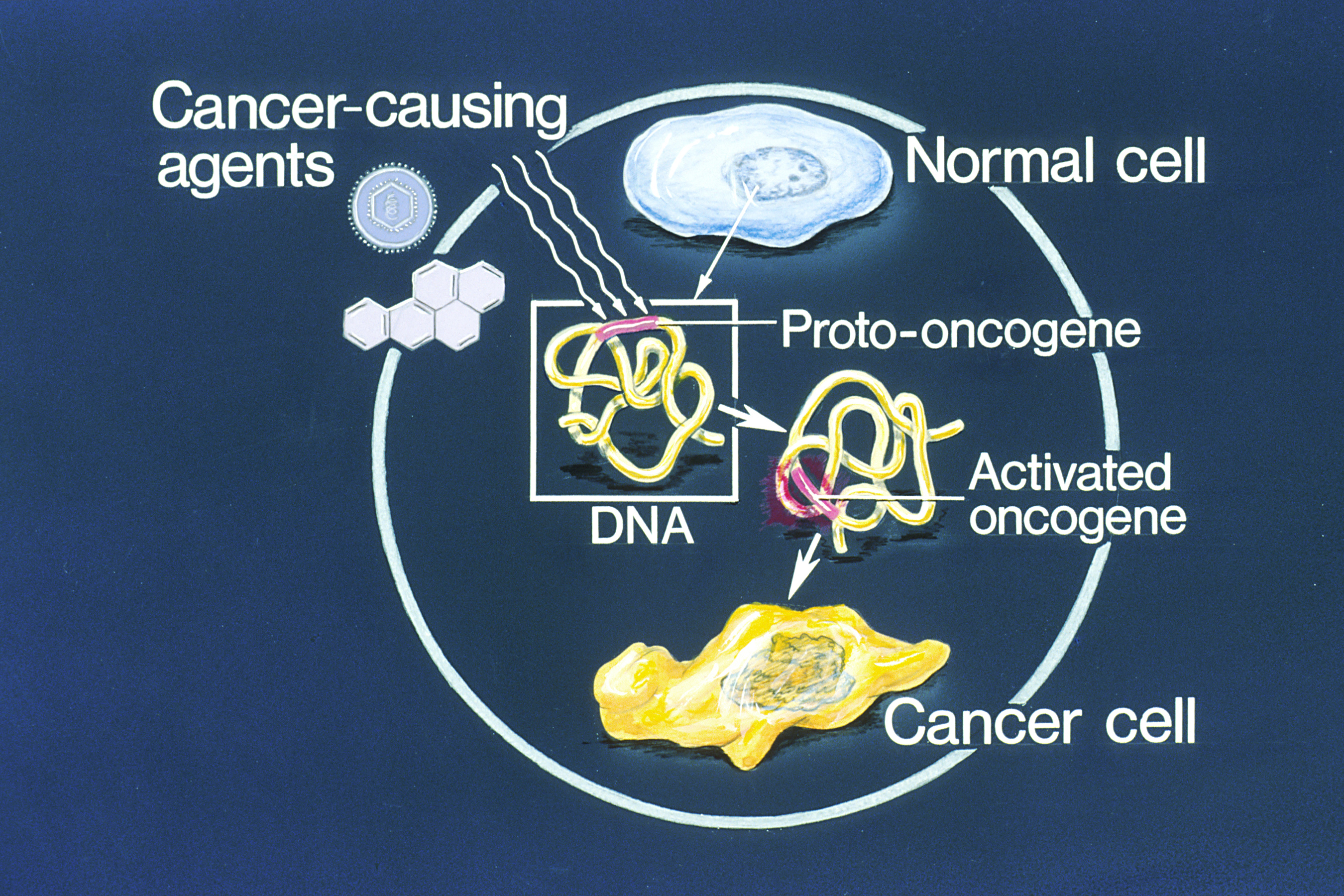
An oncogene is a gene that has the potential to cause cancer. In tumor cells, these genes are often mutated, or expressed at high levels.Kimball's Biology Pages. "Oncogenes" Free full text Most normal cells will undergo a programmed form of rapid cell death ( apoptosis) when critical functions are altered and malfunctioning. Activated oncogenes can cause those cells designated for apoptosis to survive and proliferate instead. Most oncogenes began as proto-oncogenes: normal genes involved in cell growth and proliferation or inhibition of apoptosis. If, through mutation, normal genes promoting cellular growth are up-regulated (gain-of-function mutation), they will predispose the cell to cancer; thus, t ... [...More Info...] [...Related Items...] OR: [Wikipedia] [Google] [Baidu] |
Mesoderm
The mesoderm is the middle layer of the three germ layers that develops during gastrulation in the very early development of the embryo of most animals. The outer layer is the ectoderm, and the inner layer is the endoderm.Langman's Medical Embryology, 11th edition. 2010. The mesoderm forms mesenchyme, mesothelium, non-epithelial blood cells and coelomocytes. Mesothelium lines coeloms. Mesoderm forms the muscles in a process known as myogenesis, septa (cross-wise partitions) and mesenteries (length-wise partitions); and forms part of the gonads (the rest being the gametes). Myogenesis is specifically a function of mesenchyme. The mesoderm differentiates from the rest of the embryo through intercellular signaling, after which the mesoderm is polarized by an organizing center. The position of the organizing center is in turn determined by the regions in which beta-catenin is protected from degradation by GSK-3. Beta-catenin acts as a co-factor that alters the activity of ... [...More Info...] [...Related Items...] OR: [Wikipedia] [Google] [Baidu] |
Embryonic Death
Embryo loss (also known as embryo death or embryo resorption) is the death of an embryo at any stage of its development which in humans, is between the second through eighth week after fertilization. Failed development of an embryo often results in the disintegration and assimilation of its tissue in the uterus. Loss during the stages of prenatal development after organogenesis of the fetus results in the similar process of fetal resorption. Embryo loss often happens without an awareness of pregnancy, and an estimated 40 to 60% of all embryos do not survive. Fertility clinics Within fertility clinics embryo loss is associated with a high number of implanted embryos. The keeping of embryos in tanks can also increase risks of loss in instances where technical malfunctions can occur. See also * Perinatal death Perinatal mortality (PNM) refers to the death of a fetus or neonate and is the basis to calculate the perinatal mortality rate. Variations in the precise definition of the ... [...More Info...] [...Related Items...] OR: [Wikipedia] [Google] [Baidu] |


