|
Cotyledon Arteries
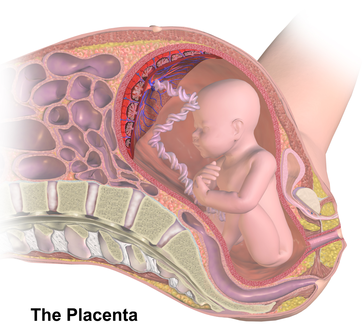
The placenta of humans, and certain other mammals contains structures known as cotyledons, which transmit fetal blood and allow exchange of oxygen and nutrients with the maternal blood. Ruminants The Artiodactyla have a cotyledonary placenta. In this form of placenta the chorionic villi form a number of separate circular structures (''cotyledons'') which are distributed over the surface of the chorionic sac. Sheep, goats and cattle have between 72 and 125 cotyledons whereas deer have 4-6 larger cotyledons. Human The form of the human placenta is generally classified as a discoid placenta. Within this the ''cotyledons'' are the approximately 15-25 separations of the decidua basalis of the placenta, separated by placental septa.Page 565 in:/ref> Each cotyledon consists of a main stem of a chorionic villus as well as its branches and sub-branches. Vasculature The cotyledons receive fetal blood from chorionic vessels Chorionic (plate) vessels, also fetal surface vessels are blood ... [...More Info...] [...Related Items...] OR: [Wikipedia] [Google] [Baidu] |
Placenta
The placenta is a temporary embryonic and later fetal organ that begins developing from the blastocyst shortly after implantation. It plays critical roles in facilitating nutrient, gas and waste exchange between the physically separate maternal and fetal circulations, and is an important endocrine organ, producing hormones that regulate both maternal and fetal physiology during pregnancy. The placenta connects to the fetus via the umbilical cord, and on the opposite aspect to the maternal uterus in a species-dependent manner. In humans, a thin layer of maternal decidual (endometrial) tissue comes away with the placenta when it is expelled from the uterus following birth (sometimes incorrectly referred to as the 'maternal part' of the placenta). Placentas are a defining characteristic of placental mammals, but are also found in marsupials and some non-mammals with varying levels of development. Mammalian placentas probably first evolved about 150 million to 200 million years ... [...More Info...] [...Related Items...] OR: [Wikipedia] [Google] [Baidu] |
Artiodactyla
The even-toed ungulates (Artiodactyla , ) are ungulates—hoofed animals—which bear weight equally on two (an even number) of their five toes: the third and fourth. The other three toes are either present, absent, vestigial, or pointing posteriorly. By contrast, odd-toed ungulates bear weight on an odd number of the five toes. Another difference between the two is that many other even-toed ungulates (with the exception of Suina) digest plant cellulose in one or more stomach chambers rather than in their intestine as the odd-toed ungulates do. Cetaceans (whales, dolphins, and porpoises) evolved from even-toed ungulates, and are therefore often classified under the same taxonomic branch because a species cannot outgrow its evolutionary ancestry; some modern taxonomists combine the two under the name Cetartiodactyla , while others opt to include cetaceans in the already-existing Artiodactyla. The roughly 270 land-based even-toed ungulate species include pigs, peccaries, hippopo ... [...More Info...] [...Related Items...] OR: [Wikipedia] [Google] [Baidu] |
Decidua Basalis
The decidua is the modified mucosal lining of the uterus (that is, modified endometrium) that forms every month, in preparation for pregnancy. It is shed off each month when there is no fertilised egg to support. The decidua is under the influence of progesterone. Endometrial cells become highly characteristic. The decidua forms the maternal part of the placenta and remains for the duration of the pregnancy. After birth the decidua is shed together with the placenta. Structure The part of the decidua that interacts with the trophoblast is the ''decidua basalis'' (also called ''decidua placentalis''), while the ''decidua capsularis'' grows over the embryo on the luminal side, enclosing it into the endometrium. The remainder of the decidua is termed the ''decidua parietalis'' or ''decidua vera'', and it will fuse with the decidua capsularis by the fourth month of gestation. Three morphologically distinct layers of the decidua basalis can then be described: * Compact outer layer ( ... [...More Info...] [...Related Items...] OR: [Wikipedia] [Google] [Baidu] |
Septum
In biology, a septum (Latin for ''something that encloses''; plural septa) is a wall, dividing a cavity or structure into smaller ones. A cavity or structure divided in this way may be referred to as septate. Examples Human anatomy * Interatrial septum, the wall of tissue that is a sectional part of the left and right atria of the heart * Interventricular septum, the wall separating the left and right ventricles of the heart * Lingual septum, a vertical layer of fibrous tissue that separates the halves of the tongue. *Nasal septum: the cartilage wall separating the nostrils of the nose * Alveolar septum: the thin wall which separates the alveoli from each other in the lungs * Orbital septum, a palpebral ligament in the upper and lower eyelids * Septum pellucidum or septum lucidum, a thin structure separating two fluid pockets in the brain * Uterine septum, a malformation of the uterus * Vaginal septum, a lateral or transverse partition inside the vagina * Intermuscular sep ... [...More Info...] [...Related Items...] OR: [Wikipedia] [Google] [Baidu] |
Chorionic Villus
Chorionic villi are villi that sprout from the chorion to provide maximal contact area with maternal blood. They are an essential element in pregnancy from a histomorphologic perspective, and are, by definition, a product of conception. Branches of the umbilical arteries carry embryonic blood to the villi. After circulating through the capillaries of the villi, blood returns to the embryo through the umbilical vein. Thus, villi are part of the border between maternal and fetal blood during pregnancy. Structure Villi can also be classified by their relations: * Floating villi float freely in the intervillous space. They exhibit a bi-layered epithelium consisting of cytotrophoblasts with overlaying syncytium (syncytiotrophoblast). * Anchoring (stem) villi stabilize mechanical integrity of the placental-maternal interface. Development The chorion undergoes rapid proliferation and forms numerous processes, the chorionic villi, which invade and destroy the uterine decidua and at ... [...More Info...] [...Related Items...] OR: [Wikipedia] [Google] [Baidu] |
Chorionic Vessels
Chorionic (plate) vessels, also fetal surface vessels are blood vessels, including both arteries and veins, that carry blood through the chorion in the fetoplacental circulation. Chorionic arteries branch off the umbilical artery, and supply the capillaries of the chorionic villi. Increased vasocontractility of chorionic arteries may contribute to preeclampsia Pre-eclampsia is a disorder of pregnancy characterized by the onset of high blood pressure and often a significant amount of protein in the urine. When it arises, the condition begins after 20 weeks of pregnancy. In severe cases of the disease .... References Embryology of cardiovascular system {{circulatory-stub ... [...More Info...] [...Related Items...] OR: [Wikipedia] [Google] [Baidu] |
Vertebrate Developmental Biology
Vertebrates () comprise all animal taxa within the subphylum Vertebrata () (chordates with backbones), including all mammals, birds, reptiles, amphibians, and fish. Vertebrates represent the overwhelming majority of the phylum Chordata, with currently about 69,963 species described. Vertebrates comprise such groups as the following: * jawless fish, which include hagfish and lampreys * jawed vertebrates, which include: ** cartilaginous fish (sharks, rays, and ratfish) ** bony vertebrates, which include: *** ray-fins (the majority of living bony fish) *** lobe-fins, which include: **** coelacanths and lungfish **** tetrapods (limbed vertebrates) Extant vertebrates range in size from the frog species ''Paedophryne amauensis'', at as little as , to the blue whale, at up to . Vertebrates make up less than five percent of all described animal species; the rest are invertebrates, which lack vertebral columns. The vertebrates traditionally include the hagfish, which do not have p ... [...More Info...] [...Related Items...] OR: [Wikipedia] [Google] [Baidu] |


