|
Coracobrachialis Muscle
The coracobrachialis muscle is the smallest of the three muscles that attach to the coracoid process of the scapula. (The other two muscles are pectoralis minor and the short head of the biceps brachii.) It is situated at the upper and medial part of the arm. Structure Coracobrachialis muscle arises from the apex of the coracoid process, in common with the short head of the biceps brachii, and from the intermuscular septum between the two muscles. It is inserted by means of a flat tendon into an impression at the middle of the medial surface and border of the body of the humerus (shaft of the humerus) between the origins of the triceps brachii and brachialis. Innervation Coracobrachialis muscle is perforated by and innervated by the musculocutaneous nerve, which arises from the anterior division of the upper trunk ( C5, C6) and middle trunk ( C7) of the brachial plexus. Development Variation Function The action of the coracobrachialis is to flex and adduct the arm at the ... [...More Info...] [...Related Items...] OR: [Wikipedia] [Google] [Baidu] |
Coracoid Process
The coracoid process (from Greek κόραξ, raven) is a small hook-like structure on the lateral edge of the superior anterior portion of the scapula (hence: coracoid, or "like a raven's beak"). Pointing laterally forward, it, together with the acromion, serves to stabilize the shoulder joint. It is palpable in the deltopectoral groove between the deltoid and pectoralis major muscles. Structure The coracoid process is a thick curved process attached by a broad base to the upper part of the neck of the scapula; it runs at first upward and medialward; then, becoming smaller, it changes its direction, and projects forward and lateralward. Anatomically it is divided into intervals of: base of coracoid process, angle of coracoid process, shaft and the apex of the coracoid process. The coracoglenoid notch is an indentation localized between the coracoid process and the glenoid. As the coracoid process projects laterally, it house underneath it the subcoracoid space. The ''ascend ... [...More Info...] [...Related Items...] OR: [Wikipedia] [Google] [Baidu] |
Brachialis
The brachialis (brachialis anticus), also known as the Teichmann muscle, is a muscle in the upper arm that flexes the elbow. It lies deeper than the biceps brachii, and makes up part of the floor of the region known as the cubital fossa (elbow pit). The brachialis is the prime mover of elbow flexion generating about 50% more power than the biceps.Saladin, Kenneth S, Stephen J. Sullivan, and Christina A. Gan. Anatomy & Physiology: The Unity of Form and Function. 2015. Print. Structure The brachialis originates from the anterior surface of the distal half of the humerus, near the insertion of the deltoid muscle, which it embraces by two angular processes. Its origin extends below to within 2.5 cm of the margin of the articular surface of the humerus at the elbow joint. Its fibers converge to a thick tendon, which is inserted into the tuberosity of the ulna and the rough depression on the anterior surface of the coronoid process of the ulna. Blood supply The brachialis is supp ... [...More Info...] [...Related Items...] OR: [Wikipedia] [Google] [Baidu] |
Avulsion Fracture
An avulsion fracture is a bone fracture which occurs when a fragment of bone tears away from the main mass of bone as a result of physical trauma. This can occur at the ligament by the application of forces external to the body (such as a fall or pull) or at the tendon by a muscular contraction that is stronger than the forces holding the bone together. Generally muscular avulsion is prevented by the neurological limitations placed on muscle contractions. Highly trained athletes can overcome this neurological inhibition of strength and produce a much greater force output capable of breaking or avulsing a bone. Types Dental avulsion Traumatic complete displacement of a tooth from its socket in alveolar bone. It is a serious dental emergency in which prompt management (within 20–40 minutes of injury) affects the prognosis of the tooth. Tuberosity avulsion of the 5th metatarsal left, Proximal fractures of 5th metatarsa The tuberosity avulsion fracture (also known as pseud ... [...More Info...] [...Related Items...] OR: [Wikipedia] [Google] [Baidu] |
Strain (injury)
A strain is an acute or chronic soft tissue injury that occurs to a muscle, tendon, or both. The equivalent injury to a ligament is a sprain. Generally, the muscle or tendon overstretches and partially tears, under more physical stress than it can withstand, often from a sudden increase in duration, intensity, or frequency of an activity. Strains most commonly occur in the foot, leg, or back. Immediate treatment typically includes five steps abbreviated as P.R.I.C.E.: protection, rest, ice, compression, elevation. Signs and symptoms Typical signs and symptoms of a strain include pain, functional loss of the involved structure, muscle weakness, contusion, and localized inflammation. A strain can range from mild overstretching to severe tears, depending on the extent of injury. Cause A strain can occur as a result of improper body mechanics with any activity (e.g., contact sports, lifting heavy objects) that can induce mechanical trauma or injury. Generally, the muscle ... [...More Info...] [...Related Items...] OR: [Wikipedia] [Google] [Baidu] |
Anatomical Terms Of Motion
Motion, the process of movement, is described using specific anatomical terms. Motion includes movement of organs, joints, limbs, and specific sections of the body. The terminology used describes this motion according to its direction relative to the anatomical position of the body parts involved. Anatomists and others use a unified set of terms to describe most of the movements, although other, more specialized terms are necessary for describing unique movements such as those of the hands, feet, and eyes. In general, motion is classified according to the anatomical plane it occurs in. ''Flexion'' and ''extension'' are examples of ''angular'' motions, in which two axes of a joint are brought closer together or moved further apart. ''Rotational'' motion may occur at other joints, for example the shoulder, and are described as ''internal'' or ''external''. Other terms, such as ''elevation'' and ''depression'', describe movement above or below the horizontal plane. Many anatomica ... [...More Info...] [...Related Items...] OR: [Wikipedia] [Google] [Baidu] |
Forearm
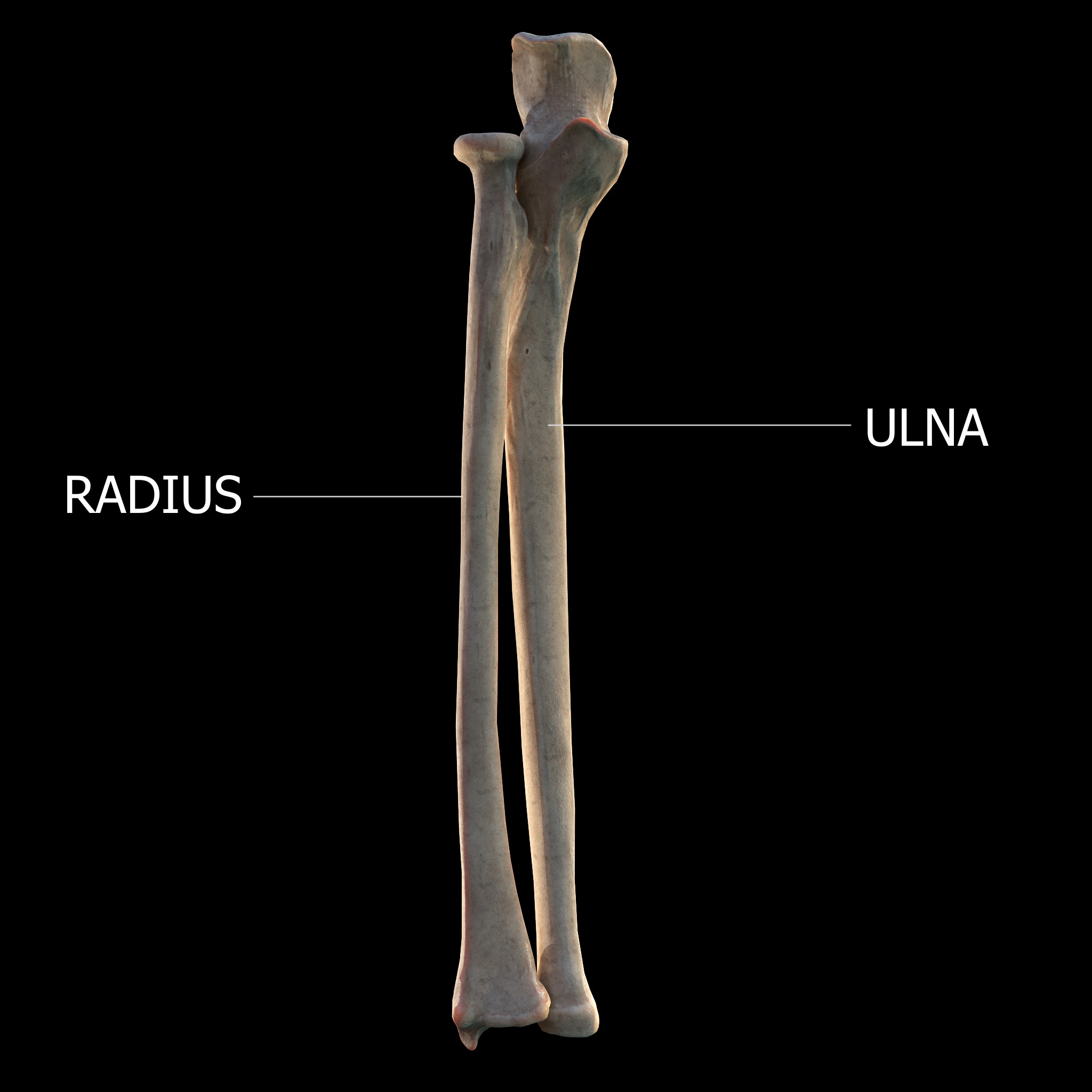
The forearm is the region of the upper limb between the elbow and the wrist. The term forearm is used in anatomy to distinguish it from the arm, a word which is most often used to describe the entire appendage of the upper limb, but which in anatomy, technically, means only the region of the upper arm, whereas the lower "arm" is called the forearm. It is homologous to the region of the leg that lies between the knee and the ankle joints, the crus. The forearm contains two long bones, the radius and the ulna, forming the two radioulnar joints. The interosseous membrane connects these bones. Ultimately, the forearm is covered by skin, the anterior surface usually being less hairy than the posterior surface. The forearm contains many muscles, including the flexors and extensors of the wrist, flexors and extensors of the digits, a flexor of the elbow (brachioradialis), and pronators and supinators that turn the hand to face down or upwards, respectively. In cross-section, the for ... [...More Info...] [...Related Items...] OR: [Wikipedia] [Google] [Baidu] |
Nerve Compression Syndrome
Nerve compression syndrome, or compression neuropathy, or nerve entrapment syndrome, is a medical condition caused by direct pressure on a nerve. It is known colloquially as a ''trapped nerve'', though this may also refer to nerve root compression (by a herniated disc, for example). Its symptoms include pain, tingling, numbness and muscle weakness. The symptoms affect just one particular part of the body, depending on which nerve is affected. Nerve conduction studies help to confirm the diagnosis. In some cases, surgery may help to relieve the pressure on the nerve but this does not always relieve all the symptoms. Nerve injury by a single episode of physical trauma is in one sense a compression neuropathy but is not usually included under this heading. Syndromes * Upper limb * Lower limb, abdomen and pelvis Signs and symptoms Tingling, numbness, and/or a burning sensation in the area of the body affected by the corresponding nerve. These experiences may occur directly fol ... [...More Info...] [...Related Items...] OR: [Wikipedia] [Google] [Baidu] |
Brachial Plexus
The brachial plexus is a network () of nerves formed by the anterior rami of the lower four cervical nerves and first thoracic nerve ( C5, C6, C7, C8, and T1). This plexus extends from the spinal cord, through the cervicoaxillary canal in the neck, over the first rib, and into the armpit, it supplies afferent and efferent nerve fibers the to chest, shoulder, arm, forearm, and hand. Structure The brachial plexus is divided into five ''roots'', three ''trunks'', six ''divisions'' (three anterior and three posterior), three ''cords'', and five ''branches''. There are five "terminal" branches and numerous other "pre-terminal" or "collateral" branches, such as the subscapular nerve, the thoracodorsal nerve, and the long thoracic nerve, that leave the plexus at various points along its length. A common structure used to identify part of the brachial plexus in cadaver dissections is the M or W shape made by the musculocutaneous nerve, lateral cord, median nerve, medial cord, and ... [...More Info...] [...Related Items...] OR: [Wikipedia] [Google] [Baidu] |
Cervical Spinal Nerve 7
The cervical spinal nerve 7 (C7) is a spinal nerve of the cervical segment. Nervous System -- Groups of Nerves It originates from the spinal column from above the cervical vertebra 7
In tetrapods, cervical vertebrae (singular: vertebra) are the vertebrae of the neck, immediately below the skull. Truncal vertebrae (divided into thoracic and lumbar vertebrae in mammals) lie caudal (toward the tail) of cervical vertebrae. In ... (C7).
It runs through the interspace between thC6 and C7 ... [...More Info...] [...Related Items...] OR: [Wikipedia] [Google] [Baidu] |
Cervical Spinal Nerve 6
The cervical spinal nerve 6 (C6) is a spinal nerve of the cervical segment. It originates from the spinal column from above the cervical vertebra 6 (C6). The C6 nerve root shares a common branch from C5, and has a role in innervating many muscles of the rotator cuff and distal arm, including: *Subclavius *Supraspinatus *Infraspinatus * Biceps Brachii * Brachialis * Deltoid *Teres Minor *Brachioradialis *Serratus Anterior *Subscapularis *Pectoralis Major *Coracobrachialis *Teres Major * Supinator *Extensor Carpi Radialis Brevis * Extensor Carpi Radialis Longus * Latissimus Dorsi Damage to the C6 motor neuron, by way of impingement, ischemia, trauma, or degeneration of nerve tissue, can cause denervation Denervation is any loss of nerve supply regardless of the cause. If the nerves lost to denervation are part of the neuronal communication to a specific function in the body then altered or a loss of physiological functioning can occur. Denervati ... of one or more of the asso ... [...More Info...] [...Related Items...] OR: [Wikipedia] [Google] [Baidu] |
Cervical Spinal Nerve 5
The cervical spinal nerve 5 (C5) is a spinal nerve of the cervical segment. Nervous System -- Groups of Nerves It originates from the spinal column from above the (C5). It contributes to the phrenic nerve, , and before joining |
Upper Trunk
The upper (superior) trunk is part of the brachial plexus. It is formed by joining of the ventral rami of the fifth (C5) and sixth (C6) cervical nerves. The upper trunk divides into an anterior and posterior division. The branches of the upper trunk from proximal to distal are: * subclavian nerve (C5-C6) *suprascapular nerve (C5-C6) *anterior division of upper trunk (C5-C6, forms part of lateral cord) *posterior division of upper trunk (C5-C6, forms part of posterior cord) The axillary, radial, musculocutaneous and median nerve The median nerve is a nerve in humans and other animals in the upper limb. It is one of the five main nerves originating from the brachial plexus. The median nerve originates from the lateral and medial cords of the brachial plexus, and has contr ...s all contain axons derived from the upper trunk. Additional images File:Slide1cord.JPG, Brachial plexus.Deep dissection. File:Slide1ecc.JPG, Brachial plexus.Deep dissection.Anterolateral view {{Authori ... [...More Info...] [...Related Items...] OR: [Wikipedia] [Google] [Baidu] |


