|
Congenital Muscular Dystrophy
Congenital muscular dystrophies are autosomal recessively-inherited muscle diseases. They are a group of heterogeneous disorders characterized by muscle weakness which is present at birth and the different changes on muscle biopsy that ranges from myopathic to overtly dystrophic due to the age at which the biopsy takes place.update 2012 Signs and symptoms Most infants with CMD will display some progressive muscle weakness or muscle wasting (atrophy), although there can be different degrees and symptoms of severeness of progression. The weakness is indicated as ''hypotonia'', or lack of muscle tone, which can make an infant seem unstable. Children may be slow with their motor skills; such as rolling over, sitting up or walking, or may not even reach these milestones of life. Some of the rarer forms of CMD can result in significant learning disabilities. Genetics Congenital muscular dystrophies (CMDs) are autosomal recessively inherited, except in some cases of de novo g ... [...More Info...] [...Related Items...] OR: [Wikipedia] [Google] [Baidu] |
Autosomal
An autosome is any chromosome that is not a sex chromosome. The members of an autosome pair in a diploid cell have the same morphology, unlike those in allosomal (sex chromosome) pairs, which may have different structures. The DNA in autosomes is collectively known as atDNA or auDNA. For example, humans have a diploid genome that usually contains 22 pairs of autosomes and one allosome pair (46 chromosomes total). The autosome pairs are labeled with numbers (1–22 in humans) roughly in order of their sizes in base pairs, while allosomes are labelled with their letters. By contrast, the allosome pair consists of two X chromosomes in females or one X and one Y chromosome in males. Unusual combinations of XYY, XXY, XXX, XXXX, XXXXX or XXYY, among other Salome combinations, are known to occur and usually cause developmental abnormalities. Autosomes still contain sexual determination genes even though they are not sex chromosomes. For example, the SRY gene on the Y chromosome e ... [...More Info...] [...Related Items...] OR: [Wikipedia] [Google] [Baidu] |
Collagen VI
Collagen VI (ColVI) is a type of collagen primarily associated with the extracellular matrix of skeletal muscle. ColVI maintains regularity in muscle function and stabilizes the cell membrane. It is synthesized by a complex, multistep pathway that leads to the formation of a unique network of linked microfilaments located in the extracellular matrix (ECM). ColVI plays a vital role in numerous cell types, including chondrocytes, neurons, myocytes, fibroblasts, and cardiomyocytes. ColVI molecules are made up of three alpha chains: α1(VI), α2(VI), and α3(VI). It is encoded by 6 genes: COL6A1, COL6A2, COL6A3, COL6A4, COL6A5, and COL6A6.Collagen VI at a glance Matilde Cescon, Francesca Gattazzo, Peiwen Chen, Paolo Bonaldo J Cell Sci 2015 128: 3525-3531; doi: 10.1242/jcs.169748 The chain lengths of α1(VI) and α2(VI) are about 1,000 amino acids. The chain length of α3(VI) is roughly a third larger than those of α1(VI) and α2(VI), and it consists of several spliced variants w ... [...More Info...] [...Related Items...] OR: [Wikipedia] [Google] [Baidu] |
POMT2
Protein O-mannosyl-transferase 2 is an enzyme that in humans is encoded by the ''POMT2'' gene In biology, the word gene (from , ; "... Wilhelm Johannsen coined the word gene to describe the Mendelian units of heredity..." meaning ''generation'' or ''birth'' or ''gender'') can have several different meanings. The Mendelian gene is a b .... Function POMT2 encodes an integral membrane protein of the endoplasmic reticulum (ER) that shares significant sequence similarity with a family of protein O-mannosyltransferases of S. cerevisiae. For additional background information, see POMT1 (MIM 607423). upplied by OMIMref name="entrez" /> References Further reading * * * * * * * * * * * External links GeneReviews/NCBI/NIH/UW entry on Congenital Muscular Dystrophy Overview {{gene-14-stub ... [...More Info...] [...Related Items...] OR: [Wikipedia] [Google] [Baidu] |
POMT1
Protein O-mannosyl-transferase 1 is an enzyme that in humans is encoded by the ''POMT1'' gene. It is a member of the dolichyl-phosphate-mannose-protein mannosyltransferase In enzymology, a dolichyl-phosphate-mannose-protein mannosyltransferase () is an enzyme that catalyzes the chemical reaction :dolichyl phosphate D-mannose + protein \rightleftharpoons dolichyl phosphate + O-D-mannosylprotein Thus, the two subs ...s. Function O-mannosylation is an important protein modification in eukaryotes that is initiated by an evolutionarily conserved family of protein O-mannosyltransferases. POMT1 shares sequence similarity with protein O-mannosyltransferases of S. cerevisiae. In yeast, these enzymes are located in the endoplasmic reticulum (ER) and are required for cell integrity and cell wall rigidity. POMT1 also shows similarity to the Drosophila 'rotated abdomen' (rt) gene, which when mutated causes defects in myogenesis and muscle structure. upplied by OMIMref name="entrez" /> ... [...More Info...] [...Related Items...] OR: [Wikipedia] [Google] [Baidu] |
Walker–Warburg Syndrome
Walker–Warburg syndrome (WWS), also called Warburg syndrome, Chemke syndrome, HARD syndrome (Hydrocephalus, Agyria and Retinal Dysplasia), Pagon syndrome, cerebroocular dysgenesis (COD) or cerebroocular dysplasia-muscular dystrophy syndrome (COD-MD), is a rare form of autosomal recessive congenital muscular dystrophy. It is associated with brain (lissencephaly, hydrocephalus, cerebellar malformations) and eye abnormalities. This condition has a worldwide distribution. The overall incidence is unknown but a survey in North-eastern Italy has reported an incidence rate of 1.2 per 100,000 live births. It is the most severe form of congenital muscular dystrophy with most children dying before the age of three years. Presentation The clinical manifestations present at birth are generalized hypotonia, muscle weakness, developmental delay with intellectual disability and occasional seizures. The congenital muscular dystrophy is characterized by hypoglycosylation of α-dystroglycan. Those ... [...More Info...] [...Related Items...] OR: [Wikipedia] [Google] [Baidu] |
Cytoskeleton
The cytoskeleton is a complex, dynamic network of interlinking protein filaments present in the cytoplasm of all cells, including those of bacteria and archaea. In eukaryotes, it extends from the cell nucleus to the cell membrane and is composed of similar proteins in the various organisms. It is composed of three main components, microfilaments, intermediate filaments and microtubules, and these are all capable of rapid growth or disassembly dependent on the cell's requirements. A multitude of functions can be performed by the cytoskeleton. Its primary function is to give the cell its shape and mechanical resistance to deformation, and through association with extracellular connective tissue and other cells it stabilizes entire tissues. The cytoskeleton can also contract, thereby deforming the cell and the cell's environment and allowing cells to migrate. Moreover, it is involved in many cell signaling pathways and in the uptake of extracellular material (endocytosis), ... [...More Info...] [...Related Items...] OR: [Wikipedia] [Google] [Baidu] |
Dystroglycan
Dystroglycan is a protein that in humans is encoded by the ''DAG1'' gene. Dystroglycan is one of the dystrophin-associated glycoproteins, which is encoded by a 5.5 kb transcript in ''Homo sapiens'' on chromosome 3. There are two exons that are separated by a large intron. The spliced exons code for a protein product that is finally cleaved into two non-covalently associated subunits, lpha(N-terminal) and eta(C-terminal). Function In skeletal muscle the dystroglycan complex works as a transmembrane linkage between the extracellular matrix and the cytoskeleton. lphadystroglycan is extracellular and binds to merosin lpha2 laminin in the basement membrane, while etadystroglycan is a transmembrane protein and binds to dystrophin, which is a large rod-like cytoskeletal protein, absent in Duchenne muscular dystrophy patients. Dystrophin binds to intracellular actin cables. In this way, the dystroglycan complex, which links the extracellular matrix to the intracellular actin cable ... [...More Info...] [...Related Items...] OR: [Wikipedia] [Google] [Baidu] |
Glycosylation
Glycosylation is the reaction in which a carbohydrate (or 'glycan'), i.e. a glycosyl donor, is attached to a hydroxyl or other functional group of another molecule (a glycosyl acceptor) in order to form a glycoconjugate. In biology (but not always in chemistry), glycosylation usually refers to an enzyme-catalysed reaction, whereas glycation (also 'non-enzymatic glycation' and 'non-enzymatic glycosylation') may refer to a non-enzymatic reaction (though in practice, 'glycation' often refers more specifically to Maillard-type reactions). Glycosylation is a form of co-translational and post-translational modification. Glycans serve a variety of structural and functional roles in membrane and secreted proteins. The majority of proteins synthesized in the rough endoplasmic reticulum undergo glycosylation. Glycosylation is also present in the cytoplasm and nucleus as the ''O''-GlcNAc modification. Aglycosylation is a feature of engineered antibodies to bypass glycosylation. Fi ... [...More Info...] [...Related Items...] OR: [Wikipedia] [Google] [Baidu] |
Congenital Myopathy
Congenital myopathy is a very broad term for any muscle disorder present at birth. This defect primarily affects skeletal muscle fibres and causes muscular weakness and/or hypotonia. Congenital myopathies account for one of the top neuromuscular disorders in the world today, comprising approximately 6 in 100,000 live births every year. As a whole, congenital myopathies can be broadly classified as follows: * A distinctive abnormality in skeletal muscle fibres on the cellular level; observable via light microscope * Symptoms of muscle weakness and hypotonia * Is a congenital disorder, meaning it occurs during development and symptoms present themselves at birth or in early life. * Is a genetic disorder. Classification Myopathies with inclusion bodies and abnormal protein accumulation Congenital myopathies with inclusion bodies and protein accumulation is a broad category, and some congenital myopathies that fall within this group are well understood, such as nemaline myopathy (see b ... [...More Info...] [...Related Items...] OR: [Wikipedia] [Google] [Baidu] |
Muscle Atrophy
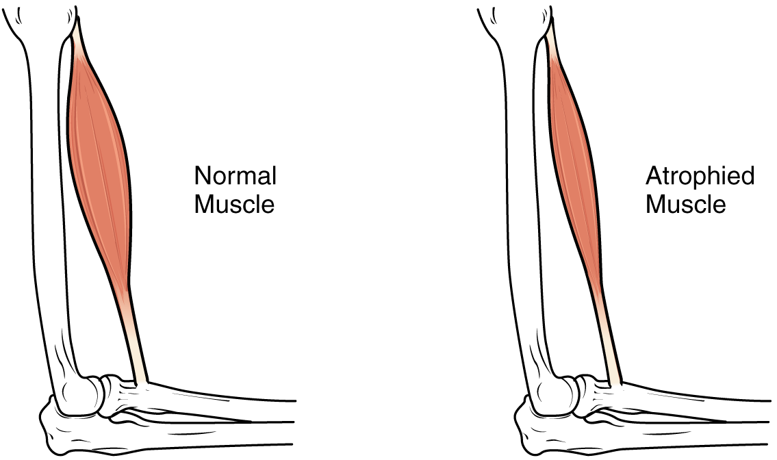
Muscle atrophy is the loss of skeletal muscle mass. It can be caused by immobility, aging, malnutrition, medications, or a wide range of injuries or diseases that impact the musculoskeletal or nervous system. Muscle atrophy leads to muscle weakness and causes disability. Disuse causes rapid muscle atrophy and often occurs during injury or illness that requires immobilization of a limb or bed rest. Depending on the duration of disuse and the health of the individual, this may be fully reversed with activity. Malnutrition first causes fat loss but may progress to muscle atrophy in prolonged starvation and can be reversed with nutritional therapy. In contrast, cachexia is a wasting syndrome caused by an underlying disease such as cancer that causes dramatic muscle atrophy and cannot be completely reversed with nutritional therapy. Sarcopenia is age-related muscle atrophy and can be slowed by exercise. Finally, diseases of the muscles such as muscular dystrophy or myopathies can caus ... [...More Info...] [...Related Items...] OR: [Wikipedia] [Google] [Baidu] |
Scoliosis
Scoliosis is a condition in which a person's spine has a sideways curve. The curve is usually "S"- or "C"-shaped over three dimensions. In some, the degree of curve is stable, while in others, it increases over time. Mild scoliosis does not typically cause problems, but more severe cases can affect breathing and movement. Pain is usually present in adults, and can worsen with age. The cause of most cases is unknown, but it is believed to involve a combination of genetic and environmental factors. Risk factors include other affected family members. It can also occur due to another condition such as muscle spasms, cerebral palsy, Marfan syndrome, and tumors such as neurofibromatosis. Diagnosis is confirmed with X-rays. Scoliosis is typically classified as either structural in which the curve is fixed, or functional in which the underlying spine is normal. Treatment depends on the degree of curve, location, and cause. Minor curves may simply be watched periodically. Trea ... [...More Info...] [...Related Items...] OR: [Wikipedia] [Google] [Baidu] |
Skeletal Muscle
Skeletal muscles (commonly referred to as muscles) are organs of the vertebrate muscular system and typically are attached by tendons to bones of a skeleton. The muscle cells of skeletal muscles are much longer than in the other types of muscle tissue, and are often known as muscle fibers. The muscle tissue of a skeletal muscle is striated – having a striped appearance due to the arrangement of the sarcomeres. Skeletal muscles are voluntary muscles under the control of the somatic nervous system. The other types of muscle are cardiac muscle which is also striated and smooth muscle which is non-striated; both of these types of muscle tissue are classified as involuntary, or, under the control of the autonomic nervous system. A skeletal muscle contains multiple fascicles – bundles of muscle fibers. Each individual fiber, and each muscle is surrounded by a type of connective tissue layer of fascia. Muscle fibers are formed from the fusion of developmental myoblas ... [...More Info...] [...Related Items...] OR: [Wikipedia] [Google] [Baidu] |


