|
Congenital Endothelial Dystrophy Type 2
Congenital hereditary corneal dystrophy (CHED) is a form of corneal endothelial dystrophy that presents at birth. CHED was previously subclassified into two subtypes: CHED1 and CHED2. However in 2015, the International Classification of Corneal Dystrophies (IC3D) renamed the condition "CHED1" to become posterior polymorphous corneal dystrophy, and renamed the condition "CHED2" to become, simply, CHED. Consequently, the scope of this article is restricted to the condition currently referred to as ''CHED'' Signs and symptoms CHED presents congenitally, but has a stationary course. The cornea exhibits a variable degree of clouding: from a diffuse haze, to a "ground glass" appearance, with occasional focal gray spots. The cornea thickens to between two and three times is normal thickness. Rarely, sub-epithelial band keratopathy and elevated intraocular pressure occur. Patients have blurred vision and nystagmus, however it is rare for the condition to be associated with either epi ... [...More Info...] [...Related Items...] OR: [Wikipedia] [Google] [Baidu] |
Corneal Dystrophy
Corneal dystrophy is a group of rare hereditary disorders characterised by bilateral abnormal deposition of substances in the transparent front part of the eye called the cornea. Signs and symptoms Corneal dystrophy may not significantly affect vision in the early stages. However, it does require proper evaluation and treatment for restoration of optimal vision. Corneal dystrophies usually manifest themselves during the first or second decade but sometimes later. It appears as grayish white lines, circles, or clouding of the cornea. Corneal dystrophy can also have a crystalline appearance. There are over 20 corneal dystrophies that affect all parts of the cornea. These diseases share many traits: * They are usually inherited. * They affect the right and left eyes equally. * They are not caused by outside factors, such as injury or diet. * Most progress gradually. * Most usually begin in one of the five corneal layers and may later spread to nearby layers. * Most do not affect oth ... [...More Info...] [...Related Items...] OR: [Wikipedia] [Google] [Baidu] |
Posterior Polymorphous Corneal Dystrophy
Posterior polymorphous corneal dystrophy (PPCD; sometimes also ''Schlichting dystrophy'') is a type of corneal dystrophy, characterised by changes in Descemet's membrane and endothelial layer. Symptoms mainly consist of decreased vision due to corneal edema. In some cases they are present from birth, other patients are asymptomatic. Histopathological analysis shows that the cells of endothelium have some characteristics of epithelial cells and have become multilayered. The disease was first described in 1916 by Koeppe as ''keratitis bullosa interna''. Genetics PPCD type 2 is linked to the mutations in COL8A2, and PPCD type 3 mutations in ZEB1 gene, but the underlying genetic disturbance in PPCD type 1 is unknown. Pathophysiology Vacuoles are demonstrated in the posterior parts of the cornea. The vesicles are located on the endothelial surface. The corneal endothelium is normally a single layer of cells that lose their mitotic potential after development is complete. In posterior ... [...More Info...] [...Related Items...] OR: [Wikipedia] [Google] [Baidu] |
Band Keratopathy
Band keratopathy is a corneal disease derived from the appearance of calcium on the central cornea. This is an example of metastatic calcification, which by definition, occurs in the presence of hypercalcemia. Signs and symptoms Signs and symptoms of band keratopathy include eye pain and decreased visual acuity. Causes Band keratopathy has several causes. These causes include uveitis, interstitial keratitis, superficial keratitis, phthisis, sarcoidosis, trauma, intraocular silicone oil, systemic diseases ( high levels of calcium in the blood, vitamin D intoxication, Fanconi's Syndrome, low levels of phosphorus in the blood, gout, milk-alkali syndrome, myotonic dystrophy, and chronic mercury exposure). Pathology Band keratopathy is seen when there is calcification of the epithelial basement membrane, Bowman's membrane, and the anterior stroma with destruction of Bowman's membrane. The calcium salts are intracellular when the process is due to alteration of systemic calcium me ... [...More Info...] [...Related Items...] OR: [Wikipedia] [Google] [Baidu] |
Intraocular Pressure
Intraocular pressure (IOP) is the fluid pressure inside the eye. Tonometry is the method eye care professionals use to determine this. IOP is an important aspect in the evaluation of patients at risk of glaucoma. Most tonometers are calibrated to measure pressure in millimeters of mercury ( mmHg). Physiology Intraocular pressure is determined by the production and drainage of aqueous humour by the ciliary body and its drainage via the trabecular meshwork and uveoscleral outflow. The reason for this is because the vitreous humour in the posterior segment has a relatively fixed volume and thus does not affect intraocular pressure regulation. An important quantitative relationship (Goldmann's equation) is as follows: :P_o = \frac + P_v Where: * P_o is the IOP in millimeters of mercury (mmHg) * F the rate of aqueous humour formation in microliters per minute (μL/min) * U the resorption of aqueous humour through the uveoscleral route (μL/min) * C is the facility of outflow in micr ... [...More Info...] [...Related Items...] OR: [Wikipedia] [Google] [Baidu] |
Epiphora (medicine)
Epiphora is an overflow of tears onto the face, other than caused by normal crying. It is a clinical sign or condition that constitutes insufficient tear film drainage from the eyes, in that tears will drain down the face rather than through the nasolacrimal system. Cause Causes of epiphora are any that cause either overproduction of tears or decreased drainage of tears, resulting in tearing onto the cheek. This can be due to ocular irritation and inflammation (including trichiasis and entropion) or an obstructed tear outflow tract which is divided according to its anatomical location (i.e. ectropion, punctal, canalicular or nasolacrimal duct obstruction). The latter is often due to aging (a spontaneous process), conjunctivochalasis, infection (i.e. dacryocystitis), rhinitis, and in neonates or infants, failure of the nasolacrimal duct to open. Another cause could be poor reconstruction of the nasolacrimal duct system after trauma to the area. Cause of trauma could be facial f ... [...More Info...] [...Related Items...] OR: [Wikipedia] [Google] [Baidu] |
Photophobia
Photophobia is a medical symptom of abnormal intolerance to visual perception of light. As a medical symptom photophobia is not a morbid fear or phobia, but an experience of discomfort or pain to the eyes due to light exposure or by presence of actual physical sensitivity of the eyes, though the term is sometimes additionally applied to abnormal or irrational fear of light such as heliophobia. The term ''photophobia'' comes from the Greek language, Greek φῶς (''phōs''), meaning "light", and φόβος (''phóbos''), meaning "fear". Causes Patients may develop photophobia as a result of several different medical conditions, related to the human eye, eye, the nervous system, genetic, or other causes. Photophobia may manifest itself in an increased response to light starting at any step in the visual system, such as: *Too much light entering the eye. Too much light can enter the eye if it is damaged, such as with corneal abrasion and retinal damage, or if its pupil(s) is unabl ... [...More Info...] [...Related Items...] OR: [Wikipedia] [Google] [Baidu] |
SLC4A11
Sodium bicarbonate transporter-like protein 11 is a protein that in humans is encoded by the ''SLC4A11'' gene. See also * Solute carrier family * Congenital endothelial dystrophy type 2 Congenital hereditary corneal dystrophy (CHED) is a form of corneal endothelial dystrophy that presents at birth. CHED was previously subclassified into two subtypes: CHED1 and CHED2. However in 2015, the International Classification of Corneal ... References Further reading * * * * * * * * * * * * * * Solute carrier family {{membrane-protein-stub ... [...More Info...] [...Related Items...] OR: [Wikipedia] [Google] [Baidu] |
Descemet's Membrane
Descemet's membrane ( or the Descemet membrane) is the basement membrane that lies between the corneal proper substance, also called stroma, and the endothelial layer of the cornea. It is composed of different kinds of collagen (Type IV and VIII) than the stroma. The endothelial layer is located at the posterior of the cornea. Descemet's membrane, as the basement membrane for the endothelial layer, is secreted by the single layer of squamous epithelial cells that compose the endothelial layer of the cornea. Structure Its thickness ranges from 3 μm at birth to 8–10 μm in adults.Johnson DH, Bourne WM, Campbell RJ: The ultrastructure of Descemet's membrane. I. Changes with age in normal cornea. Arch Ophthalmol 100:1942, 1982 The corneal endothelium is a single layer of squamous cells covering the surface of the cornea that faces the anterior chamber. Clinical significance Significant damage to the membrane may require a corneal transplant. Damage caused by the hereditary co ... [...More Info...] [...Related Items...] OR: [Wikipedia] [Google] [Baidu] |
Basement Membrane
The basement membrane is a thin, pliable sheet-like type of extracellular matrix that provides cell and tissue support and acts as a platform for complex signalling. The basement membrane sits between Epithelium, epithelial tissues including mesothelium and endothelium, and the underlying connective tissue. Structure As seen with the electron microscope, the basement membrane is composed of two layers, the basal lamina and the reticular lamina. The underlying connective tissue attaches to the basal lamina with collagen VII anchoring fibrils and fibrillin microfibrils. The basal lamina layer can further be subdivided into two layers based on their visual appearance in electron microscopy. The lighter-colored layer closer to the epithelium is called the lamina lucida, while the denser-colored layer closer to the connective tissue is called the lamina densa. The Electron microscope, electron-dense lamina densa layer is about 30–70 nanometers thick and consists of an underlying ... [...More Info...] [...Related Items...] OR: [Wikipedia] [Google] [Baidu] |
Corneal Stroma
The stroma of the cornea (or substantia propria) is a fibrous, tough, unyielding, perfectly transparent and the thickest layer of the cornea of the eye. It is between Bowman's membrane anteriorly, and Descemet's membrane posteriorly. At its centre, human corneal stroma is composed of about 200 flattened ''lamellæ'' (layers of collagen fibrils), superimposed one on another. They are each about 1.5-2.5 μm in thickness. The anterior lamellæ interweave more than posterior lamellæ. The fibrils of each lamella are parallel with one another, but at different angles to those of adjacent lamellæ. The lamellæ are produced by keratocytes (corneal connective tissue cells), which occupy about 10% of the substantia propria. Apart from the cells, the major non-aqueous constituents of the stroma are collagen fibrils and proteoglycans. The collagen fibrils are made of a mixture of type I and type V collagens. These molecules are tilted by about 15 degrees to the fibril axis, and becaus ... [...More Info...] [...Related Items...] OR: [Wikipedia] [Google] [Baidu] |
Corneal Transplantation
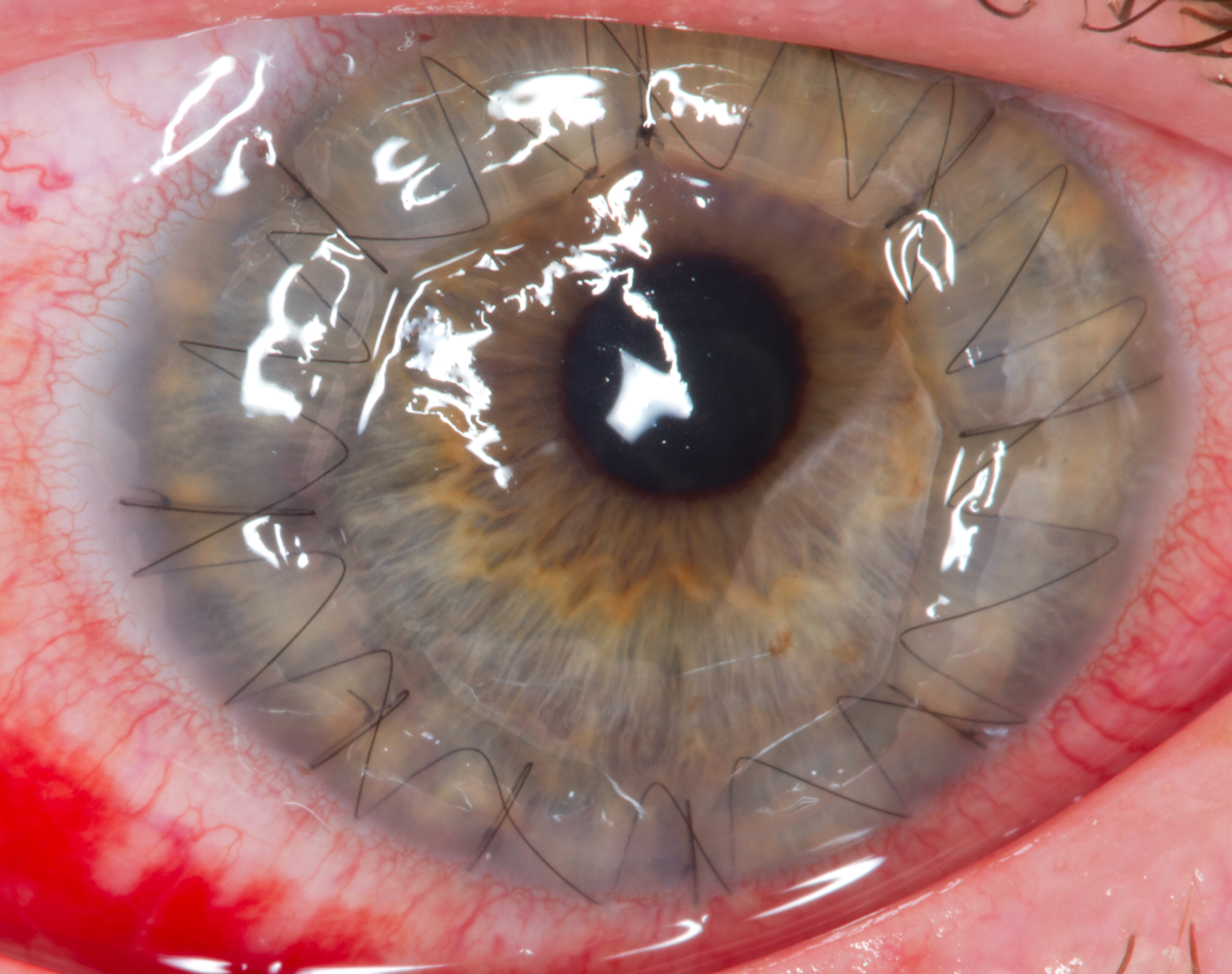
Corneal transplantation, also known as corneal grafting, is a surgical procedure where a damaged or diseased cornea is replaced by donated corneal tissue (the graft). When the entire cornea is replaced it is known as penetrating keratoplasty and when only part of the cornea is replaced it is known as lamellar keratoplasty. Keratoplasty simply means surgery to the cornea. The graft is taken from a recently deceased individual with no known diseases or other factors that may affect the chance of survival of the donated tissue or the health of the recipient. The cornea is the transparent front part of the eye that covers the iris, pupil and anterior chamber. The surgical procedure is performed by ophthalmologists, physicians who specialize in eyes, and is often done on an outpatient basis. Donors can be of any age, as is shown in the case of Janis Babson, who donated her eyes after dying at the age of 10. Corneal transplantation is performed when medicines, keratoconus conservat ... [...More Info...] [...Related Items...] OR: [Wikipedia] [Google] [Baidu] |
Posterior Polymorphous Corneal Dystrophy
Posterior polymorphous corneal dystrophy (PPCD; sometimes also ''Schlichting dystrophy'') is a type of corneal dystrophy, characterised by changes in Descemet's membrane and endothelial layer. Symptoms mainly consist of decreased vision due to corneal edema. In some cases they are present from birth, other patients are asymptomatic. Histopathological analysis shows that the cells of endothelium have some characteristics of epithelial cells and have become multilayered. The disease was first described in 1916 by Koeppe as ''keratitis bullosa interna''. Genetics PPCD type 2 is linked to the mutations in COL8A2, and PPCD type 3 mutations in ZEB1 gene, but the underlying genetic disturbance in PPCD type 1 is unknown. Pathophysiology Vacuoles are demonstrated in the posterior parts of the cornea. The vesicles are located on the endothelial surface. The corneal endothelium is normally a single layer of cells that lose their mitotic potential after development is complete. In posterior ... [...More Info...] [...Related Items...] OR: [Wikipedia] [Google] [Baidu] |


