|
Cone Dystrophy
A cone dystrophy is an inherited ocular disorder characterized by the loss of cone cells, the photoreceptors responsible for both central and color vision. Presentation The most common symptoms of cone dystrophy are vision loss (age of onset ranging from the late teens to the sixties), sensitivity to bright lights, and poor color vision. Therefore, patients see better at dusk. Visual acuity usually deteriorates gradually, but it can deteriorate rapidly to 20/200; later, in more severe cases, it drops to "counting fingers" vision. Color vision testing using color test plates (HRR series) reveals many errors on both red-green and blue-yellow plates. Dystrophy of the rods and cones Dystrophy of the light-sensing cells of the eye may also occur in the rods as well, or in both the cones and the rods. A type of rod-cone dystrophy—where rod function decline is typically earlier or more pronounced than cone dystrophy—has been identified as a relatively common characteristi ... [...More Info...] [...Related Items...] OR: [Wikipedia] [Google] [Baidu] |
Heredity
Heredity, also called inheritance or biological inheritance, is the passing on of traits from parents to their offspring; either through asexual reproduction or sexual reproduction, the offspring cells or organisms acquire the genetic information of their parents. Through heredity, variations between individuals can accumulate and cause species to evolve by natural selection. The study of heredity in biology is genetics. Overview In humans, eye color is an example of an inherited characteristic: an individual might inherit the "brown-eye trait" from one of the parents. Inherited traits are controlled by genes and the complete set of genes within an organism's genome is called its genotype. The complete set of observable traits of the structure and behavior of an organism is called its phenotype. These traits arise from the interaction of its genotype with the environment. As a result, many aspects of an organism's phenotype are not inherited. For example, suntanned ski ... [...More Info...] [...Related Items...] OR: [Wikipedia] [Google] [Baidu] |
Ophthalmoscopy
Ophthalmoscopy, also called funduscopy, is a test that allows a health professional to see inside the fundus of the eye and other structures using an ophthalmoscope (or funduscope). It is done as part of an eye examination and may be done as part of a routine physical examination. It is crucial in determining the health of the retina, optic disc, and vitreous humor. The pupil is a hole through which the eye's interior will be viewed. Opening the pupil wider (dilating it) is a simple and effective way to better see the structures behind it. Therefore, dilation of the pupil (mydriasis) is often accomplished with medicated eye drops before funduscopy. However, although dilated fundus examination is ideal, undilated examination is more convenient and is also helpful (albeit not as comprehensive), and it is the most common type in primary care. An alternative or complement to ophthalmoscopy is to perform a fundus photography, where the image can be analysed later by a professio ... [...More Info...] [...Related Items...] OR: [Wikipedia] [Google] [Baidu] |
Correlation
In statistics, correlation or dependence is any statistical relationship, whether causal or not, between two random variables or bivariate data. Although in the broadest sense, "correlation" may indicate any type of association, in statistics it usually refers to the degree to which a pair of variables are ''linearly'' related. Familiar examples of dependent phenomena include the correlation between the height of parents and their offspring, and the correlation between the price of a good and the quantity the consumers are willing to purchase, as it is depicted in the so-called demand curve. Correlations are useful because they can indicate a predictive relationship that can be exploited in practice. For example, an electrical utility may produce less power on a mild day based on the correlation between electricity demand and weather. In this example, there is a causal relationship, because extreme weather causes people to use more electricity for heating or cooling. Howev ... [...More Info...] [...Related Items...] OR: [Wikipedia] [Google] [Baidu] |
Eicosapentaenoic Acid
Eicosapentaenoic acid (EPA; also icosapentaenoic acid) is an omega-3 fatty acid. In physiological literature, it is given the name 20:5(n-3). It also has the trivial name timnodonic acid. In chemical structure, EPA is a carboxylic acid with a 20-carbon chain and five '' cis'' double bonds; the first double bond is located at the third carbon from the omega end. EPA is a polyunsaturated fatty acid (PUFA) that acts as a precursor for prostaglandin-3 (which inhibits platelet aggregation), thromboxane-3, and leukotriene-5 eicosanoids. EPA is both a precursor and the hydrolytic breakdown product of eicosapentaenoyl ethanolamide (EPEA: C22 H35 NO2; 20:5,n-3). Although studies of fish oil supplements, which contain both docosahexaenoic acid (DHA) and EPA, have failed to support claims of preventing heart attacks or strokes, a recent multi-year study of Vascepa ( ethyl eicosapentaenoate, the ethyl ester of the free fatty acid), a prescription drug containing only EPA, was sh ... [...More Info...] [...Related Items...] OR: [Wikipedia] [Google] [Baidu] |
Docosahexaenoic Acid
Docosahexaenoic acid (DHA) is an omega-3 fatty acid that is a primary structural component of the human brain, cerebral cortex, skin, and retina. In physiological literature, it is given the name 22:6(n-3). It can be synthesized from alpha-linolenic acid or obtained directly from maternal milk (breast milk), fatty fish, fish oil, or algae oil. DHA's structure is a carboxylic acid (-''oic acid'') with a 22- carbon chain (''docosa-'' derives from the Ancient Greek for 22) and six (''hexa-'') '' cis'' double bonds (''-en-''); with the first double bond located at the third carbon from the omega end. Its trivial name is cervonic acid (from the Latin word ''cerebrum'' for "brain"), its systematic name is ''all-cis''-docosa-4,7,10,13,16,19-hexa-enoic acid, and its shorthand name is 22:6(n−3) in the nomenclature of fatty acids. Most of the docosahexaenoic acid in fish and multi-cellular organisms with access to cold-water oceanic foods originates from photosynthetic and heterotr ... [...More Info...] [...Related Items...] OR: [Wikipedia] [Google] [Baidu] |
Omega-3 Fatty Acid
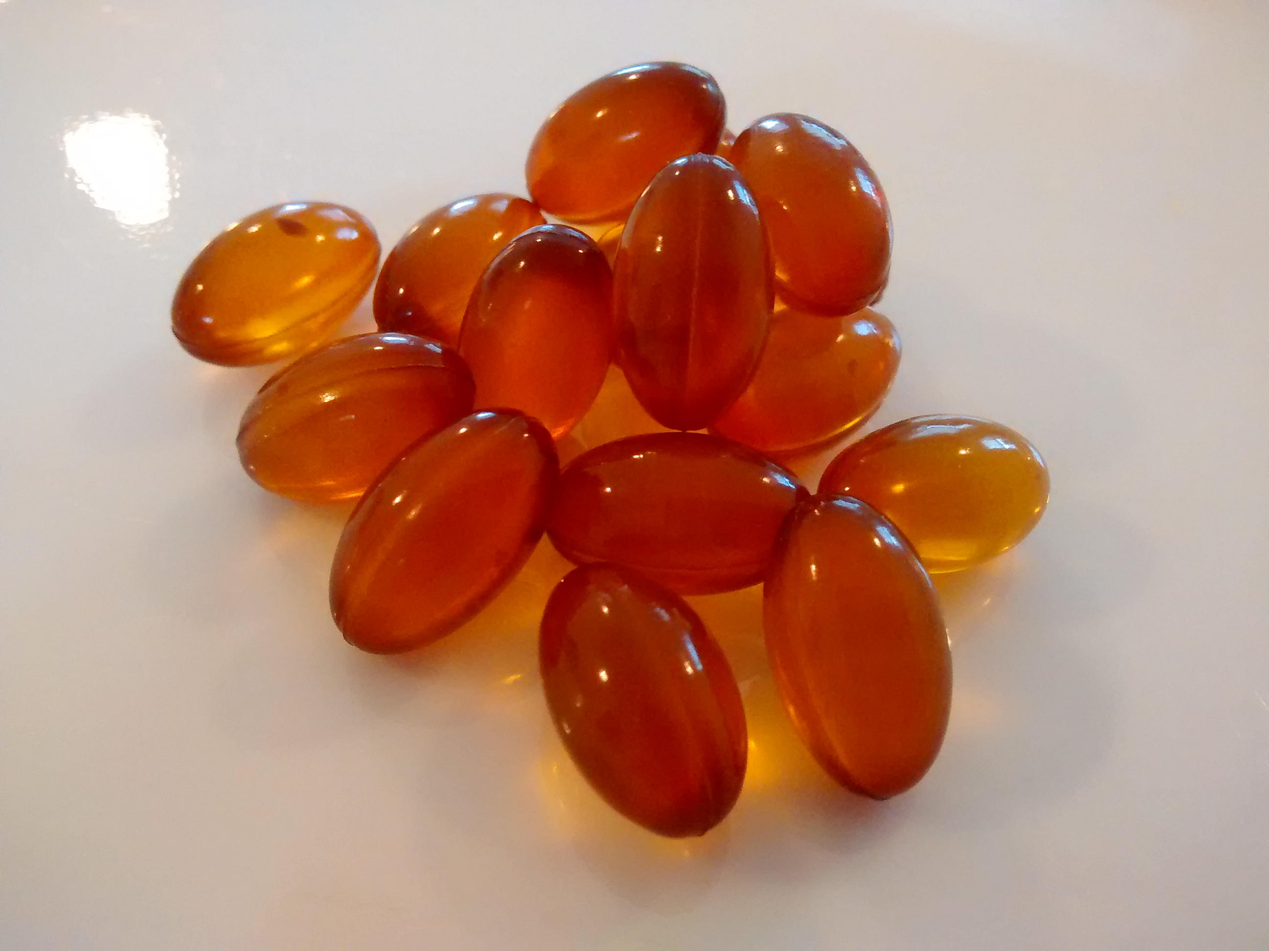
Omega−3 fatty acids, also called Omega-3 oils, ω−3 fatty acids or ''n''−3 fatty acids, are polyunsaturated fatty acids (PUFAs) characterized by the presence of a double bond, three atoms away from the terminal methyl group in their chemical structure. They are widely distributed in nature, being important constituents of animal lipid metabolism, and they play an important role in the human diet and in human physiology. The three types of omega−3 fatty acids involved in human physiology are α-linolenic acid (ALA), eicosapentaenoic acid (EPA) and docosahexaenoic acid (DHA). ALA can be found in plants, while DHA and EPA are found in algae and fish. Marine algae and phytoplankton are primary sources of omega−3 fatty acids. DHA and EPA accumulate in fish that eat these algae. Common sources of plant oils containing ALA include walnuts, edible seeds, and flaxseeds as well as hempseed oil, while sources of EPA and DHA include fish and fish oils, and algae oil. ... [...More Info...] [...Related Items...] OR: [Wikipedia] [Google] [Baidu] |
Zeaxanthin
Zeaxanthin is one of the most common carotenoids in nature, and is used in the xanthophyll cycle. Synthesized in plants and some micro-organisms, it is the pigment that gives paprika (made from bell peppers), corn, saffron, goji ( wolfberries), and many other plants and microbes their characteristic color. The name (pronounced ''zee-uh-zan'-thin'') is derived from '' Zea mays'' (common yellow maize corn, in which zeaxanthin provides the primary yellow pigment), plus ''xanthos'', the Greek word for "yellow" (see xanthophyll). Xanthophylls such as zeaxanthin are found in highest quantity in the leaves of most green plants, where they act to modulate light energy and perhaps serve as a non-photochemical quenching agent to deal with triplet chlorophyll (an excited form of chlorophyll) which is overproduced at high light levels during photosynthesis. Animals derive zeaxanthin from a plant diet. Zeaxanthin is one of the two primary xanthophyll carotenoids contained within the re ... [...More Info...] [...Related Items...] OR: [Wikipedia] [Google] [Baidu] |
Lutein
Lutein (;"Lutein" . from ''luteus'' meaning "yellow") is a and one of 600 known naturally occurring carotenoids. Lutein is synthesized only by plants, and like other xanthophylls is found in high quantities in [...More Info...] [...Related Items...] OR: [Wikipedia] [Google] [Baidu] |
Electroretinography
Electroretinography measures the electrical responses of various cell types in the retina, including the photoreceptors ( rods and cones), inner retinal cells ( bipolar and amacrine cells), and the ganglion cells. Electrodes are placed on the surface of the cornea (DTL silver/nylon fiber string or ERG jet) or on the skin beneath the eye (sensor strips) to measure retinal responses. Retinal pigment epithelium (RPE) responses are measured with an EOG test with skin-contact electrodes placed near the canthi. During a recording, the patient's eyes are exposed to standardized stimuli and the resulting signal is displayed showing the time course of the signal's amplitude (voltage). Signals are very small, and typically are measured in microvolts or nanovolts. The ERG is composed of electrical potentials contributed by different cell types within the retina, and the stimulus conditions (flash or pattern stimulus, whether a background light is present, and the colors of the stimul ... [...More Info...] [...Related Items...] OR: [Wikipedia] [Google] [Baidu] |
Scotoma
A scotoma is an area of partial alteration in the field of vision consisting of a partially diminished or entirely degenerated visual acuity that is surrounded by a field of normal – or relatively well-preserved – vision. Every normal mammalian eye has a scotoma in its field of vision, usually termed its blind spot. This is a location with no photoreceptor cells, where the retinal ganglion cell axons that compose the optic nerve exit the retina. This location is called the optic disc. There is no direct conscious awareness of visual scotomas. They are simply regions of reduced information within the visual field. Rather than recognizing an incomplete image, patients with scotomas report that things "disappear" on them. The presence of the blind spot scotoma can be demonstrated subjectively by covering one eye, carefully holding fixation with the open eye, and placing an object (such as one's thumb) in the lateral and horizontal visual field, about 15 degrees from fix ... [...More Info...] [...Related Items...] OR: [Wikipedia] [Google] [Baidu] |
Visual Field Test
A visual field test is an eye examination that can detect dysfunction in central and peripheral vision which may be caused by various medical conditions such as glaucoma, stroke, pituitary disease, brain tumours or other neurological deficits. Visual field testing can be performed clinically by keeping the subject's gaze fixed while presenting objects at various places within their visual field. Simple manual equipment can be used such as in the tangent screen test or the Amsler grid. When dedicated machinery is used it is called a perimeter. The exam may be performed by a technician in one of several ways. The test may be performed by a technician directly, with the assistance of a machine, or completely by an automated machine. Machine-based tests aid diagnostics by allowing a detailed printout of the patient's visual field. Other names for this test may include perimetry, Tangent screen exam, Automated perimetry exam, Goldmann visual field exam, or brand names such as Hens ... [...More Info...] [...Related Items...] OR: [Wikipedia] [Google] [Baidu] |
Fluorescein Angiography
Fluorescein angiography (FA), fluorescent angiography (FAG), or fundus fluorescein angiography (FFA) is a technique for examining the circulation of the retina and choroid (parts of the fundus) using a fluorescent dye and a specialized camera. Sodium fluorescein is added into the systemic circulation, the retina is illuminated with blue light at a wavelength of 490 nanometers, and an angiogram is obtained by photographing the fluorescent green light that is emitted by the dye. The fluorescein is administered intravenously in intravenous fluorescein angiography (IVFA) and orally in oral fluorescein angiography (OFA). The test is a dye tracing method. The fluorescein dye also reappears in the patient urine, causing the urine to appear darker, and sometimes orange. It can also cause discolouration of the saliva. Fluorescein angiography is one of several health care applications of this dye, all of which have a risk of severe adverse effects. See fluorescein safety in health care ... [...More Info...] [...Related Items...] OR: [Wikipedia] [Google] [Baidu] |


