|
Colpocephaly
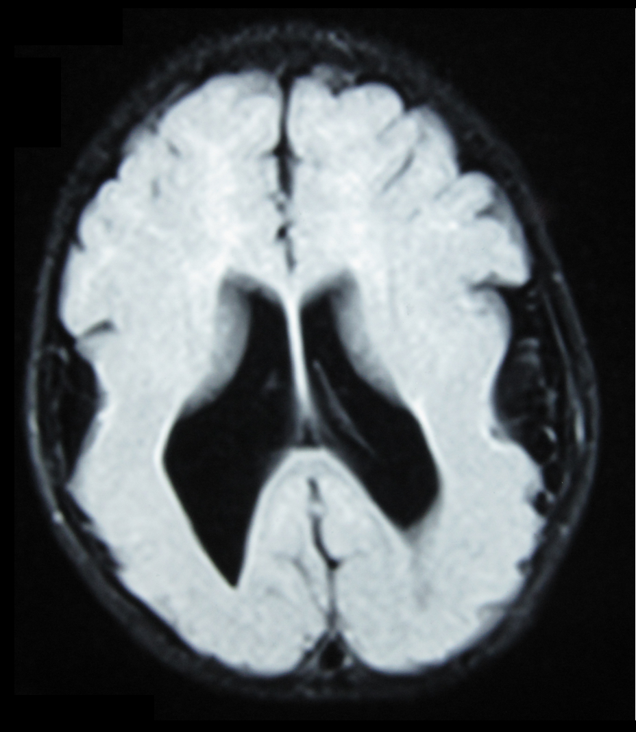
Colpocephaly is a cephalic disorder involving the disproportionate enlargement of the occipital horns of the lateral ventricles and is usually diagnosed early after birth due to seizures. It is a nonspecific finding and is associated with multiple neurological syndromes, including agenesis of the corpus callosum, Chiari malformation, lissencephaly, and microcephaly. Although the exact cause of colpocephaly is not known yet, it is commonly believed to occur as a result of neuronal migration disorders during early brain development, intrauterine disturbances, perinatal injuries, and other central nervous system disorders. Individuals with colpocephaly have various degrees of motor disabilities, visual defects, spasticity, and moderate to severe intellectual disability. No specific treatment for colpocephaly exists, but patients may undergo certain treatments to improve their motor function or intellectual disability. Symptoms There are various symptoms of colpocephaly and pati ... [...More Info...] [...Related Items...] OR: [Wikipedia] [Google] [Baidu] |
Colpocephaly W Corpus Callosum Dysgenesis MRI 3 09 Resultat
Colpocephaly is a cephalic disorder involving the disproportionate enlargement of the Posterior horn of lateral ventricle, occipital horns of the lateral ventricles and is usually diagnosed early after birth due to Epileptic seizures, seizures. It is a nonspecific finding and is associated with multiple neurological disorder, neurological syndromes, including agenesis of the corpus callosum, Chiari malformation, lissencephaly, and microcephaly. Although the exact cause of colpocephaly is not known yet, it is commonly believed to occur as a result of neuronal migration disorders during early brain development, Uterus, intrauterine disturbances, perinatal injuries, and other central nervous system disorders. Individuals with colpocephaly have various degrees of motor disabilities, visual defects, spasticity, and moderate to severe intellectual disability. No specific treatment for colpocephaly exists, but patients may undergo certain treatments to improve their motor function or intel ... [...More Info...] [...Related Items...] OR: [Wikipedia] [Google] [Baidu] |
Colpocephaly W Corpus Callosum Dysgenesis MRI 10 15 Resultat
Colpocephaly is a cephalic disorder involving the disproportionate enlargement of the occipital horns of the lateral ventricles and is usually diagnosed early after birth due to seizures. It is a nonspecific finding and is associated with multiple neurological syndromes, including agenesis of the corpus callosum, Chiari malformation, lissencephaly, and microcephaly. Although the exact cause of colpocephaly is not known yet, it is commonly believed to occur as a result of neuronal migration disorders during early brain development, intrauterine disturbances, perinatal injuries, and other central nervous system disorders. Individuals with colpocephaly have various degrees of motor disabilities, visual defects, spasticity, and moderate to severe intellectual disability. No specific treatment for colpocephaly exists, but patients may undergo certain treatments to improve their motor function or intellectual disability. Symptoms There are various symptoms of colpocephaly and patie ... [...More Info...] [...Related Items...] OR: [Wikipedia] [Google] [Baidu] |
Cephalic Disorder
Cephalic disorders () are congenital conditions that stem from damage to, or abnormal development of, the budding nervous system. Cephalic disorders are not necessarily caused by a single factor, but may be influenced by hereditary or genetic conditions, nutritional deficiencies, or by environmental exposures during pregnancy, such as medication taken by the mother, maternal infection, or exposure to radiation. Some cephalic disorders occur when the cranial sutures (the fibrous joints that connect the bones of the skull) join prematurely. Most cephalic disorders are caused by a disturbance that occurs very early in the development of the fetal nervous system. The human nervous system develops from a small, specialized plate of cells on the surface of the embryo. Early in development, this plate of cells forms the neural tube, a narrow sheath that closes between the third and fourth weeks of pregnancy to form the brain and spinal cord of the embryo. Four main processes are resp ... [...More Info...] [...Related Items...] OR: [Wikipedia] [Google] [Baidu] |
Lateral Ventricles
The lateral ventricles are the two largest ventricles of the brain and contain cerebrospinal fluid (CSF). Each cerebral hemisphere contains a lateral ventricle, known as the left or right ventricle, respectively. Each lateral ventricle resembles a C-shaped cavity that begins at an inferior horn in the temporal lobe, travels through a body in the parietal lobe and frontal lobe, and ultimately terminates at the interventricular foramina where each lateral ventricle connects to the single, central third ventricle. Along the path, a posterior horn extends backward into the occipital lobe, and an anterior horn extends farther into the frontal lobe. Structure Each lateral ventricle takes the form of an elongated curve, with an additional anterior-facing continuation emerging inferiorly from a point near the posterior end of the curve; the junction is known as the ''trigone of the lateral ventricle''. The centre of the superior curve is referred to as the ''body'', while the three ... [...More Info...] [...Related Items...] OR: [Wikipedia] [Google] [Baidu] |
Posterior Horn Of Lateral Ventricle
The lateral ventricles are the two largest ventricles of the brain and contain cerebrospinal fluid (CSF). Each cerebral hemisphere contains a lateral ventricle, known as the left or right ventricle, respectively. Each lateral ventricle resembles a C-shaped cavity that begins at an inferior horn in the temporal lobe, travels through a body in the parietal lobe and frontal lobe, and ultimately terminates at the interventricular foramina where each lateral ventricle connects to the single, central third ventricle. Along the path, a posterior horn extends backward into the occipital lobe, and an anterior horn extends farther into the frontal lobe. Structure Each lateral ventricle takes the form of an elongated curve, with an additional anterior-facing continuation emerging inferiorly from a point near the posterior end of the curve; the junction is known as the ''trigone of the lateral ventricle''. The centre of the superior curve is referred to as the ''body'', while the three ... [...More Info...] [...Related Items...] OR: [Wikipedia] [Google] [Baidu] |
Agenesis Of The Corpus Callosum
Agenesis of the corpus callosum (ACC) is a rare birth defect in which there is a complete or partial absence of the corpus callosum. It occurs when the development of the corpus callosum, the band of white matter connecting the two hemispheres in the brain, in the embryo is disrupted. The result of this is that the fibers that would otherwise form the corpus callosum are instead longitudinally oriented along the ipsilateral ventricular wall and form structures called Probst bundles. In addition to agenesis, other degrees of callosal defects exist, including hypoplasia (underdevelopment or thinness), hypogenesis (partial agenesis) or dysgenesis (malformation). ACC is found in many syndromes and can often present alongside hypoplasia of the cerebellar vermis. When this is the case, there can also be an enlarged fourth ventricle or hydrocephalus; this is called Dandy–Walker malformation. Signs and symptoms Signs and symptoms of ACC and other callosal disorders vary greatly ... [...More Info...] [...Related Items...] OR: [Wikipedia] [Google] [Baidu] |
Chiari Malformation
Chiari malformation (CM) is a structural defect in the cerebellum, characterized by a downward displacement of one or both cerebellar tonsils through the foramen magnum (the opening at the base of the skull). CMs can cause headaches, difficulty swallowing, vomiting, dizziness, neck pain, unsteady gait, poor hand coordination, numbness and tingling of the hands and feet, and speech problems. Less often, people may experience ringing or buzzing in the ears, weakness, slow heart rhythm, or fast heart rhythm, curvature of the spine ( scoliosis) related to spinal cord impairment, abnormal breathing, such as central sleep apnea, characterized by periods of breathing cessation during sleep, and, in severe cases, paralysis. This can sometimes lead to non-communicating hydrocephalus as a result of obstruction of cerebrospinal fluid (CSF) outflow. The cerebrospinal fluid outflow is caused by phase difference in outflow and influx of blood in the vasculature of the brain. The malforma ... [...More Info...] [...Related Items...] OR: [Wikipedia] [Google] [Baidu] |
Agenesis Of The Corpus Callosum
Agenesis of the corpus callosum (ACC) is a rare birth defect in which there is a complete or partial absence of the corpus callosum. It occurs when the development of the corpus callosum, the band of white matter connecting the two hemispheres in the brain, in the embryo is disrupted. The result of this is that the fibers that would otherwise form the corpus callosum are instead longitudinally oriented along the ipsilateral ventricular wall and form structures called Probst bundles. In addition to agenesis, other degrees of callosal defects exist, including hypoplasia (underdevelopment or thinness), hypogenesis (partial agenesis) or dysgenesis (malformation). ACC is found in many syndromes and can often present alongside hypoplasia of the cerebellar vermis. When this is the case, there can also be an enlarged fourth ventricle or hydrocephalus; this is called Dandy–Walker malformation. Signs and symptoms Signs and symptoms of ACC and other callosal disorders vary greatly ... [...More Info...] [...Related Items...] OR: [Wikipedia] [Google] [Baidu] |
Cisterna Magna
The cisterna magna (or cerebellomedullar cistern) is one of three principal openings in the subarachnoid space between the arachnoid and pia mater layers of the meninges surrounding the brain. The openings are collectively referred to as the subarachnoid cisterns. The cisterna magna is located between the cerebellum and the dorsal surface of the medulla oblongata. Cerebrospinal fluid produced in the fourth ventricle drains into the cisterna magna via the lateral apertures and median aperture. The two other principal cisterns are the ''pontine cistern'' located between the pons and the medulla and the ''interpeduncular cistern'' located between the cerebral peduncles. While the most commonly used clinical method for obtaining cerebrospinal fluid is a lumbar puncture Lumbar puncture (LP), also known as a spinal tap, is a medical procedure in which a needle is inserted into the spinal canal, most commonly to collect cerebrospinal fluid (CSF) for diagnostic testing. The main ... [...More Info...] [...Related Items...] OR: [Wikipedia] [Google] [Baidu] |
Lissencephaly
Lissencephaly (, meaning "smooth brain") is a set of rare brain disorders whereby the whole or parts of the surface of the brain appear smooth. It is caused by defective neuronal migration during the 12th to 24th weeks of gestation resulting in a lack of development of brain folds (gyri) and grooves (sulci). It is a form of cephalic disorder. Terms such as ''agyria'' (no gyri) and ''pachygyria'' (broad gyri) are used to describe the appearance of the surface of the brain. Children with lissencephaly generally have significant developmental delays, but these vary greatly from child to child depending on the degree of brain malformation and seizure control. Life expectancy can be shortened, generally due to respiratory problems. Symptoms and signs Affected children display severe psychomotor impairment, failure to thrive, seizures, and muscle spasticity or hypotonia. Other symptoms of the disorder may include unusual facial appearance, difficulty swallowing, and anomalies of the ... [...More Info...] [...Related Items...] OR: [Wikipedia] [Google] [Baidu] |
Periventricular Leukomalacia
Periventricular leukomalacia (PVL) is a form of white-matter brain injury, characterized by the necrosis (more often coagulation) of white matter near the lateral ventricles. It can affect newborns and (less commonly) fetuses; premature infants are at the greatest risk of neonatal encephalopathy which may lead to this condition. Affected individuals generally exhibit motor control problems or other developmental delays, and they often develop cerebral palsy or epilepsy later in life. The white matter in preterm born children is particularly vulnerable during the third trimester of pregnancy when white matter developing takes place and the myelination process starts around 30 weeks of gestational age. This pathology of the brain was described under various names ("encephalodystrophy", "ischemic necrosis", "periventricular infarction", "coagulation necrosis", "leukomalacia," "softening of the brain", "infarct periventricular white matter", "necrosis of white matter", "diffuse sym ... [...More Info...] [...Related Items...] OR: [Wikipedia] [Google] [Baidu] |
Hydrocephalus
Hydrocephalus is a condition in which an accumulation of cerebrospinal fluid (CSF) occurs within the brain. This typically causes increased intracranial pressure, pressure inside the skull. Older people may have headaches, double vision, poor balance, urinary incontinence, personality changes, or mental impairment. In babies, it may be seen as a rapid increase in head size. Other symptoms may include vomiting, sleepiness, seizures, and Parinaud's syndrome, downward pointing of the eyes. Hydrocephalus can occur due to birth defects or be acquired later in life. Associated birth defects include neural tube defects and those that result in aqueductal stenosis. Other causes include meningitis, brain tumors, traumatic brain injury, intraventricular hemorrhage, and subarachnoid hemorrhage. The four types of hydrocephalus are communicating, noncommunicating, ''ex vacuo'', and normal pressure hydrocephalus, normal pressure. Diagnosis is typically made by physical examination and medic ... [...More Info...] [...Related Items...] OR: [Wikipedia] [Google] [Baidu] |


