|
Color Opponency
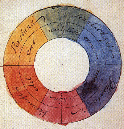
The opponent process is a color theory that states that the human visual system interprets information about color by processing signals from photoreceptor cells in an antagonistic manner. The opponent-process theory suggests that there are three opponent channels, each comprising an opposing color pair: red versus green, blue versus yellow, and black versus white (luminance). The theory was first proposed in 1892 by the German physiologist Ewald Hering. Color theory Complementary colors When staring at a bright color for a while (e.g. red), then looking away at a white field, an afterimage is perceived, such that the original color will evoke its complementary colors, complementary color (green, in the case of red input). When complementary colors are combined or mixed, they "cancel each other out" and become neutral (white or gray). That is, complementary colors are never perceived as a mixture; there is no "greenish red" or "yellowish blue", despite Impossible color#Colors ou ... [...More Info...] [...Related Items...] OR: [Wikipedia] [Google] [Baidu] |
Color Theory
Color theory, or more specifically traditional color theory, is a historical body of knowledge describing the behavior of colors, namely in color mixing, color contrast effects, color harmony, color schemes and color symbolism. Modern color theory is generally referred to as color science. While there is no clear distinction in scope, traditional color theory tends to be more subjective and have artistic applications, while color science tends to be more objective and have functional applications, such as in chemistry, astronomy or color reproduction. Color theory dates back at least as far as Aristotle's treatise ''On Colors'' and Bharata_(sage), Bharata's Natya_Shastra, Nāṭya Shāstra. A formalization of "color theory" began in the 18th century, initially within a partisan controversy over Isaac Newton's theory of color (''Opticks'', 1704) and the nature of primary colors. By the end of the 19th century, a schism had formed between traditional color theory and color science ... [...More Info...] [...Related Items...] OR: [Wikipedia] [Google] [Baidu] |
Young–Helmholtz Theory
The Young–Helmholtz theory (based on the work of Thomas Young and Hermann von Helmholtz in the 19th century), also known as the trichromatic theory, is a theory of trichromatic color vision – the manner in which the visual system gives rise to the phenomenological experience of color. In 1802, Young postulated the existence of three types of photoreceptors (now known as cone cells) in the eye, with different but overlapping response to different wavelengths of visible light. Hermann von Helmholtz developed the theory further in 1850: that the three types of cone photoreceptors could be classified as short-preferring ( violet), middle-preferring (green), and long-preferring ( red), according to their response to the wavelengths of light striking the retina. The relative strengths of the signals detected by the three types of cones are interpreted by the brain as a visible color. For instance, yellow light uses different proportions of red and green, but little blue, so any ... [...More Info...] [...Related Items...] OR: [Wikipedia] [Google] [Baidu] |
Parvocellular Layer
In neuroanatomy, the lateral geniculate nucleus (LGN; also called the lateral geniculate body or lateral geniculate complex) is a structure in the thalamus and a key component of the mammalian visual pathway. It is a small, ovoid, ventral projection of the thalamus where the thalamus connects with the optic nerve. There are two LGNs, one on the left and another on the right side of the thalamus. In humans, both LGNs have six layers of neurons (grey matter) alternating with optic fibers (white matter). The LGN receives information directly from the ascending retinal ganglion cells via the optic tract and from the reticular activating system. Neurons of the LGN send their axons through the optic radiation, a direct pathway to the primary visual cortex. In addition, the LGN receives many strong feedback connections from the primary visual cortex. In humans as well as other mammals, the two strongest pathways linking the eye to the brain are those projecting to the dorsal part of the ... [...More Info...] [...Related Items...] OR: [Wikipedia] [Google] [Baidu] |
Retinal Ganglion Cell
A retinal ganglion cell (RGC) is a type of neuron located near the inner surface (the ganglion cell layer) of the retina of the eye. It receives visual information from photoreceptor cell, photoreceptors via two intermediate neuron types: Bipolar cell of the retina, bipolar cells and retina amacrine cells. Retina amacrine cells, particularly narrow field cells, are important for creating functional subunits within the ganglion cell layer and making it so that ganglion cells can observe a small dot moving a small distance. Retinal ganglion cells collectively transmit image-forming and non-image forming visual information from the retina in the form of action potential to several regions in the thalamus, hypothalamus, and mesencephalon, or midbrain. Retinal ganglion cells vary significantly in terms of their size, connections, and responses to visual stimulation but they all share the defining property of having a long axon that extends into the brain. These axons form the optic ner ... [...More Info...] [...Related Items...] OR: [Wikipedia] [Google] [Baidu] |
Retina Bipolar Cell
As a part of the retina, bipolar cells exist between photoreceptors (rod cells and cone cells) and ganglion cells. They act, directly or indirectly, to transmit signals from the photoreceptors to the ganglion cells. Structure Bipolar cells are so-named as they have a central body from which two sets of processes arise. They can synapse with either rods or cones (rod/cone mixed input BCs have been found in teleost fish but not mammals), and they also accept synapses from horizontal cells. The bipolar cells then transmit the signals from the photoreceptors or the horizontal cells, and pass it on to the ganglion cells directly or indirectly (via amacrine cells). Unlike most neurons, bipolar cells communicate via graded potentials, rather than action potentials. Function Bipolar cells receive synaptic input from either rods or cones, or both rods and cones, though they are generally designated rod bipolar or cone bipolar cells. There are roughly 10 distinct forms of cone bipola ... [...More Info...] [...Related Items...] OR: [Wikipedia] [Google] [Baidu] |
Thalamus
The thalamus (: thalami; from Greek language, Greek Wikt:θάλαμος, θάλαμος, "chamber") is a large mass of gray matter on the lateral wall of the third ventricle forming the wikt:dorsal, dorsal part of the diencephalon (a division of the forebrain). Nerve fibers project out of the thalamus to the cerebral cortex in all directions, known as the thalamocortical radiations, allowing hub (network science), hub-like exchanges of information. It has several functions, such as the relaying of sensory neuron, sensory and motor neuron, motor signals to the cerebral cortex and the regulation of consciousness, sleep, and alertness. Anatomically, the thalami are paramedian symmetrical structures (left and right), within the vertebrate brain, situated between the cerebral cortex and the midbrain. It forms during embryonic development as the main product of the diencephalon, as first recognized by the Swiss embryologist and anatomist Wilhelm His Sr. in 1893. Anatomy The thalami ar ... [...More Info...] [...Related Items...] OR: [Wikipedia] [Google] [Baidu] |
Lateral Geniculate Nucleus
In neuroanatomy, the lateral geniculate nucleus (LGN; also called the lateral geniculate body or lateral geniculate complex) is a structure in the thalamus and a key component of the mammalian visual pathway. It is a small, ovoid, Anatomical terms of location#Dorsal_and_ventral, ventral projection of the thalamus where the thalamus connects with the optic nerve. There are two LGNs, one on the left and another on the right side of the thalamus. In humans, both LGNs have six layers of neurons (grey matter) alternating with optic fibers (white matter). The LGN receives information directly from the ascending retinal ganglion cells via the optic tract and from the reticular activating system. Neurons of the LGN send their axons through the optic radiation, a direct pathway to the primary visual cortex. In addition, the LGN receives many strong feedback connections from the primary visual cortex. In humans as well as other mammals, the two strongest pathways linking the eye to the bra ... [...More Info...] [...Related Items...] OR: [Wikipedia] [Google] [Baidu] |
Opponent Process Contrast Sensitivity Functions
The Opponent may refer to: * ''The Opponent'' (1988 film), a 1988 film starring Daniel Greene * ''The Opponent'' (2000 film), a 2000 film starring Erika Eleniak See also * * * * * Opposition (other) Opposition may refer to: Arts and media * ''Opposition'' (Altars EP), 2011 EP by Christian metalcore band Altars * The Opposition (band), a London post-punk band * '' The Opposition with Jordan Klepper'', a late-night television series on Comed ... * Antagonist (other) * Villain (other) {{DEFAULTSORT:Opponent, The ... [...More Info...] [...Related Items...] OR: [Wikipedia] [Google] [Baidu] |
Rod Cell
Rod cells are photoreceptor cells in the retina of the eye that can function in lower light better than the other type of visual photoreceptor, cone cells. Rods are usually found concentrated at the outer edges of the retina and are used in peripheral vision. On average, there are approximately 92 million rod cells (vs ~4.6 million cones) in the human retina. Rod cells are more sensitive than cone cells and are almost entirely responsible for night vision. However, rods have little role in color vision, which is the main reason why colors are much less apparent in dim light. Structure Rods are a little longer and leaner than cones but have the same basic structure. Opsin-containing disks lie at the end of the cell adjacent to the retinal pigment epithelium, which in turn is attached to the inside of the eye. The stacked-disc structure of the detector portion of the cell allows for very high efficiency. Rods are much more common than cones, with about 120 million rod cells ... [...More Info...] [...Related Items...] OR: [Wikipedia] [Google] [Baidu] |
CIE 1931 Color Space
In 1931, the International Commission on Illumination (CIE) published the CIE 1931 color spaces which define the relationship between the visible spectrum and human color vision. The CIE color spaces are mathematical models that comprise a "standard observer", which is a static idealization of the color vision of a normal human. A useful application of the CIEXYZ colorspace is that a mixture of two colors in some proportion lies on the straight line between those two colors. One disadvantage is that it is not perceptually uniform. This disadvantage is remedied in subsequent color models such as CIELUV and CIELAB, but these and modern color models still use the CIE 1931 color spaces as a foundation. The CIE (from the French name " Commission Internationale de l'éclairage" - International Commission on Illumination) developed and maintains many of the standards in use today relating to colorimetry. The CIE color spaces were created using data from a series of experiments, where ... [...More Info...] [...Related Items...] OR: [Wikipedia] [Google] [Baidu] |
LMS Color Space
LMS (long, medium, short), is a color space which represents the response of the three types of Cone cell, cones of the human eye, named for their responsivity (sensitivity) peaks at long, medium, and short wavelengths. The numerical range is generally not specified, except that the lower end is generally bounded by zero. It is common to use the LMS color space when performing chromatic adaptation (estimating the appearance of a sample under a different illuminant). It is also useful in the study of color blindness, when one or more cone types are defective. Definition The cone response functions \bar(\lambda), \bar(\lambda),\bar(\lambda) are the color matching functions (CMFs) for the LMS color space. The chromaticity coordinates (L, M, S) for a spectral distribution J(\lambda) are defined as: : L = \int^\infty_0 J(\lambda)\bar(\lambda)d\lambda : M = \int^\infty_0 J(\lambda)\bar(\lambda)d\lambda : S = \int^\infty_0 J(\lambda)\bar(\lambda)d\lambda The cone response function ... [...More Info...] [...Related Items...] OR: [Wikipedia] [Google] [Baidu] |


