|
Collateral Sulcus
The collateral fissure (or sulcus) is on the tentorial surface of the hemisphere and extends from near the occipital pole to within a short distance of the temporal pole. Behind, it lies below and lateral to the calcarine fissure, from which it is separated by the lingual gyrus; in front, it is situated between the parahippocampal gyrus and the anterior part of the fusiform gyrus. Additional images File:Gray738.png, Coronal section through posterior cornua of lateral ventricle The lateral ventricles are the two largest ventricles of the brain and contain cerebrospinal fluid (CSF). Each cerebral hemisphere contains a lateral ventricle, known as the left or right ventricle, respectively. Each lateral ventricle resemble .... (Collateral fissure labeled at bottom center.) File:Hippocampal Limbic Connections Functions - Sanjoy Sanyal (Cropped from 5m28s to 6m30s) Collateral sulcus.webm, Human brain dissection video (62 sec). Demonstrating location of collateral sulcus. Re ... [...More Info...] [...Related Items...] OR: [Wikipedia] [Google] [Baidu] |
Limbic Lobe
The limbic lobe is an arc-shaped region of cortex on the medial surface of each cerebral hemisphere of the mammalian brain, consisting of parts of the frontal, parietal and temporal lobes. The term is ambiguous, with some authors including the paraterminal gyrus, the subcallosal area, the cingulate gyrus, the parahippocampal gyrus, the dentate gyrus, the hippocampus and the subiculum; while the Terminologia Anatomica includes the cingulate sulcus, the cingulate gyrus, the isthmus of cingulate gyrus, the fasciolar gyrus, the parahippocampal gyrus, the parahippocampal sulcus, the dentate gyrus, the fimbrodentate sulcus, the fimbria of hippocampus, the collateral sulcus, and the rhinal sulcus, and omits the hippocampus. History Broca named the limbic lobe in 1878, identifying it with the cingulate and parahippocampal gyri, and associating it with the sense of smell - Treviranus having earlier noted that, between species, the size of the parahippocampal gyrus varies with the ... [...More Info...] [...Related Items...] OR: [Wikipedia] [Google] [Baidu] |
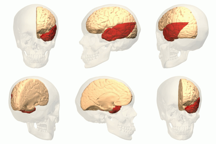
Temporal Lobe
The temporal lobe is one of the four major lobes of the cerebral cortex in the brain of mammals. The temporal lobe is located beneath the lateral fissure on both cerebral hemispheres of the mammalian brain. The temporal lobe is involved in processing sensory input into derived meanings for the appropriate retention of visual memory, language comprehension, and emotion association. ''Temporal'' refers to the head's temples. Structure The temporal lobe consists of structures that are vital for declarative or long-term memory. Declarative (denotative) or explicit memory is conscious memory divided into semantic memory (facts) and episodic memory (events). Medial temporal lobe structures that are critical for long-term memory include the hippocampus, along with the surrounding hippocampal region consisting of the perirhinal, parahippocampal, and entorhinal neocortical regions. The hippocampus is critical for memory formation, and the surrounding medial temporal cortex is curre ... [...More Info...] [...Related Items...] OR: [Wikipedia] [Google] [Baidu] |
Occipital Pole
The vertebrate cerebrum (brain) is formed by two cerebral hemispheres that are separated by a groove, the longitudinal fissure. The brain can thus be described as being divided into left and right cerebral hemispheres. Each of these hemispheres has an outer layer of grey matter, the cerebral cortex, that is supported by an inner layer of white matter. In eutherian (placental) mammals, the hemispheres are linked by the corpus callosum, a very large bundle of nerve fibers. Smaller commissures, including the anterior commissure, the posterior commissure and the fornix, also join the hemispheres and these are also present in other vertebrates. These commissures transfer information between the two hemispheres to coordinate localized functions. There are three known poles of the cerebral hemispheres: the '' occipital pole'', the '' frontal pole'', and the ''temporal pole''. The central sulcus is a prominent fissure which separates the parietal lobe from the frontal lobe and the prima ... [...More Info...] [...Related Items...] OR: [Wikipedia] [Google] [Baidu] |
Temporal Pole
The vertebrate cerebrum (brain) is formed by two cerebral hemispheres that are separated by a groove, the longitudinal fissure. The brain can thus be described as being divided into left and right cerebral hemispheres. Each of these hemispheres has an outer layer of grey matter, the cerebral cortex, that is supported by an inner layer of white matter. In eutherian (placental) mammals, the hemispheres are linked by the corpus callosum, a very large bundle of nerve fibers. Smaller commissures, including the anterior commissure, the posterior commissure and the fornix, also join the hemispheres and these are also present in other vertebrates. These commissures transfer information between the two hemispheres to coordinate localized functions. There are three known poles of the cerebral hemispheres: the '' occipital pole'', the '' frontal pole'', and the '' temporal pole''. The central sulcus is a prominent fissure which separates the parietal lobe from the frontal lobe and the pr ... [...More Info...] [...Related Items...] OR: [Wikipedia] [Google] [Baidu] |
Calcarine Fissure
The calcarine sulcus (or calcarine fissure) is an anatomical landmark located at the caudal end of the medial surface of the brain of humans and other primates. Its name comes from the Latin "calcar" meaning "spur". It is very deep, and known as a complete sulcus. Structure The calcarine sulcus begins near the occipital pole in two converging rami. It runs forward to a point a little below the splenium of the corpus callosum. Here, it is joined at an acute angle by the medial part of the parieto-occipital sulcus. The anterior part of this sulcus gives rise to the prominence of the calcar avis in the posterior cornu of the lateral ventricle. The cuneus is above the calcarine sulcus, while the lingual gyrus is below it. Development In humans, the calcarine sulcus usually becomes visible between 20 weeks and 28 weeks of gestation. Function The calcarine sulcus is associated with visual cortex. It is where the primary visual cortex (V1) is concentrated. The central visu ... [...More Info...] [...Related Items...] OR: [Wikipedia] [Google] [Baidu] |
Lingual Gyrus
The lingual gyrus, also known as the ''medial'' occipitotemporal gyrus, is a brain structure that is linked to processing vision, especially related to letters. It is thought to also play a role in analysis of logical conditions (i.e., logical order of events) and encoding visual memories. It is named after its shape, which is somewhat similar to a tongue. Contrary to the name, the region has little to do with speech. It is believed that a hypermetabolism of the lingual gyrus is associated with visual snow. Location The lingual gyrus of the occipital lobe lies between the calcarine sulcus and the posterior part of the collateral sulcus; behind, it reaches the occipital pole; in front, it is continued on to the tentorial surface of the temporal lobe, and joins the parahippocampal gyrus. Function Role in vision This region is believed to play an important role in vision and dreaming. Visual memory dysfunction and visuo- limbic disconnection have been shown in cases where the l ... [...More Info...] [...Related Items...] OR: [Wikipedia] [Google] [Baidu] |
Parahippocampal Gyrus
The parahippocampal gyrus (or hippocampal gyrus') is a grey matter cortical region of the brain that surrounds the hippocampus and is part of the limbic system. The region plays an important role in memory encoding and retrieval. It has been involved in some cases of hippocampal sclerosis. Asymmetry has been observed in schizophrenia. Structure The anterior part of the gyrus includes the perirhinal and entorhinal cortices. The term parahippocampal cortex is used to refer to an area that encompasses both the posterior parahippocampal gyrus and the medial portion of the fusiform gyrus. Function Scene recognition The parahippocampal place area (PPA) is a sub-region of the parahippocampal cortex that lies medially in the inferior temporo-occipital cortex. PPA plays an important role in the encoding and recognition of environmental scenes (rather than faces). fMRI studies indicate that this region of the brain becomes highly active when human subjects view topographical scene ... [...More Info...] [...Related Items...] OR: [Wikipedia] [Google] [Baidu] |
Fusiform Gyrus
The fusiform gyrus, also known as the ''lateral occipitotemporal gyrus'','' ''is part of the temporal lobe and occipital lobe in Brodmann area 37. The fusiform gyrus is located between the lingual gyrus and parahippocampal gyrus above, and the inferior temporal gyrus below. Though the functionality of the fusiform gyrus is not fully understood, it has been linked with various neural pathways related to recognition. Additionally, it has been linked to various neurological phenomena such as synesthesia, dyslexia, and prosopagnosia. Anatomy Anatomically, the fusiform gyrus is the largest macro-anatomical structure within the ventral temporal cortex, which mainly includes structures involved in high-level vision. The term fusiform gyrus (lit. "spindle-shaped convolution") refers to the fact that the shape of the gyrus is wider at its centre than at its ends. This term is based on the description of the gyrus by Emil Huschke in 1854. (see also section on history). The fus ... [...More Info...] [...Related Items...] OR: [Wikipedia] [Google] [Baidu] |
Lateral Ventricle
The lateral ventricles are the two largest ventricles of the brain and contain cerebrospinal fluid (CSF). Each cerebral hemisphere contains a lateral ventricle, known as the left or right ventricle, respectively. Each lateral ventricle resembles a C-shaped cavity that begins at an inferior horn in the temporal lobe, travels through a body in the parietal lobe and frontal lobe, and ultimately terminates at the interventricular foramina where each lateral ventricle connects to the single, central third ventricle. Along the path, a posterior horn extends backward into the occipital lobe, and an anterior horn extends farther into the frontal lobe. Structure Each lateral ventricle takes the form of an elongated curve, with an additional anterior-facing continuation emerging inferiorly from a point near the posterior end of the curve; the junction is known as the ''trigone of the lateral ventricle''. The centre of the superior curve is referred to as the ''body'', while the three ... [...More Info...] [...Related Items...] OR: [Wikipedia] [Google] [Baidu] |
Cerebrum
The cerebrum, telencephalon or endbrain is the largest part of the brain containing the cerebral cortex (of the two cerebral hemispheres), as well as several subcortical structures, including the hippocampus, basal ganglia, and olfactory bulb. In the human brain, the cerebrum is the uppermost region of the central nervous system. The cerebrum develops prenatally from the forebrain (prosencephalon). In mammals, the dorsal telencephalon, or pallium, develops into the cerebral cortex, and the ventral telencephalon, or subpallium, becomes the basal ganglia. The cerebrum is also divided into approximately symmetric left and right cerebral hemispheres. With the assistance of the cerebellum, the cerebrum controls all voluntary actions in the human body. Structure The cerebrum is the largest part of the brain. Depending upon the position of the animal it lies either in front or on top of the brainstem. In humans, the cerebrum is the largest and best-developed of the five majo ... [...More Info...] [...Related Items...] OR: [Wikipedia] [Google] [Baidu] |
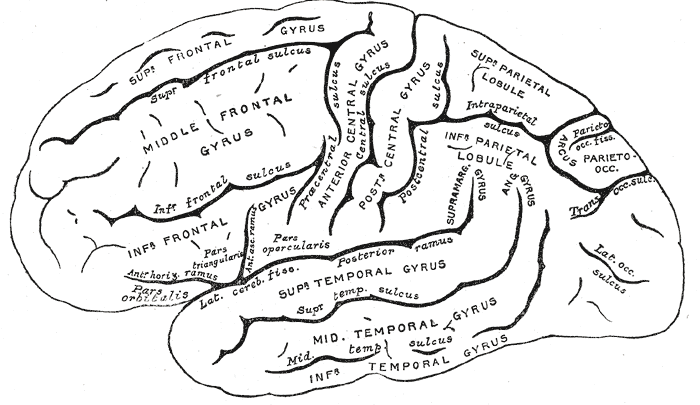
Sulci (neuroanatomy)
In neuroanatomy, a sulcus ( Latin: "furrow", pl. ''sulci'') is a depression or groove in the cerebral cortex. It surrounds a gyrus (pl. gyri), creating the characteristic folded appearance of the brain in humans and other mammals. The larger sulci are usually called fissures. Structure Sulci, the grooves, and gyri, the folds or ridges, make up the folded surface of the cerebral cortex. Larger or deeper sulci are termed fissures, and in many cases the two terms are interchangeable. The folded cortex creates a larger surface area for the brain in humans and other mammals. When looking at the human brain, two-thirds of the surface are hidden in the grooves. The sulci and fissures are both grooves in the cortex, but they are differentiated by size. A sulcus is a shallower groove that surrounds a gyrus. A fissure is a large furrow that divides the brain into lobes and also into the two hemispheres as the longitudinal fissure. Importance of expanded surface area As the ... [...More Info...] [...Related Items...] OR: [Wikipedia] [Google] [Baidu] |

_-_inferiror_view.png)

