|
Coenenchyme
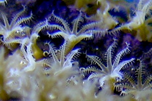
Coenenchyme is the common tissue that surrounds and links the polyps in octocorals. It consists of mesoglea penetrated by tubes (''solenia'') and canals of the gastrodermis and contains sclerites, microscopic mineralised spicules of silica or of calcium carbonate. The outer layer of the coenenchyme is made of columnar or squamous epithelial cells, and can be covered in microvilli. The stiff projecting portion of coenenchyme that surrounds each polyp is usually reinforced by modified sclerites and is called the calyx Calyx or calyce (plural "calyces"), from the Latin ''calix'' which itself comes from the Ancient Greek ''κάλυξ'' (''kálux'') meaning "husk" or "pod", may refer to: Biology * Calyx (anatomy), collective name for several cup-like structures ..., a term borrowed from botany. The solenia circulate nutrients throughout the coenenchyme. Coenosarc is an alternative name. References {{reflist Cnidarian anatomy ... [...More Info...] [...Related Items...] OR: [Wikipedia] [Google] [Baidu] |
Octocorallia
Octocorallia (also known as Alcyonaria) is a class of Anthozoa comprising around 3,000 species of water-based organisms formed of colonial polyps with 8-fold symmetry. It includes the blue coral, soft corals, sea pens, and gorgonians (sea fans and sea whips) within three orders: Alcyonacea, Helioporacea, and Pennatulacea. These organisms have an internal skeleton secreted by mesoglea and polyps with eight tentacles and eight mesentaries. As with all Cnidarians these organisms have a complex life cycle including a motile phase when they are considered plankton and later characteristic sessile phase. Octocorals have existed at least since the Ordovician period, as shown by Maurits Lindström's findings in the 1970s, however recent work has shown a possible Cambrian origin. Biology Octocorals resemble the stony corals in general appearance and in the size of their polyps, but lack the distinctive stony skeleton. Also unlike the stony corals, each polyp has only eight tentacle ... [...More Info...] [...Related Items...] OR: [Wikipedia] [Google] [Baidu] |
Polyp (zoology)
A polyp in zoology Zoology ()The pronunciation of zoology as is usually regarded as nonstandard, though it is not uncommon. is the branch of biology that studies the animal kingdom, including the structure, embryology, evolution, classification, habits, and ... is one of two forms found in the phylum Cnidaria, the other being the medusa (biology), medusa. Polyps are roughly cylindrical in shape and elongated at the axis of the vase-shaped body. In solitary polyps, the aboral (opposite to oral) end is attached to the substrate (biology), substrate by means of a disc-like holdfast (biology), holdfast called a pedal disc, while in colony (biology), colonies of polyps it is connected to other polyps, either directly or indirectly. The oral end contains the mouth, and is surrounded by a circlet of tentacles. Classes In the class (biology), class Anthozoa, comprising the sea anemones and corals, the individual is always a polyp; in the class Hydrozoa, however, the indi ... [...More Info...] [...Related Items...] OR: [Wikipedia] [Google] [Baidu] |
Mesoglea
Mesoglea refers to the extracellular matrix found in cnidarians like coral or jellyfish that functions as a hydrostatic skeleton. It is related to but distinct from mesohyl, which generally refers to extracellular material found in sponges. Description The mesoglea is mostly water. Other than water, the mesoglea is composed of several substances including fibrous proteins, like collagen and heparan sulphate proteoglycans. The mesoglea is mostly acellular, but in both cnidaria and ctenophora the mesoglea contains muscle bundles and nerve fibres. Other nerve and muscle cells lie just under the epithelial layers. The mesoglea also contains wandering amoebocytes that play a role in phagocytosing debris and bacteria. These cells also fight infections by producing antibacterial chemicals. The mesoglea may be thinner than either of the cell layers in smaller coelenterates like a hydra or may make up the bulk of the body in larger jellyfish. The mesoglea serves as an internal skelet ... [...More Info...] [...Related Items...] OR: [Wikipedia] [Google] [Baidu] |
Gastrodermis
The gastrodermis is the inner layer of cells that serves as a lining membrane of the gastrovascular cavity of Cnidarians. The term is also used for the analogous inner epithelial layer of Ctenophore Ctenophora (; ctenophore ; ) comprise a phylum of marine invertebrates, commonly known as comb jellies, that inhabit sea waters worldwide. They are notable for the groups of cilia they use for swimming (commonly referred to as "combs"), and ...s. It has been shown that the gastrodermis is among the sites where early signals of heat stress are expressed in corals. References Digestive system {{Cell-biology-stub ... [...More Info...] [...Related Items...] OR: [Wikipedia] [Google] [Baidu] |
Sclerite
A sclerite ( Greek , ', meaning " hard") is a hardened body part. In various branches of biology the term is applied to various structures, but not as a rule to vertebrate anatomical features such as bones and teeth. Instead it refers most commonly to the hardened parts of arthropod exoskeletons and the internal spicules of invertebrates such as certain sponges and soft corals. In paleontology, a scleritome is the complete set of sclerites of an organism, often all that is known from fossil invertebrates. Sclerites in combination Sclerites may occur practically isolated in an organism, such as the sting of a cone shell. Also, they can be more or less scattered, such as tufts of defensive sharp, mineralised bristles as in many marine Polychaetes. Or, they can occur as structured, but unconnected or loosely connected arrays, such as the mineral "teeth" in the radula of many Mollusca, the valves of Chitons, the beak of Cephalopod, or the articulated exoskeletons of Arthropoda. ... [...More Info...] [...Related Items...] OR: [Wikipedia] [Google] [Baidu] |
Sponge Spicule
Spicules are structural elements found in most sponges. The meshing of many spicules serves as the sponge's skeleton and thus it provides structural support and potentially defense against predators. Sponge spicules are made of calcium carbonate or silica. Large spicules visible to the naked eye are referred to as megascleres, while smaller, microscopic ones are termed microscleres. The composition, size, and shape of spicules are major characters in sponge systematics and taxonomy. Overview Sponges are a species-rich clade of the earliest-diverging (most basal) animals. They are distributed globally, with diverse ecologies and functions, and a record spanning at least the entire Phanerozoic. Most sponges produce skeletons formed by spicules, structural elements that develop in a wide variety of sizes and three dimensional shapes. Among the four sub-clades of Porifera, three ( Demospongiae, Hexactinellida, and Homoscleromorpha) produce skeletons of amorphous silica and ... [...More Info...] [...Related Items...] OR: [Wikipedia] [Google] [Baidu] |
Microvilli
Microvilli (singular: microvillus) are microscopic cellular membrane protrusions that increase the surface area for diffusion and minimize any increase in volume, and are involved in a wide variety of functions, including absorption, secretion, cellular adhesion, and mechanotransduction. Structure Microvilli are covered in plasma membrane, which encloses cytoplasm and microfilaments. Though these are cellular extensions, there are little or no cellular organelles present in the microvilli. Each microvillus has a dense bundle of cross-linked actin filaments, which serves as its structural core. 20 to 30 tightly bundled actin filaments are cross-linked by bundling proteins fimbrin (or plastin-1), villin and espin to form the core of the microvilli. In the enterocyte microvillus, the structural core is attached to the plasma membrane along its length by lateral arms made of myosin 1a and Ca2+ binding protein calmodulin. Myosin 1a functions through a binding site for filamentous ... [...More Info...] [...Related Items...] OR: [Wikipedia] [Google] [Baidu] |
Calyx (anatomy)
Calyx is a term used in animal anatomy for some cuplike areas or structures. Etymology Latin, from ''calyx'' (from Ancient Greek ''κάλυξ, ''case of a bud, husk"). Cnidarians The spicules containing the basal portion of the upper tentacular part of the polyp of some soft corals (also called ''calice''). Entoprocta A body part of the Entoprocta from which tentacles arise and the mouth and anus are located. Echinoderms The body disk that is covered with a leathery tegumen containing calcareous plates (in crinoids and ophiuroids the main part of the body where the viscera are located). Humans Either a minor calyx in the kidney, a conglomeration of two or three minor calyces to form a major calyx, or the Calyx of Held, a particularly large synapse in the mammalian auditory central nervous system, named by H. Held in his 1893 article ''Die centrale Gehörleitung'', due to its flower-petal-like shape. Satzler, K., L. F. Sohl, et al. (2002). "Three-dimensional reconstru ... [...More Info...] [...Related Items...] OR: [Wikipedia] [Google] [Baidu] |
Coenosarc
In corals, the coenosarc is the living tissue overlying the stony skeletal material of the coral. It secretes the coenosteum, the layer of skeletal material lying between the corallites (the stony cups in which the polyps sit). The coensarc is composed of mesogloea between two thin layers of epidermis and is continuous with the body wall of the polyps. The coenosarc contains the gastrovascular canal system that links the polyps and allow them to share nutrients and symbiotic zooxanthellae Zooxanthellae is a colloquial term for single-celled dinoflagellates that are able to live in symbiosis with diverse marine invertebrates including demosponges, corals, jellyfish, and nudibranchs. Most known zooxanthellae are in the genus '' .... References {{reflist Cnidarian anatomy ... [...More Info...] [...Related Items...] OR: [Wikipedia] [Google] [Baidu] |


