|
Clostridial Cytotoxin Family
The Clostridial Cytotoxin (CCT) FamilyTC# 1.C.57 is a member of the RTX-toxin superfamily. There are currently 13 classified members belonging to the CCT family. A representative list of these proteins is available in thTransporter Classification Database Homologues are found in a variety of Gram-positive and Gram-negative bacteria. ''Clostridium difficile'' cytotoxins ''Clostridium difficile'', the causative agent of nosocomial antibiotic-associated diarrhea and pseudomembranous colitis, possesses two main virulence factors: the large clostridial cytotoxins A (TcdATC# 1.C.57.1.2 and B (TcdBTC# 1.C.57.1.1. Action by large clostridial toxins (LCTs) from Clostridium difficile includes four steps: (1) receptor-mediated endocytosis, (2) translocation of a catalytic glucosyltransferase domain across the membrane, (3) release of the enzymatic part by auto-proteolysis, and (4) inactivation of Rho family proteins. Cleavage of toxin B and all other large clostridial cytotoxins, is an au ... [...More Info...] [...Related Items...] OR: [Wikipedia] [Google] [Baidu] |
RTX Toxin
The RTX toxin superfamily is a group of cytolysins and cytotoxins produced by bacteria. There are over 1000 known members with a variety of functions. The RTX family is defined by two common features: characteristic repeats in the toxin protein sequences, and extracellular secretion by the type I secretion systems (T1SS). The name RTX (repeats in toxin) refers to the glycine and aspartate-rich repeats located at the C-terminus of the toxin proteins, which facilitate export by a dedicated T1SS encoded within the ''rtx'' operon. Structure and function RTX proteins range from 40 to over 600 kDa in size and all contain C-terminally located glycine and aspartate-rich repeat sequences of nine amino acids. The repeats contain the common sequence structure , (where X represents any amino acid), but the number of repeats varies within RTX protein family members. These consensus regions function as sites for Ca2+ binding, which facilitate folding of the RTX protein following export via an ... [...More Info...] [...Related Items...] OR: [Wikipedia] [Google] [Baidu] |
Gram-positive Bacteria
In bacteriology, gram-positive bacteria are bacteria that give a positive result in the Gram stain test, which is traditionally used to quickly classify bacteria into two broad categories according to their type of cell wall. Gram-positive bacteria take up the crystal violet stain used in the test, and then appear to be purple-coloured when seen through an optical microscope. This is because the thick peptidoglycan layer in the bacterial cell wall retains the stain after it is washed away from the rest of the sample, in the decolorization stage of the test. Conversely, gram-negative bacteria cannot retain the violet stain after the decolorization step; alcohol used in this stage degrades the outer membrane of gram-negative cells, making the cell wall more porous and incapable of retaining the crystal violet stain. Their peptidoglycan layer is much thinner and sandwiched between an inner cell membrane and a bacterial outer membrane, causing them to take up the counterstain (sa ... [...More Info...] [...Related Items...] OR: [Wikipedia] [Google] [Baidu] |
Gram-negative Bacteria
Gram-negative bacteria are bacteria that do not retain the crystal violet stain used in the Gram staining method of bacterial differentiation. They are characterized by their cell envelopes, which are composed of a thin peptidoglycan cell wall sandwiched between an inner cytoplasmic cell membrane and a bacterial outer membrane. Gram-negative bacteria are found in virtually all environments on Earth that support life. The gram-negative bacteria include the model organism ''Escherichia coli'', as well as many pathogenic bacteria, such as ''Pseudomonas aeruginosa'', '' Chlamydia trachomatis'', and ''Yersinia pestis''. They are a significant medical challenge as their outer membrane protects them from many antibiotics (including penicillin), detergents that would normally damage the inner cell membrane, and lysozyme, an antimicrobial enzyme produced by animals that forms part of the innate immune system. Additionally, the outer leaflet of this membrane comprises a complex lipopol ... [...More Info...] [...Related Items...] OR: [Wikipedia] [Google] [Baidu] |
Hospital-acquired Infection
A hospital-acquired infection, also known as a nosocomial infection (from the Greek , meaning "hospital"), is an infection that is acquired in a hospital or other health care facility. To emphasize both hospital and nonhospital settings, it is sometimes instead called a healthcare–associated infection. Such an infection can be acquired in hospital, nursing home, rehabilitation facility, outpatient clinic, diagnostic laboratory or other clinical settings. Infection is spread to the susceptible patient in the clinical setting by various means. Health care staff also spread infection, in addition to contaminated equipment, bed linens, or air droplets. The infection can originate from the outside environment, another infected patient, staff that may be infected, or in some cases, the source of the infection cannot be determined. In some cases the microorganism originates from the patient's own skin microbiota, becoming opportunistic after surgery or other procedures that compromise ... [...More Info...] [...Related Items...] OR: [Wikipedia] [Google] [Baidu] |
Clostridium Difficile Colitis
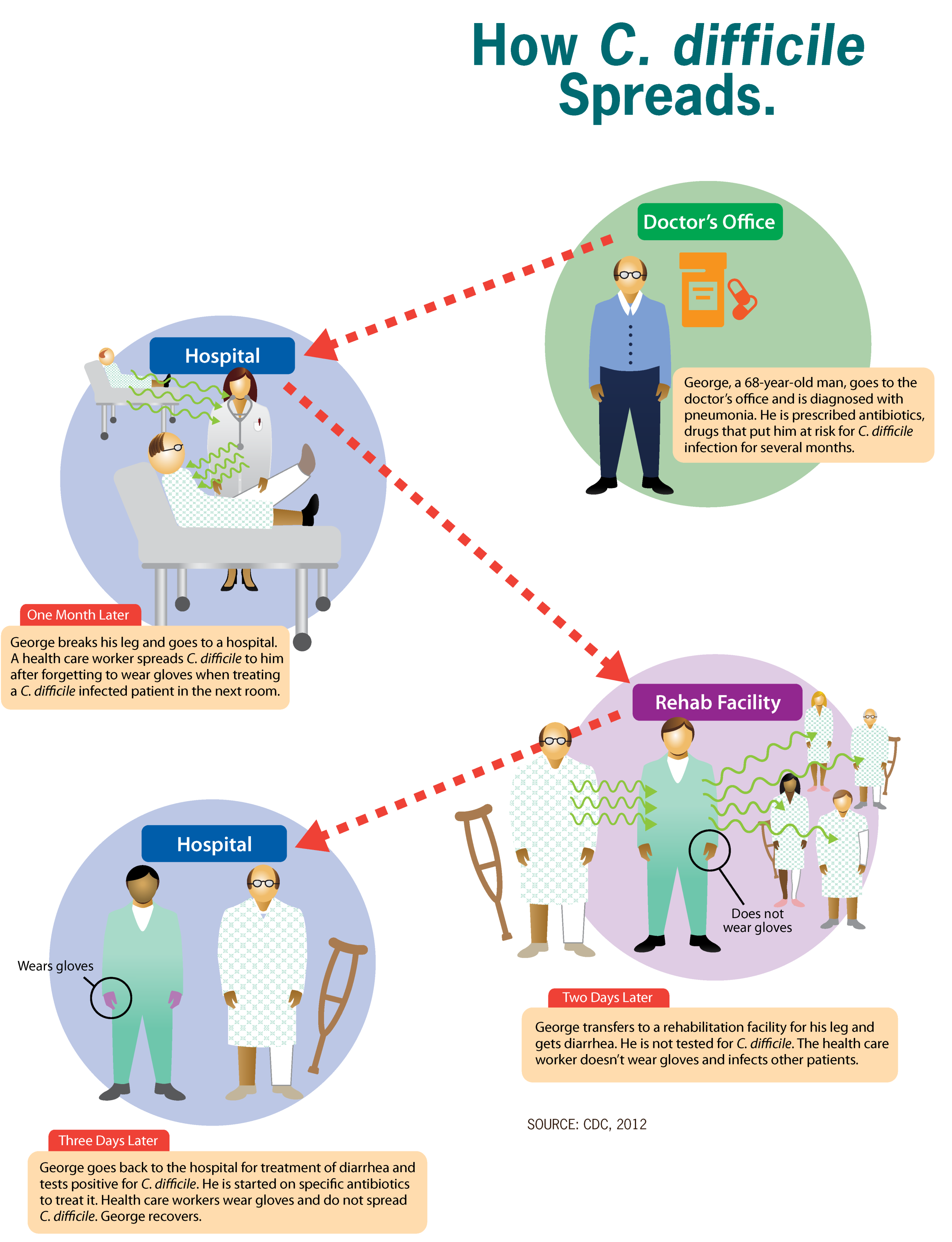
''Clostridioides difficile'' infection (CDI or C-diff), also known as ''Clostridium difficile'' infection, is a symptomatic infection due to the spore-forming bacterium ''Clostridioides difficile''. Symptoms include watery diarrhea, fever, nausea, and abdominal pain. It makes up about 20% of cases of antibiotic-associated diarrhea. Antibiotics can contribute to detrimental changes in gut microbiota; specifically, they decrease short-chain fatty acid absorption which results in osmotic, or watery, diarrhea. Complications may include pseudomembranous colitis, toxic megacolon, perforation of the colon, and sepsis. ''Clostridioides difficile'' infection is spread by bacterial spores found within feces. Surfaces may become contaminated with the spores with further spread occurring via the hands of healthcare workers. Risk factors for infection include antibiotic or proton pump inhibitor use, hospitalization, other health problems, and older age. Diagnosis is by stool culture or ... [...More Info...] [...Related Items...] OR: [Wikipedia] [Google] [Baidu] |
Clostridium Difficile (bacteria)
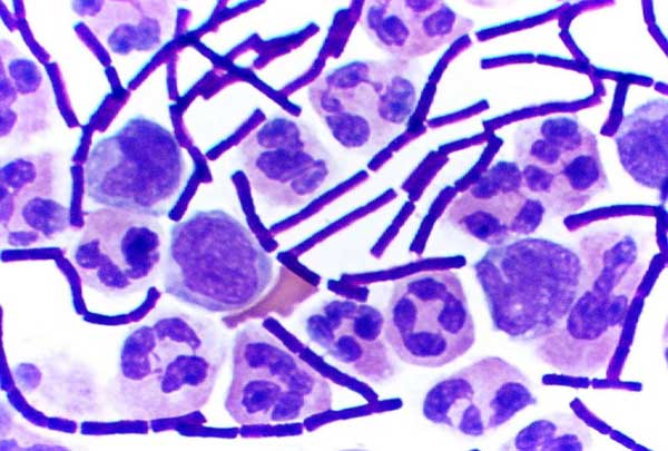
''Clostridioides difficile'' ( syn. ''Clostridium difficile'') is a bacterium that is well known for causing serious diarrheal infections, and may also cause colon cancer. Also known as ''C. difficile'', or ''C. diff'' (), is Gram-positive species of spore-forming bacteria. ''Clostridioides'' spp. are anaerobic, motile bacteria, ubiquitous in nature and especially prevalent in soil. Its vegetative cells are rod-shaped, pleomorphic, and occur in pairs or short chains. Under the microscope, they appear as long, irregular (often drumstick- or spindle-shaped) cells with a bulge at their terminal ends (forms subterminal spores). Under Gram staining, ''C. difficile'' cells are Gram-positive and show optimum growth on blood agar at human body temperatures in the absence of oxygen. ''C. difficile'' is catalase- and superoxide dismutase-negative, and produces up to three types of toxins: enterotoxin A, cytotoxin B and Clostridioides difficile transferase (CDT). Under stress condition ... [...More Info...] [...Related Items...] OR: [Wikipedia] [Google] [Baidu] |
Endocytosis
Endocytosis is a cellular process in which substances are brought into the cell. The material to be internalized is surrounded by an area of cell membrane, which then buds off inside the cell to form a vesicle containing the ingested material. Endocytosis includes pinocytosis (cell drinking) and phagocytosis (cell eating). It is a form of active transport. History The term was proposed by De Duve in 1963. Phagocytosis was discovered by Élie Metchnikoff in 1882. Pathways Endocytosis pathways can be subdivided into four categories: namely, receptor-mediated endocytosis (also known as clathrin-mediated endocytosis), caveolae, pinocytosis, and phagocytosis Phagocytosis () is the process by which a cell uses its plasma membrane to engulf a large particle (≥ 0.5 μm), giving rise to an internal compartment called the phagosome. It is one type of endocytosis. A cell that performs phagocytosis is .... *Clathrin-mediated endocytosis is mediated by the production of smal ... [...More Info...] [...Related Items...] OR: [Wikipedia] [Google] [Baidu] |
Glucosyltransferase
Glucosyltransferases are a type of glycosyltransferase that enable the transfer of glucose. Examples include: * glycogen synthase * glycogen phosphorylase Glycogen phosphorylase is one of the phosphorylase enzymes (). Glycogen phosphorylase catalyzes the rate-limiting step in glycogenolysis in animals by releasing glucose-1-phosphate from the terminal alpha-1,4-glycosidic bond. Glycogen phosphory ... They are categorized under EC number 2.4.1. References External links * EC 2.4 {{2.4-enzyme-stub ... [...More Info...] [...Related Items...] OR: [Wikipedia] [Google] [Baidu] |
Diphtheria Toxin
Diphtheria toxin is an exotoxin secreted by '' Corynebacterium diphtheriae'', the pathogenic bacterium that causes diphtheria. The toxin gene is encoded by a prophageA prophage is a virus that has inserted itself into the genome of the host bacterium. called corynephage β. The toxin causes the disease in humans by gaining entry into the cell cytoplasm and inhibiting protein synthesis. Structure Diphtheria toxin is a single polypeptide chain of 535 amino acids consisting of two subunits linked by disulfide bridges, known as an A-B toxin. Binding to the cell surface of the B subunit (the less stable of the two subunits) allows the A subunit (the more stable part of the protein) to penetrate the host cell. The crystal structure of the diphtheria toxin homodimer has been determined to 2.5 Ångstrom resolution. The structure reveals a Y-shaped molecule consisting of three domains. Fragment A contains the catalytic C domain, and fragment B consists of the T and R domains: ... [...More Info...] [...Related Items...] OR: [Wikipedia] [Google] [Baidu] |
Endosome
Endosomes are a collection of intracellular sorting organelles in eukaryotic cells. They are parts of endocytic membrane transport pathway originating from the trans Golgi network. Molecules or ligands internalized from the plasma membrane can follow this pathway all the way to lysosomes for degradation or can be recycled back to the cell membrane in the endocytic cycle. Molecules are also transported to endosomes from the trans Golgi network and either continue to lysosomes or recycle back to the Golgi apparatus. Endosomes can be classified as early, sorting, or late depending on their stage post internalization. Endosomes represent a major sorting compartment of the endomembrane system in cells. Function Endosomes provide an environment for material to be sorted before it reaches the degradative lysosome. For example, low-density lipoprotein (LDL) is taken into the cell by binding to the LDL receptor at the cell surface. Upon reaching early endosomes, the LDL dissociates ... [...More Info...] [...Related Items...] OR: [Wikipedia] [Google] [Baidu] |
Clostridium Sordellii
''Paeniclostridium sordellii'' is a rare anaerobic, gram-positive, spore-forming rod with peritrichous flagella that is capable of causing pneumonia, endocarditis, arthritis, peritonitis, and myonecrosis. ''C. sordellii'' bacteremia and sepsis occur rarely. Most cases of sepsis from ''C. sordellii'' occur in patients with underlying conditions. Severe toxic shock syndrome among previously healthy persons has been described in a small number of ''C. sordellii'' cases, most often associated with gynecologic infections in women and infection of the umbilical stump in newborns. It has also been described in post-partum females, medically induced abortions, injection drug users and trauma cases. So far, all documented post-partum females who contracted ''C. sordellii'' septicaemia have died, and all but one woman who contracted the bacterium post-abortion have died . __TOC__ Infection The source of the bacteria has not been determined but it has been documented that about 0.5% to 10 ... [...More Info...] [...Related Items...] OR: [Wikipedia] [Google] [Baidu] |
Clostridium Difficile Toxin B
Clostridium difficile toxin B is a cytotoxin produced by the bacteria ''Clostridioides difficile (bacteria), Clostridioides difficile'', formerly known as ''Clostridium difficile''. It is one of two major kinds of toxins produced by ''C. difficile'', the other being an related enterotoxin (Clostridium difficile toxin A, Toxin A). Both are very potent and lethal. Structure Toxin B (TcdB) is a cytotoxin that has a molecular weight of 270 kDa and an isoelectric point, pl, of 4.1. Toxin B has four different structural domains: catalytic, cysteine protease, protein translocation, translocation, and receptor binding. The N-terminal glucosyltransferase catalytic domain includes amino acid residues 1–544 while the cysteine protease domain includes residues 545–801. Additionally, the translocation region incorporates amino acid residues from 802 to 1664 while the receptor binding region is part of the C-terminal region and includes amino acid residues from 1665 to 2366. The gl ... [...More Info...] [...Related Items...] OR: [Wikipedia] [Google] [Baidu] |


