|
Clinical Cardiac Electrophysiology
{{unreferenced, date=February 2009 Clinical cardiac electrophysiology (also referred to as cardiac electrophysiology, arrhythmia services, or electrophysiology), is a branch of the medical specialty of cardiology and is concerned with the study and treatment of rhythm disorders of the heart. Cardiologists with expertise in this area are usually referred to as electrophysiologists. Electrophysiologists are trained in the mechanism, function, and performance of the electrical activities of the heart. Electrophysiologists work closely with other cardiologists and cardiac surgeons to assist or guide therapy for heart rhythm disturbances (arrhythmias). They are trained to perform interventional and surgical procedures to treat cardiac arrhythmia. The training required to become an electrophysiologist is lengthy and requires eight years after medical school (in the U.S.), entailing three years of internal medicine residency, three years of clinical cardiology fellowship, and two year ... [...More Info...] [...Related Items...] OR: [Wikipedia] [Google] [Baidu] |
Electrophysiology
Electrophysiology (from Greek , ''ēlektron'', "amber" Electron#Etymology">etymology of "electron" , ''physis'', "nature, origin"; and , ''-logia'') is the branch of physiology that studies the electrical properties of biological cells and tissues. It involves measurements of voltage changes or electric current or manipulations on a wide variety of scales from single ion channel proteins to whole organs like the heart. In neuroscience, it includes measurements of the electrical activity of neurons, and, in particular, action potential activity. Recordings of large-scale electric signals from the nervous system, such as electroencephalography, may also be referred to as electrophysiological recordings. They are useful for electrodiagnosis and monitoring. Definition and scope Classical electrophysiological techniques Principle and mechanisms Electrophysiology is the branch of physiology that pertains broadly to the flow of ions ( ion current) in biological tissues and, in pa ... [...More Info...] [...Related Items...] OR: [Wikipedia] [Google] [Baidu] |
Signal-averaged Electrocardiogram
Signal-averaged electrocardiography (SAECG) is a special electrocardiographic technique, in which multiple electric signals from the heart are averaged to remove interference and reveal small variations in the QRS complex, usually the so-called "late potentials". These may represent a predisposition towards potentially dangerous ventricular tachyarrhythmias. Technique Procedure A resting electrocardiogram (ECG) is recorded in the supine position using an ECG machine equipped with SAECG software; this can be done by a physician, nurse, or medical technician. Unlike standard basal ECG recording, which requires only a few seconds, SAECG recording requires a few minutes (usually about 7-10 minutes), as the machine must record multiple subsequent QRS potentials to remove interference due to skeletal muscle and to obtain a statistically significant average trace. For this reason, it is important for the patient to lie as still as possible during the recording. Results SAECG ... [...More Info...] [...Related Items...] OR: [Wikipedia] [Google] [Baidu] |
Cardiac Arrhythmia
Arrhythmias, also known as cardiac arrhythmias, heart arrhythmias, or dysrhythmias, are irregularities in the Cardiac cycle, heartbeat, including when it is too fast or too slow. A resting heart rate that is too fast – above 100 beats per minute in adults – is called tachycardia, and a resting heart rate that is too slow – below 60 beats per minute – is called bradycardia. Some types of arrhythmias have no symptoms. Symptoms, when present, may include palpitations or feeling a pause between heartbeats. In more serious cases, there may be presyncope, lightheadedness, Syncope (medicine), passing out, shortness of breath or chest pain. While most cases of arrhythmia are not serious, some predispose a person to complications such as stroke or heart failure. Others may result in cardiac arrest, sudden death. Arrhythmias are often categorized into four groups: premature heart beat, extra beats, supraventricular tachycardias, ventricular arrhythmias and bradyarrhythmias. Extr ... [...More Info...] [...Related Items...] OR: [Wikipedia] [Google] [Baidu] |
Cardiology
Cardiology () is a branch of medicine that deals with disorders of the heart and the cardiovascular system. The field includes medical diagnosis and treatment of congenital heart defects, coronary artery disease, heart failure, valvular heart disease and electrophysiology. Physicians who specialize in this field of medicine are called cardiologists, a specialty of internal medicine. Pediatric cardiologists are pediatricians who specialize in cardiology. Physicians who specialize in cardiac surgery are called cardiothoracic surgeons or cardiac surgeons, a specialty of general surgery. Specializations All cardiologists study the disorders of the heart, but the study of adult and child heart disorders each require different training pathways. Therefore, an adult cardiologist (often simply called "cardiologist") is inadequately trained to take care of children, and pediatric cardiologists are not trained to treat adult heart disease. Surgical aspects are not included in ca ... [...More Info...] [...Related Items...] OR: [Wikipedia] [Google] [Baidu] |
Implantable Loop Recorder
An implantable loop recorder (ILR), also known as an insertable cardiac monitor (ICM), is a small device that is implanted under the skin of the chest for cardiac monitoring, to record the heart's electrical activity for an extended period).Implantable Loop Recorder (ILR) System with illustrations, Operation The ILR monitors the electrical activity of the heart, continuously storing information in its circular memory (hence the name "loop" ...[...More Info...] [...Related Items...] OR: [Wikipedia] [Google] [Baidu] |
Ventricular Tachycardia
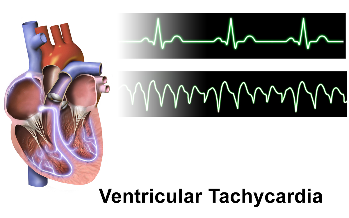
Ventricular tachycardia (V-tach or VT) is a fast heart rate arising from the lower chambers of the heart. Although a few seconds of VT may not result in permanent problems, longer periods are dangerous; and multiple episodes over a short period of time are referred to as an electrical storm. Short periods may occur without symptoms, or present with lightheadedness, palpitations, or chest pain. Ventricular tachycardia may result in ventricular fibrillation (VF) and turn into cardiac arrest. This conversion of the VT into VF is called the degeneration of the VT. It is found initially in about 7% of people in cardiac arrest. Ventricular tachycardia can occur due to coronary heart disease, aortic stenosis, cardiomyopathy, electrolyte problems, or a heart attack. Diagnosis is by an electrocardiogram (ECG) showing a rate of greater than 120 beats per minute and at least three wide QRS complexes in a row. It is classified as non-sustained versus sustained based on whether ... [...More Info...] [...Related Items...] OR: [Wikipedia] [Google] [Baidu] |
Multifocal Atrial Tachycardia
Multifocal (or multiform) atrial tachycardia (MAT) is an abnormal heart rhythm, specifically a type of supraventricular tachycardia, that is particularly common in older people and is associated with exacerbations of chronic obstructive pulmonary disease (COPD). Normally, the heart rate is controlled by a cluster of cells called the sinoatrial node (SA node). When a number of different clusters of cells outside the SA node take over control of the heart rate, and the rate exceeds 100 beats per minute, this is called multifocal atrial tachycardia (if the heart rate is ≤100, this is technically not a tachycardia and it is then termed multifocal atrial rhythm). "Multiform" refers to the observation of variable P wave shapes, while "multifocal" refers to the underlying cause. Although these terms are used interchangeably, some sources prefer "multiform" since it does not presume any underlying mechanism. Causes MAT usually arises because of an underlying medical condition. It ... [...More Info...] [...Related Items...] OR: [Wikipedia] [Google] [Baidu] |
Fluoroscopy
Fluoroscopy () is an imaging technique that uses X-rays to obtain real-time moving images of the interior of an object. In its primary application of medical imaging, a fluoroscope () allows a physician to see the internal structure and function of a patient, so that the pumping action of the heart or the motion of swallowing, for example, can be watched. This is useful for both diagnosis and therapy and occurs in general radiology, interventional radiology, and image-guided surgery. In its simplest form, a fluoroscope consists of an X-ray source and a fluorescent screen, between which a patient is placed. However, since the 1950s most fluoroscopes have included X-ray image intensifiers and cameras as well, to improve the image's visibility and make it available on a remote display screen. For many decades, fluoroscopy tended to produce live pictures that were not recorded, but since the 1960s, as technology improved, recording and playback became the norm. Fluoroscopy i ... [...More Info...] [...Related Items...] OR: [Wikipedia] [Google] [Baidu] |
Catheters
In medicine, a catheter (/ˈkæθətər/) is a thin tube made from medical grade materials serving a broad range of functions. Catheters are medical devices that can be inserted in the body to treat diseases or perform a surgical procedure. Catheters are manufactured for specific applications, such as cardiovascular, urological, gastrointestinal, neurovascular and ophthalmic procedures. The process of inserting a catheter is ''catheterization''. In most uses, a catheter is a thin, flexible tube (''soft'' catheter) though catheters are available in varying levels of stiffness depending on the application. A catheter left inside the body, either temporarily or permanently, may be referred to as an "indwelling catheter" (for example, a peripherally inserted central catheter). A permanently inserted catheter may be referred to as a "permcath" (originally a trademark). Catheters can be inserted into a body cavity, duct, or vessel, brain, skin or adipose tissue. Functionally, they a ... [...More Info...] [...Related Items...] OR: [Wikipedia] [Google] [Baidu] |
AV Nodal Reentrant Tachycardia
AV-nodal reentrant tachycardia (AVNRT) is a type of abnormal fast heart rhythm. It is a type of supraventricular tachycardia (SVT), meaning that it originates from a location within the heart above the bundle of His. AV nodal reentrant tachycardia is the most common regular supraventricular tachycardia. It is more common in women than men (approximately 75% of cases occur in females). The main symptom is palpitations. Treatment may be with specific physical maneuvers, medications, or, rarely, synchronized cardioversion. Frequent attacks may require radiofrequency ablation, in which the abnormally conducting tissue in the heart is destroyed. AVNRT occurs when a reentrant circuit forms within or just next to the atrioventricular node. The circuit usually involves two anatomical pathways: the fast pathway and the slow pathway, which are both in the right atrium. The slow pathway (which is usually targeted for ablation) is located inferior and slightly posterior to the AV node, ... [...More Info...] [...Related Items...] OR: [Wikipedia] [Google] [Baidu] |
.png)


.png)