|
Circular Folds
The circular folds (also known as valves of Kerckring, valves of Kerchkring, plicae circulares, ''plicae circulae, and'' ''valvulae conniventes'') are large valvular flaps projecting into the lumen of the small intestine. Structure The entire small intestine has circular folds of mucous membrane. The majority extend transversely around the cylinder of the small intestine, for about one-half or two-thirds of its circumference. Some form complete circles. Others have a spiral direction. The latter usually extend a little more than once around the bowel, but occasionally two or three times. The larger folds are about 1 cm. in depth at their broadest part; but the greater number are smaller. The larger and smaller folds alternate with each other. These can increase the surface area of the small intestine threefold. These larger folds are called ''plica circularis''. Distribution They are not found at the commencement of the duodenum, but begin to appear about 2.5 or 5 ... [...More Info...] [...Related Items...] OR: [Wikipedia] [Google] [Baidu] |
Small Intestine
The small intestine or small bowel is an organ in the gastrointestinal tract where most of the absorption of nutrients from food takes place. It lies between the stomach and large intestine, and receives bile and pancreatic juice through the pancreatic duct to aid in digestion. The small intestine is about long and folds many times to fit in the abdomen. Although it is longer than the large intestine, it is called the small intestine because it is narrower in diameter. The small intestine has three distinct regions – the duodenum, jejunum, and ileum. The duodenum, the shortest, is where preparation for absorption through small finger-like protrusions called villi begins. The jejunum is specialized for the absorption through its lining by enterocytes: small nutrient particles which have been previously digested by enzymes in the duodenum. The main function of the ileum is to absorb vitamin B12, bile salts, and whatever products of digestion that were not absorbe ... [...More Info...] [...Related Items...] OR: [Wikipedia] [Google] [Baidu] |
Ileum
The ileum () is the final section of the small intestine in most higher vertebrates, including mammals, reptiles, and birds. In fish, the divisions of the small intestine are not as clear and the terms posterior intestine or distal intestine may be used instead of ileum. Its main function is to absorb vitamin B12, bile salts, and whatever products of digestion that were not absorbed by the jejunum. The ileum follows the duodenum and jejunum and is separated from the cecum by the ileocecal valve (ICV). In humans, the ileum is about 2–4 m long, and the pH is usually between 7 and 8 (neutral or slightly basic). ''Ileum ''is derived from the Greek word ''eilein'', meaning "to twist up tightly". Structure The ileum is the third and final part of the small intestine. It follows the jejunum and ends at the ileocecal junction, where the terminal ileum communicates with the cecum of the large intestine through the ileocecal valve. The ileum, along with the jejunum, is sus ... [...More Info...] [...Related Items...] OR: [Wikipedia] [Google] [Baidu] |
Microvillus
Microvilli (singular: microvillus) are microscopic cellular membrane protrusions that increase the surface area for diffusion and minimize any increase in volume, and are involved in a wide variety of functions, including absorption, secretion, cellular adhesion, and mechanotransduction. Structure Microvilli are covered in plasma membrane, which encloses cytoplasm and microfilaments. Though these are cellular extensions, there are little or no cellular organelles present in the microvilli. Each microvillus has a dense bundle of cross-linked actin filaments, which serves as its structural core. 20 to 30 tightly bundled actin filaments are cross-linked by bundling proteins fimbrin (or plastin-1), villin and espin to form the core of the microvilli. In the enterocyte microvillus, the structural core is attached to the plasma membrane along its length by lateral arms made of myosin 1a and Ca2+ binding protein calmodulin. Myosin 1a functions through a binding site for filamentous ... [...More Info...] [...Related Items...] OR: [Wikipedia] [Google] [Baidu] |
Intestinal Villi
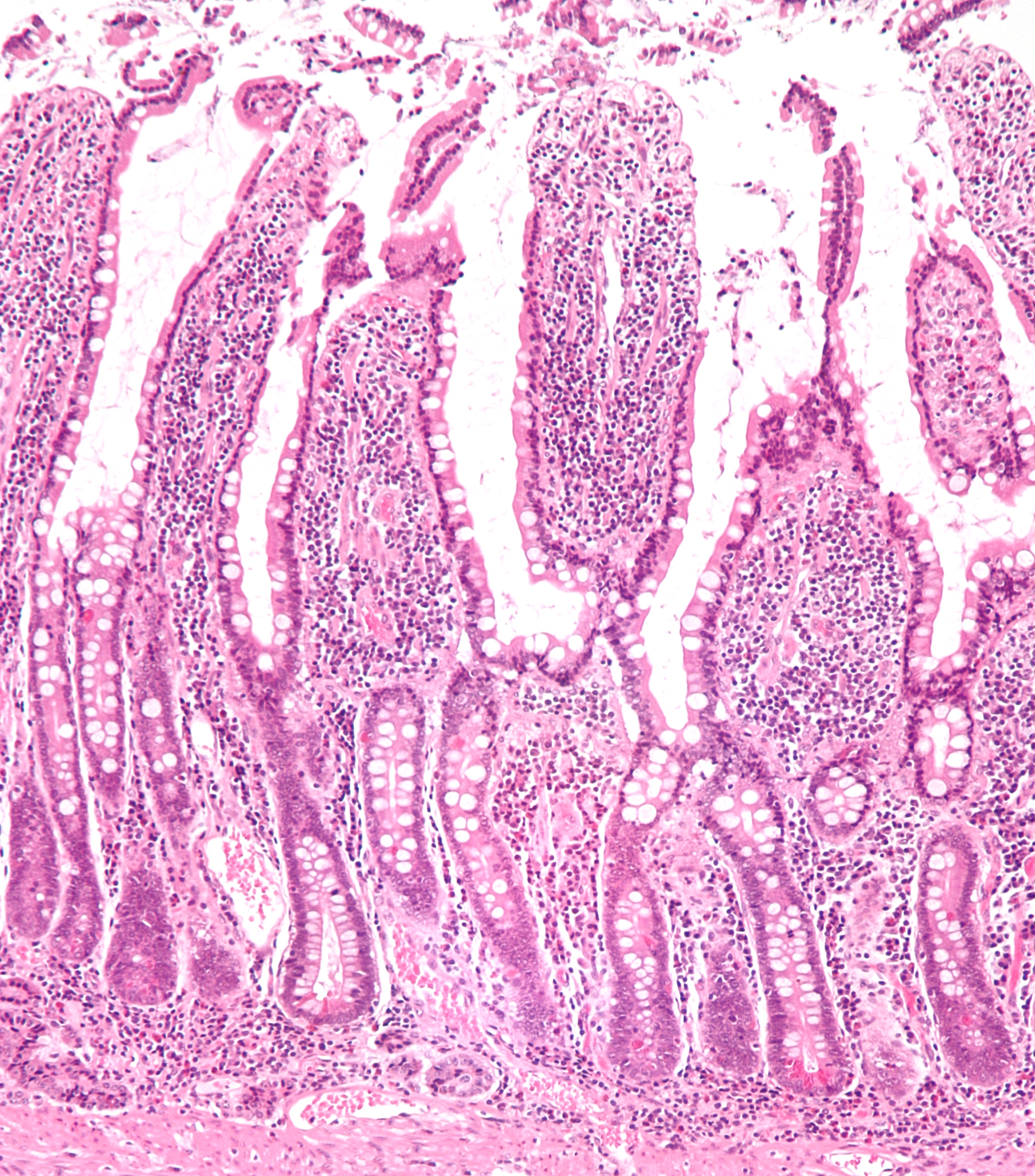
Intestinal villi (singular: villus) are small, finger-like projections that extend into the lumen of the small intestine. Each villus is approximately 0.5–1.6 mm in length (in humans), and has many microvilli projecting from the enterocytes of its epithelium which collectively form the striated or brush border. Each of these microvilli are about 1 µm in length, around 1000 times shorter than a single villus. The intestinal villi are much smaller than any of the circular folds in the intestine. Villi increase the internal surface area of the intestinal walls making available a greater surface area for absorption. An increased absorptive area is useful because digested nutrients (including monosaccharide and amino acids) pass into the semipermeable villi through diffusion, which is effective only at short distances. In other words, increased surface area (in contact with the fluid in the lumen) decreases the average distance travelled by nutrient molecules, so effective ... [...More Info...] [...Related Items...] OR: [Wikipedia] [Google] [Baidu] |
Digestion
Digestion is the breakdown of large insoluble food molecules into small water-soluble food molecules so that they can be absorbed into the watery blood plasma. In certain organisms, these smaller substances are absorbed through the small intestine into the blood stream. Digestion is a form of catabolism that is often divided into two processes based on how food is broken down: mechanical and chemical digestion. The term mechanical digestion refers to the physical breakdown of large pieces of food into smaller pieces which can subsequently be accessed by digestive enzymes. Mechanical digestion takes place in the mouth through mastication and in the small intestine through segmentation contractions. In chemical digestion, enzymes break down food into the small molecules the body can use. In the human digestive system, food enters the mouth and mechanical digestion of the food starts by the action of mastication (chewing), a form of mechanical digestion, and the wetting contact o ... [...More Info...] [...Related Items...] OR: [Wikipedia] [Google] [Baidu] |
Food
Food is any substance consumed by an organism for nutritional support. Food is usually of plant, animal, or fungal origin, and contains essential nutrients, such as carbohydrates, fats, proteins, vitamins, or minerals. The substance is ingested by an organism and assimilated by the organism's cells to provide energy, maintain life, or stimulate growth. Different species of animals have different feeding behaviours that satisfy the needs of their unique metabolisms, often evolved to fill a specific ecological niche within specific geographical contexts. Omnivorous humans are highly adaptable and have adapted to obtain food in many different ecosystems. The majority of the food energy required is supplied by the industrial food industry, which produces food with intensive agriculture and distributes it through complex food processing and food distribution systems. This system of conventional agriculture relies heavily on fossil fuels, which means that the food an ... [...More Info...] [...Related Items...] OR: [Wikipedia] [Google] [Baidu] |
Abdominal X-ray
An abdominal x-ray is an x-ray of the abdomen. It is sometimes abbreviated to AXR, or KUB (for kidneys, ureters, and urinary bladder). Indications In children, abdominal x-ray is indicated in the acute setting: *Suspected bowel obstruction or gastrointestinal perforation; Abdominal x-ray will demonstrate most cases of bowel obstruction, by showing dilated bowel loops. * Foreign body in the alimentary tract; can be identified if it is radiodense. *Suspected abdominal mass *In suspected intussusception, an abdominal x-ray does not exclude intussusception but is useful in the differential diagnosis to exclude perforation or obstruction. Yet, CT scan is the best alternative for diagnosing intra-abdominal injury. Computed tomography provides an overall better surgical strategy planning, and possibly less unnecessary laparotomies. Abdominal x-ray is therefore not recommended for adults with acute abdominal pain presenting in the emergency department. Projections The standar ... [...More Info...] [...Related Items...] OR: [Wikipedia] [Google] [Baidu] |
Colon (anatomy)
The large intestine, also known as the large bowel, is the last part of the gastrointestinal tract and of the digestive system in tetrapods. Water is absorbed here and the remaining waste material is stored in the rectum as feces before being removed by defecation. The colon is the longest portion of the large intestine, and the terms are often used interchangeably but most sources define the large intestine as the combination of the cecum, colon, rectum, and anal canal. Some other sources exclude the anal canal. In humans, the large intestine begins in the right iliac region of the pelvis, just at or below the waist, where it is joined to the end of the small intestine at the cecum, via the ileocecal valve. It then continues as the colon ascending the abdomen, across the width of the abdominal cavity as the transverse colon, and then descending to the rectum and its endpoint at the anal canal. Overall, in humans, the large intestine is about long, which is about one-fi ... [...More Info...] [...Related Items...] OR: [Wikipedia] [Google] [Baidu] |
Haustrum (anatomy)
The haustra (singular haustrum) of the colon are the small pouches caused by sacculation (sac formation), which give the colon its segmented appearance. The teniae coli run the length of the colon. Because the taenia coli are shorter than the colon, the colon becomes sacculated between the teniae coli, forming the haustra. Haustral contractions are slow segmenting, uncoordinated movements that occur approximately every 25 minutes. One haustrum distends as it fills with chyme, which stimulates muscles to contract, pushing the contents to the next haustrum. Also see peristalsis. There is a wider distance between haustra than between the circular folds of the small intestine, and the haustra don't reach around the entire circumference of the intestine, in contrast to circular folds of the small intestine that do. These differences can assist in distinguishing the small intestine from the colon on an abdominal x-ray. Clinical significance Widespread loss of haustra is a sign of ... [...More Info...] [...Related Items...] OR: [Wikipedia] [Google] [Baidu] |
Intestine
The gastrointestinal tract (GI tract, digestive tract, alimentary canal) is the tract or passageway of the digestive system that leads from the mouth to the anus. The GI tract contains all the major organs of the digestive system, in humans and other animals, including the esophagus, stomach, and intestines. Food taken in through the mouth is digested to extract nutrients and absorb energy, and the waste expelled at the anus as feces. ''Gastrointestinal'' is an adjective meaning of or pertaining to the stomach and intestines. Most animals have a "through-gut" or complete digestive tract. Exceptions are more primitive ones: sponges have small pores (ostia) throughout their body for digestion and a larger dorsal pore ( osculum) for excretion, comb jellies have both a ventral mouth and dorsal anal pores, while cnidarians and acoels have a single pore for both digestion and excretion. The human gastrointestinal tract consists of the esophagus, stomach, and intestines, an ... [...More Info...] [...Related Items...] OR: [Wikipedia] [Google] [Baidu] |
Stomach
The stomach is a muscular, hollow organ in the gastrointestinal tract of humans and many other animals, including several invertebrates. The stomach has a dilated structure and functions as a vital organ in the digestive system. The stomach is involved in the gastric phase of digestion, following chewing. It performs a chemical breakdown by means of enzymes and hydrochloric acid. In humans and many other animals, the stomach is located between the oesophagus and the small intestine. The stomach secretes digestive enzymes and gastric acid to aid in food digestion. The pyloric sphincter controls the passage of partially digested food ( chyme) from the stomach into the duodenum, where peristalsis takes over to move this through the rest of intestines. Structure In the human digestive system, the stomach lies between the oesophagus and the duodenum (the first part of the small intestine). It is in the left upper quadrant of the abdominal cavity. The top of the stomach lies ... [...More Info...] [...Related Items...] OR: [Wikipedia] [Google] [Baidu] |
Gastric Folds
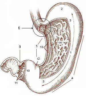
The gastric folds (or gastric rugae) are coiled sections of tissue that exist in the mucosal and submucosal layers of the stomach. They provide elasticity by allowing the stomach to expand when a bolus enters it. These folds stretch outward through the action of mechanoreceptors, which respond to the increase in pressure. This allows the stomach to expand, therefore increasing the volume of the stomach without increasing pressure. They also provide the stomach with an increased surface area for nutrient absorption during digestion. Gastric folds may be seen during esophagogastroduodenoscopy or in radiological studies. Layers The gastric folds consist of two layers: *Mucosal layer - This layer releases stomach acid. It is the innermost layer of the stomach. It is affected by the hormone histamine, which signals it to release hydrocholoric acid (HCl). * Sub-mucosal layer - This layer consists of different vessels and nerves, ganglion neurons, and adipose tissue. It is the secon ... [...More Info...] [...Related Items...] OR: [Wikipedia] [Google] [Baidu] |


