|
Circular Sulcus Of Insula
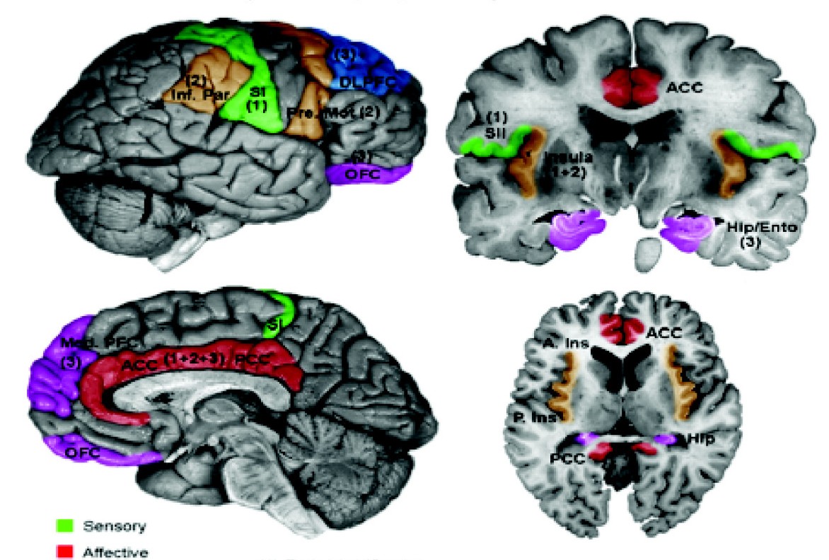
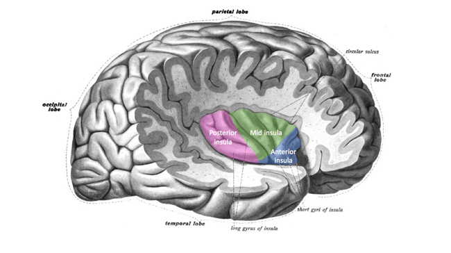
The insular cortex (also insula and insular lobe) is a portion of the cerebral cortex folded deep within the lateral sulcus (the fissure separating the temporal lobe from the parietal and frontal lobes) within each hemisphere of the mammalian brain. The insulae are believed to be involved in consciousness and play a role in diverse functions usually linked to emotion or the regulation of the body's homeostasis. These functions include compassion, empathy, taste, perception, motor control, self-awareness, cognitive functioning, interpersonal experience, and awareness of homeostatic emotions such as hunger, pain and fatigue. In relation to these, it is involved in psychopathology. The insular cortex is divided into two parts: the anterior insula and the posterior insula in which more than a dozen field areas have been identified. The cortical area overlying the insula toward the lateral surface of the brain is the operculum (meaning ''lid''). The opercula are formed from parts o ... [...More Info...] [...Related Items...] OR: [Wikipedia] [Google] [Baidu] |
Operculum (brain)
In human brain anatomy, an operculum (Latin, meaning "little lid") (pl. opercula), may refer to the frontal, temporal, or parietal operculum, which together cover the insula as the opercula of insula. It can also refer to the occipital operculum, part of the occipital lobe. The insular lobe is a portion of the cerebral cortex that has invaginated to lie deep within the lateral sulcus. It sits like an island (the meaning of ''insular'') almost surrounded by the groove of the circular sulcus and covered over and obscured by the insular opercula. A part of the parietal lobe, the frontoparietal operculum, covers the upper part of the insular lobe from the front to the back. The opercula lie on the precentral and postcentral gyri (on either side of the central sulcus). The part of the parietal operculum that forms the ceiling of the lateral sulcus functions as the secondary somatosensory cortex. Development Normally, the insular opercula begin to develop between the 20th an ... [...More Info...] [...Related Items...] OR: [Wikipedia] [Google] [Baidu] |
Motor Control
Motor control is the regulation of movement in organisms that possess a nervous system. Motor control includes reflexes as well as directed movement. To control movement, the nervous system must integrate multimodal sensory information (both from the external world as well as proprioception Proprioception ( ), also referred to as kinaesthesia (or kinesthesia), is the sense of self-movement, force, and body position. It is sometimes described as the "sixth sense". Proprioception is mediated by proprioceptors, mechanosensory neurons ...) and elicit the necessary signals to recruit muscles to carry out a goal. This pathway spans many disciplines, including multisensory integration, signal processing, Motor coordination, coordination, biomechanics, and cognition, and the computational challenges are often discussed under the term sensorimotor control. Successful motor control is crucial to interacting with the world to carry out goals as well as for posture, balance, and stabil ... [...More Info...] [...Related Items...] OR: [Wikipedia] [Google] [Baidu] |
Operculum (brain)
In human brain anatomy, an operculum (Latin, meaning "little lid") (pl. opercula), may refer to the frontal, temporal, or parietal operculum, which together cover the insula as the opercula of insula. It can also refer to the occipital operculum, part of the occipital lobe. The insular lobe is a portion of the cerebral cortex that has invaginated to lie deep within the lateral sulcus. It sits like an island (the meaning of ''insular'') almost surrounded by the groove of the circular sulcus and covered over and obscured by the insular opercula. A part of the parietal lobe, the frontoparietal operculum, covers the upper part of the insular lobe from the front to the back. The opercula lie on the precentral and postcentral gyri (on either side of the central sulcus). The part of the parietal operculum that forms the ceiling of the lateral sulcus functions as the secondary somatosensory cortex. Development Normally, the insular opercula begin to develop between the 20th an ... [...More Info...] [...Related Items...] OR: [Wikipedia] [Google] [Baidu] |
Sulcus (neuroanatomy)
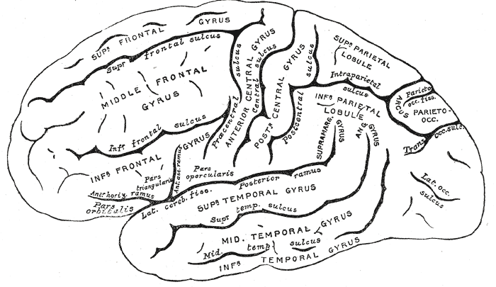
In neuroanatomy, a sulcus (Latin: "furrow", pl. ''sulci'') is a depression or groove in the cerebral cortex. It surrounds a gyrus (pl. gyri), creating the characteristic folded appearance of the brain in humans and other mammals. The larger sulci are usually called fissures. Structure Sulci, the grooves, and gyri, the folds or ridges, make up the folded surface of the cerebral cortex. Larger or deeper sulci are termed fissures, and in many cases the two terms are interchangeable. The folded cortex creates a larger surface area for the brain in humans and other mammals. When looking at the human brain, two-thirds of the surface are hidden in the grooves. The sulci and fissures are both grooves in the cortex, but they are differentiated by size. A sulcus is a shallower groove that surrounds a gyrus. A fissure is a large furrow that divides the brain into lobes and also into the two hemispheres as the longitudinal fissure. Importance of expanded surface area As the surfa ... [...More Info...] [...Related Items...] OR: [Wikipedia] [Google] [Baidu] |
Diffusion MRI
Diffusion-weighted magnetic resonance imaging (DWI or DW-MRI) is the use of specific MRI sequences as well as software that generates images from the resulting data that uses the diffusion of water molecules to generate contrast in MR images. It allows the mapping of the diffusion process of molecules, mainly water, in biological tissues, in vivo and non-invasively. Molecular diffusion in tissues is not random, but reflects interactions with many obstacles, such as macromolecules, fibers, and membranes. Water molecule diffusion patterns can therefore reveal microscopic details about tissue architecture, either normal or in a diseased state. A special kind of DWI, diffusion tensor imaging (DTI), has been used extensively to map white matter tractography in the brain. Introduction In diffusion weighted imaging (DWI), the intensity of each image element ( voxel) reflects the best estimate of the rate of water diffusion at that location. Because the mobility of water is driven by ... [...More Info...] [...Related Items...] OR: [Wikipedia] [Google] [Baidu] |
Spinothalamic Tract
The spinothalamic tract is a part of the anterolateral system or the ventrolateral system, a sensory pathway to the thalamus. From the ventral posterolateral nucleus in the thalamus, sensory information is relayed upward to the somatosensory cortex of the postcentral gyrus. The spinothalamic tract consists of two adjacent pathways: anterior and lateral. The anterior spinothalamic tract carries information about crude touch. The lateral spinothalamic tract conveys pain and temperature. In the spinal cord, the spinothalamic tract has somatotopic organization. This is the segmental organization of its cervical, thoracic, lumbar, and sacral components, which is arranged from most medial to most lateral respectively. The pathway crosses over ( decussates) at the level of the spinal cord, rather than in the brainstem like the dorsal column-medial lemniscus pathway and lateral corticospinal tract. It is one of the three tracts which make up the anterolateral system. Stru ... [...More Info...] [...Related Items...] OR: [Wikipedia] [Google] [Baidu] |
Secondary Somatosensory Cortex
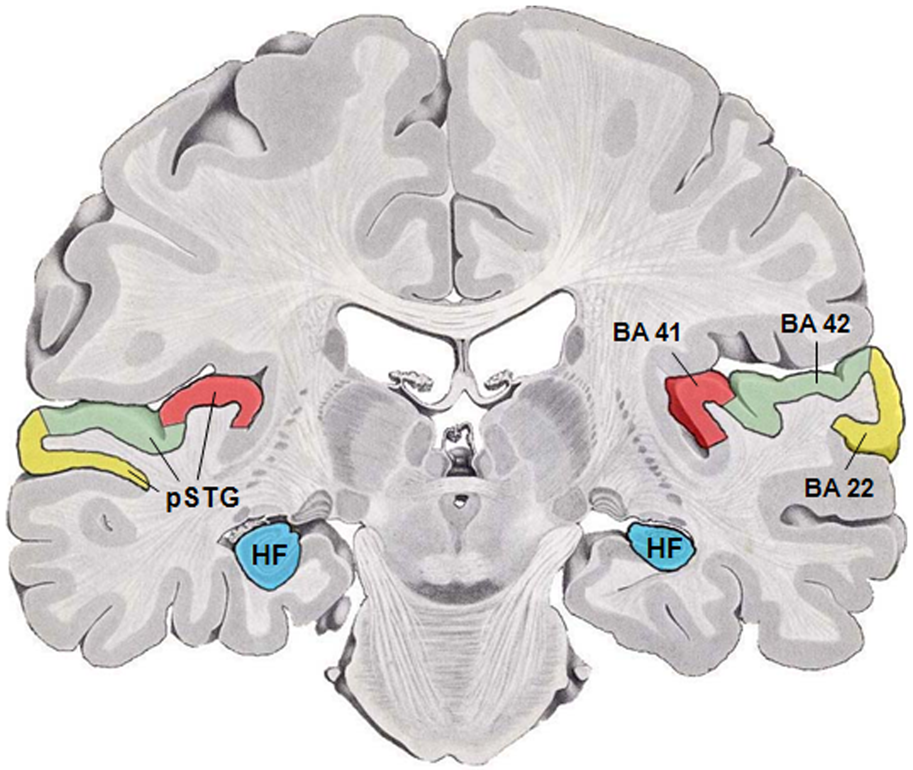
The human secondary somatosensory cortex (S2, SII) is a region of cortex in the parietal operculum on the ceiling of the lateral sulcus. Region S2 was first described by Adrian in 1940, who found that feeling in cats' feet was not only represented in the primary somatosensory cortex (S1) but also in a second region adjacent to S1. In 1954, Penfield and Jasper evoked somatosensory sensations in human patients during neurosurgery by electrically stimulating the ceiling of the lateral sulcus, which lies adjacent to S1, and their findings were confirmed in 1979 by Woolsey et al. using evoked potentials and electrical stimulation. Experiments involving ablation of the second somatosensory cortex in primates indicate that this cortical area is involved in remembering the differences between tactile shapes and textures. Functional neuroimaging studies have found S2 activation in response to light touch, pain, visceral sensation, and tactile attention. In monkeys, apes and hominids, ... [...More Info...] [...Related Items...] OR: [Wikipedia] [Google] [Baidu] |
Amygdala
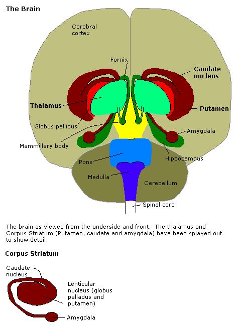
The amygdala (; plural: amygdalae or amygdalas; also '; Latin from Greek, , ', 'almond', 'tonsil') is one of two almond-shaped clusters of nuclei located deep and medially within the temporal lobes of the brain's cerebrum in complex vertebrates, including humans. Shown to perform a primary role in the processing of memory, decision making, and emotional responses (including fear, anxiety, and aggression), the amygdalae are considered part of the limbic system. The term "amygdala" was first introduced by Karl Friedrich Burdach in 1822. Structure The regions described as amygdala nuclei encompass several structures of the cerebrum with distinct connectional and functional characteristics in humans and other animals. Among these nuclei are the basolateral complex, the cortical nucleus, the medial nucleus, the central nucleus, and the intercalated cell clusters. The basolateral complex can be further subdivided into the lateral, the basal, and the accessory ba ... [...More Info...] [...Related Items...] OR: [Wikipedia] [Google] [Baidu] |
Central Nucleus Of The Amygdala
The central nucleus of the amygdala (CeA or aCeN) is a nucleus within the amygdala. It "serves as the major output nucleus of the amygdala and participates in receiving and processing pain information." CeA "connects with brainstem areas that control the expression of innate behaviors and associated physiological responses." CeA is responsible for "autonomic components of emotions (e.g., changes in heart rate, blood pressure, and respiration) primarily through output pathways to the lateral hypothalamus and brain stem." The CeA is also responsible for "conscious perception of emotion primarily through the ventral amygdalofugal output pathway to the anterior cingulate cortex, orbitofrontal cortex, and prefrontal cortex." Amygdala subdividisions and outputs The regions described as amygdala nuclei encompass several structures with distinct connectional and functional characteristics in humans and other animals. Among these nuclei are the basolateral complex, the cortical nuc ... [...More Info...] [...Related Items...] OR: [Wikipedia] [Google] [Baidu] |
Ventral Nuclear Group
The ventral nuclear group is a collection of nuclei on the ventral side of the thalamus. According to MeSH, it consists of the following: * ventral anterior nucleus * ventral lateral nucleus * ventral posterior nucleus – this is made up of two nuclei: the ventral posterolateral nucleus and the ventral posteromedial nucleus See also *Anterolateral region of the motor thalamus Anterolateral region of the motor thalamus is a composite substructure of the ventral nuclear group of the thalamus based on connectivity and function. It includes the ventral anterior nucleus and the medial part of the ventral lateral nucleus whic ... References Thalamus {{Neuroanatomy-stub ... [...More Info...] [...Related Items...] OR: [Wikipedia] [Google] [Baidu] |
Psychopathology
Psychopathology is the study of abnormal cognition, behaviour, and experiences which differs according to social norms and rests upon a number of constructs that are deemed to be the social norm at any particular era. Biological psychopathology is the study of the biological etiology of abnormal cognitions, behaviour and experiences. Child psychopathology is a specialisation applied to children and adolescents. Animal psychopathology is a specialisation applied to non-human animals. This concept is linked to the philosophical ideas first outlined by Galton (1869) and is linked to the appliance of eugenical ideations around what constitutes the human. History Early explanations for mental illnesses were influenced by religious belief and superstition. Psychological conditions that are now classified as mental disorders were initially attributed to possessions by evil spirits, demons, and the devil. This idea was widely accepted up until the sixteenth and seventeenth centur ... [...More Info...] [...Related Items...] OR: [Wikipedia] [Google] [Baidu] |
Homeostatic Emotion
Homeostatic feeling (or homeostatic affect, or homeostatic emotion, or primordial emotion) is a class of feelings (e.g., thirst, pain, fatigue) evoked by an internal body state. Some homeostatic feelings drive specific behavior (drinking, withdrawing and resting in these examples) aimed at maintaining the body in its ideal state. Derek Denton uses the term "primordial emotion" and defines it as "the subjective element of the instincts, which are the genetically programmed behaviour patterns which contrive homeostasis. They include thirst, hunger for air, hunger for food, pain, hunger for specific minerals etc. There are two constituents of a primordial emotionthe specific sensation which when severe may be imperious, and the compelling intention for gratification by a consummatory act." Denton includes sexual desire in this class and distinguishes two classes of feelings: #these ''primordial'' emotions ("imperious states of arousal and compelling intentions to act" driven b ... [...More Info...] [...Related Items...] OR: [Wikipedia] [Google] [Baidu] |
