|
Chronic Paroxysmal Hemicrania
Chronic paroxysmal hemicrania (CPH) is a severe debilitating unilateral headache usually affecting the area around the eye. It normally consists of multiple severe, yet short, headache attacks affecting only one side of the cranium. It is more commonly diagnosed in women than in men, but, unlike a migraine, has no neurological symptoms associated with it. CPH headaches are treated through the use of non-steroidal anti-inflammatory drugs, with indomethacin found to be usually effective in eliminating symptoms. Paroxysmal hemicrania is classified by the characteristic (high) frequency and (short) duration of attacks experienced by patients that is somewhat similar to cluster headaches, despite some important differences explained below. Episodic paroxysmal hemicrania attacks occur at least twice a year and last anywhere from seven days to a year with pain free periods of a month or longer separating them. Chronic paroxysmal hemicrania attacks occur over the course of more than a year ... [...More Info...] [...Related Items...] OR: [Wikipedia] [Google] [Baidu] |
Neurology
Neurology (from el, wikt:νεῦρον, νεῦρον (neûron), "string, nerve" and the suffix wikt:-logia, -logia, "study of") is the branch of specialty (medicine), medicine dealing with the diagnosis and treatment of all categories of conditions and disease involving the brain, the spinal cord and the peripheral nerves. Neurological practice relies heavily on the field of neuroscience, the scientific study of the nervous system. A neurologist is a physician specializing in neurology and trained to investigate, diagnose and treat neurological disorders. Neurologists treat a myriad of neurologic conditions, including stroke, seizures, movement disorders such as Parkinson's disease, autoimmune neurologic disorders such as multiple sclerosis, headache disorders like migraine and dementias such as Alzheimer's disease. Neurologists may also be involved in clinical research, clinical trials, and basic research, basic or translational research. While neurology is a nonsurgical sp ... [...More Info...] [...Related Items...] OR: [Wikipedia] [Google] [Baidu] |
Pancoast Tumor
A Pancoast tumor is a tumor of the apex of the lung. It is a type of lung cancer defined primarily by its location situated at the top end of either the right or left lung. It typically spreads to nearby tissues such as the ribs and vertebrae. Most Pancoast tumors are non-small-cell lung cancers. The growing tumor can cause compression of a brachiocephalic vein, subclavian artery, phrenic nerve, recurrent laryngeal nerve, vagus nerve, or, characteristically, compression of a sympathetic ganglion (the stellate ganglion), resulting in a range of symptoms known as Horner's syndrome. Pancoast tumors are named for Henry Pancoast, an American radiologist, who described them in 1924 and 1932. Signs and symptoms Aside from constitutional symptoms of cancer such as malaise, fever, weight loss and fatigue, Pancoast tumor can include a complete Horner's syndrome in severe cases: miosis (constriction of the pupils), anhidrosis (lack of sweating), ptosis (drooping of the eyelid), and pseud ... [...More Info...] [...Related Items...] OR: [Wikipedia] [Google] [Baidu] |
Facial Nerve
The facial nerve, also known as the seventh cranial nerve, cranial nerve VII, or simply CN VII, is a cranial nerve that emerges from the pons of the brainstem, controls the muscles of facial expression, and functions in the conveyance of taste sensations from the anterior two-thirds of the tongue. The nerve typically travels from the pons through the facial canal in the temporal bone and exits the skull at the stylomastoid foramen. It arises from the brainstem from an area posterior to the cranial nerve VI (abducens nerve) and anterior to cranial nerve VIII (vestibulocochlear nerve). The facial nerve also supplies preganglionic parasympathetic fibers to several head and neck ganglia. The facial and intermediate nerves can be collectively referred to as the nervus intermediofacialis. The path of the facial nerve can be divided into six segments: # intracranial (cisternal) segment # meatal (canalicular) segment (within the internal auditory canal) # labyrinthine segment ... [...More Info...] [...Related Items...] OR: [Wikipedia] [Google] [Baidu] |
Neuropathy
Peripheral neuropathy, often shortened to neuropathy, is a general term describing disease affecting the peripheral nerves, meaning nerves beyond the brain and spinal cord. Damage to peripheral nerves may impair sensation, movement, gland, or organ function depending on which nerves are affected; in other words, neuropathy affecting motor, sensory, or autonomic nerves result in different symptoms. More than one type of nerve may be affected simultaneously. Peripheral neuropathy may be acute (with sudden onset, rapid progress) or chronic (symptoms begin subtly and progress slowly), and may be reversible or permanent. Common causes include systemic diseases (such as diabetes or leprosy), hyperglycemia-induced glycation, vitamin deficiency, medication (e.g., chemotherapy, or commonly prescribed antibiotics including metronidazole and the fluoroquinolone class of antibiotics (such as ciprofloxacin, levofloxacin, moxifloxacin)), traumatic injury, ischemia, radiation therapy, exces ... [...More Info...] [...Related Items...] OR: [Wikipedia] [Google] [Baidu] |
Ptosis (eyelid)
Ptosis, also known as blepharoptosis, is a drooping or falling of the upper eyelid. The drooping may be worse after being awake longer when the individual's muscles are tired. This condition is sometimes called "lazy eye", but that term normally refers to the condition amblyopia. If severe enough and left untreated, the drooping eyelid can cause other conditions, such as amblyopia or astigmatism. This is why it is especially important for this disorder to be treated in children at a young age, before it can interfere with vision development. The term is from Greek 'fall, falling'. Signs and symptoms Signs and symptoms typically seen in this condition include: * The eyelid(s) may appear to droop. * Droopy eyelids can give the face a false appearance of being fatigued, disinterested, or even sinister. * The eyelid may not protect the eye as effectively, allowing it to dry out. * Sagging upper eyelids can partially block the person's field of view. * Obstructed vision may cause ... [...More Info...] [...Related Items...] OR: [Wikipedia] [Google] [Baidu] |
Miosis
Miosis, or myosis (), is excessive constriction of the pupil. citing: Mosby's Medical Dictionary, 8th ed. The opposite condition, , is the dilation of the pupil. Anisocoria is the condition of one being more dilated than the other. Causes Age * Senile miosis (a reduction in the size of a person's pupil in old age)Diseases * ...[...More Info...] [...Related Items...] OR: [Wikipedia] [Google] [Baidu] |
Oedema
Edema, also spelled oedema, and also known as fluid retention, dropsy, hydropsy and swelling, is the build-up of fluid in the body's tissue. Most commonly, the legs or arms are affected. Symptoms may include skin which feels tight, the area may feel heavy, and joint stiffness. Other symptoms depend on the underlying cause. Causes may include venous insufficiency, heart failure, kidney problems, low protein levels, liver problems, deep vein thrombosis, infections, angioedema, certain medications, and lymphedema. It may also occur after prolonged sitting or standing and during menstruation or pregnancy. The condition is more concerning if it starts suddenly, or pain or shortness of breath is present. Treatment depends on the underlying cause. If the underlying mechanism involves sodium retention, decreased salt intake and a diuretic may be used. Elevating the legs and support stockings may be useful for edema of the legs. Older people are more commonly affected. The word is ... [...More Info...] [...Related Items...] OR: [Wikipedia] [Google] [Baidu] |
Rhinorrhoea
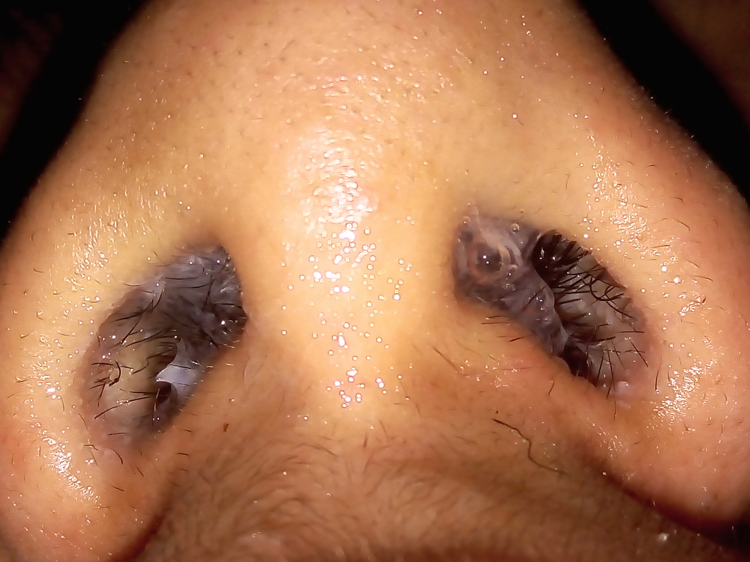
Rhinorrhea, rhinorrhoea, or informally runny nose is the free discharge of a thin mucus fluid from the nose; it is a common condition. It is a common symptom of allergies (hay fever) or certain viral infections, such as the common cold or COVID-19. It can be a side effect of crying, exposure to cold temperatures, cocaine abuse, or drug withdrawal, such as from methadone or other opioids. Treatment for rhinorrhea is not usually undertaken, but there are a number of medical treatments and preventive techniques. The term was coined in 1866 from the Greek ''rhino-'' ("of the nose") and ''-rhoia'' ("discharge" or "flow"). Signs and symptoms Rhinorrhea is characterized by an excess amount of mucus produced by the mucous membranes that line the nasal cavities. The membranes create mucus faster than it can be processed, causing a backup of mucus in the nasal cavities. As the cavity fills up, it blocks off the air passageway, causing difficulty breathing through the nose. Air caught in n ... [...More Info...] [...Related Items...] OR: [Wikipedia] [Google] [Baidu] |
Lacrimation
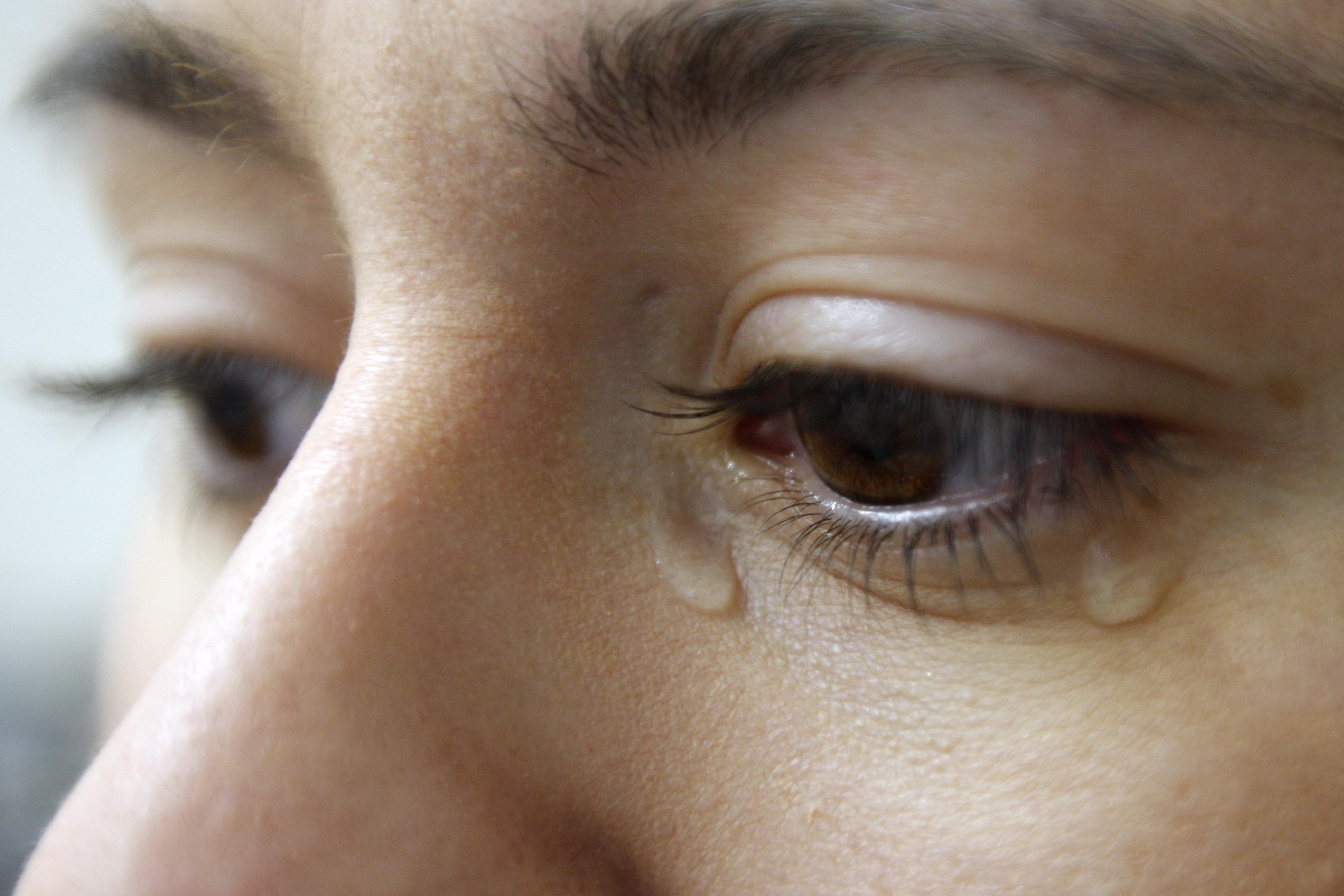
Tears are a clear liquid secreted by the lacrimal glands (tear gland) found in the eyes of all land mammals. Tears are made up of water, electrolytes, proteins, lipids, and mucins that form layers on the surface of eyes. The different types of tears—basal, reflex, and emotional—vary significantly in composition. The functions of tears include lubricating the eyes (basal tears), removing irritants (reflex tears), and also aiding the immune system. Tears also occur as a part of the body's natural pain response. Emotional secretion of tears may serve a biological function by excreting stress-inducing hormones built up through times of emotional distress. Tears have symbolic significance among humans. Physiology Chemical composition Tears are made up of three layers: lipid, aqueous, and mucous. Tears are composed of water, salts, antibodies, and lysozymes (antibacterial enzymes); though composition varies among different tear types. The composition of tears caused by an ... [...More Info...] [...Related Items...] OR: [Wikipedia] [Google] [Baidu] |
Conjunctiva
The conjunctiva is a thin mucous membrane that lines the inside of the eyelids and covers the sclera (the white of the eye). It is composed of non-keratinized, stratified squamous epithelium with goblet cells, stratified columnar epithelium and stratified cuboidal epithelium (depending on the zone). The conjunctiva is highly vascularised, with many microvessels easily accessible for imaging studies. Structure The conjunctiva is typically divided into three parts: Blood supply Blood to the bulbar conjunctiva is primarily derived from the ophthalmic artery. The blood supply to the palpebral conjunctiva (the eyelid) is derived from the external carotid artery. However, the circulations of the bulbar conjunctiva and palpebral conjunctiva are linked, so both bulbar conjunctival and palpebral conjunctival vessels are supplied by both the ophthalmic artery and the external carotid artery, to varying extents. Nerve supply Sensory innervation of the conjunctiva is divided into ... [...More Info...] [...Related Items...] OR: [Wikipedia] [Google] [Baidu] |
Cranial Nerve VII
The facial nerve, also known as the seventh cranial nerve, cranial nerve VII, or simply CN VII, is a cranial nerve that emerges from the pons of the brainstem, controls the muscles of facial expression, and functions in the conveyance of taste sensations from the anterior two-thirds of the tongue. The nerve typically travels from the pons through the facial canal in the temporal bone and exits the skull at the stylomastoid foramen. It arises from the brainstem from an area posterior to the cranial nerve VI (abducens nerve) and anterior to cranial nerve VIII (vestibulocochlear nerve). The facial nerve also supplies preganglionic parasympathetic fibers to several head and neck ganglia. The facial and intermediate nerves can be collectively referred to as the nervus intermediofacialis. The path of the facial nerve can be divided into six segments: # intracranial (cisternal) segment # meatal (canalicular) segment (within the internal auditory canal) # labyrinthine segment ... [...More Info...] [...Related Items...] OR: [Wikipedia] [Google] [Baidu] |


