|
Chromosome 5q Deletion Syndrome
Chromosome 5q deletion syndrome is an acquired, hematological disorder characterized by loss of part of the long arm ( q arm, band 5q33.1) of human chromosome 5 in bone marrow myelocyte cells. This chromosome abnormality is most commonly associated with the myelodysplastic syndrome. It should not be confused with "partial trisomy 5q", though both conditions have been observed in the same family. This should not be confused with the germ line cri du chat (5p deletion) syndrome which is a deletion of the short arm of the 5th chromosome. Presentation The 5q-syndrome is characterized by macrocytic anemia, often a moderate thrombocytosis, erythroblastopenia, megakaryocyte hyperplasia with nuclear hypolobation, and an isolated interstitial deletion of chromosome 5. The 5q- syndrome is found predominantly in females of advanced age. Causes Several genes in the deleted region appear to play a role in the pathogenesis of 5q-syndrome. Haploinsufficiency of RPS14 plays a central role, ... [...More Info...] [...Related Items...] OR: [Wikipedia] [Google] [Baidu] |
Monosomy
Monosomy is a form of aneuploidy with the presence of only one chromosome from a pair. Partial monosomy occurs when a portion of one chromosome in a pair is missing. Human monosomy Human conditions due to monosomy: * Turner syndrome – People with Turner syndrome typically have one X chromosome instead of the usual two X chromosomes. Turner syndrome is the only full monosomy that is seen in humans — all other cases of full monosomy are lethal and the individual will not survive development. * Cri du chat syndrome – (French for "cry of the cat" after the persons' malformed larynx) a partial monosomy caused by a deletion of the end of the short arm of chromosome 5 * 1p36 deletion syndrome – a partial monosomy caused by a deletion at the end of the short arm of chromosome 1 * 17q12 microdeletion syndrome - a partial monosomy caused by a deletion of part of the long arm of chromosome 17 See also * Anaphase lag * Miscarriage Miscarriage, also known in medical terms as a ... [...More Info...] [...Related Items...] OR: [Wikipedia] [Google] [Baidu] |
Haploinsufficiency
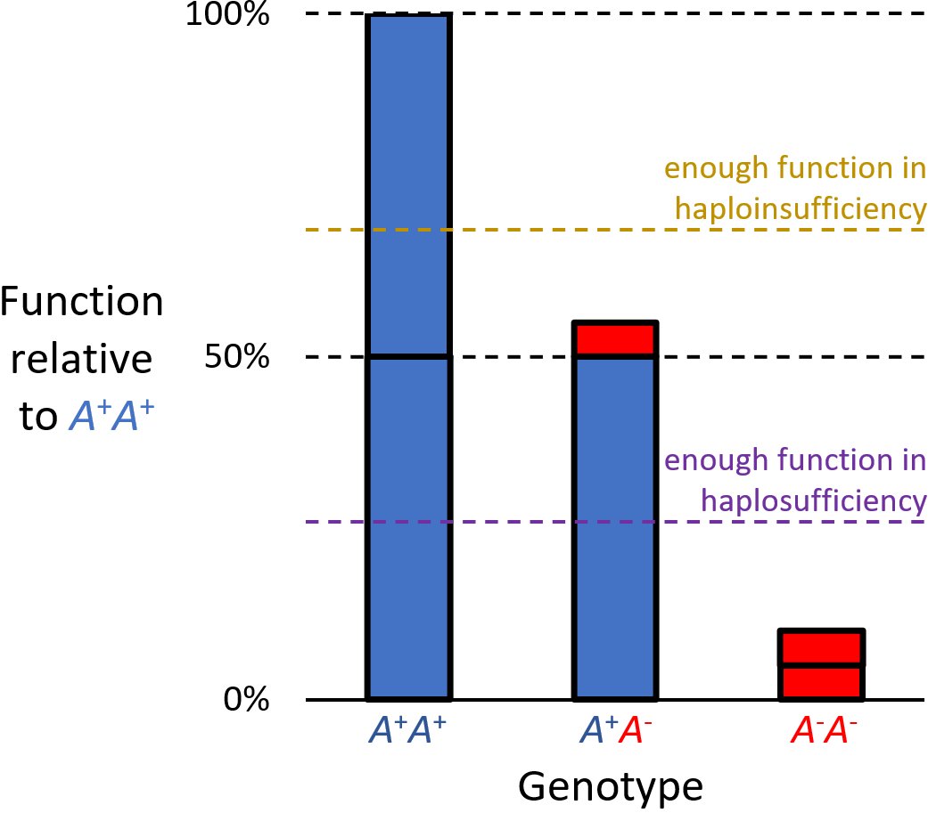
Haploinsufficiency in genetics describes a model of dominant gene action in diploid organisms, in which a single copy of the wild-type allele at a locus in heterozygous combination with a variant allele is insufficient to produce the wild-type phenotype. Haploinsufficiency may arise from a ''de novo'' or inherited loss-of-function mutation in the variant allele, such that it yields little or no gene product (often a protein). Although the other, standard allele still produces the standard amount of product, the total product is insufficient to produce the standard phenotype. This heterozygous genotype may result in a non- or sub-standard, deleterious, and (or) disease phenotype. Haploinsufficiency is the standard explanation for dominant deleterious alleles. In the alternative case of haplosufficiency, the loss-of-function allele behaves as above, but the single standard allele in the heterozygous genotype produces sufficient gene product to produce the same, standard phenotype ... [...More Info...] [...Related Items...] OR: [Wikipedia] [Google] [Baidu] |
Megakaryocytes
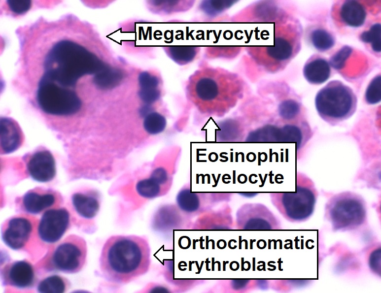
A megakaryocyte (''mega-'' + '' karyo-'' + '' -cyte'', "large-nucleus cell") is a large bone marrow cell with a lobated nucleus responsible for the production of blood thrombocytes (platelets), which are necessary for normal blood clotting. In humans, megakaryocytes usually account for 1 out of 10,000 bone marrow cells, but can increase in number nearly 10-fold during the course of certain diseases. Owing to variations in combining forms and spelling, synonyms include megalokaryocyte and megacaryocyte. Structure In general, megakaryocytes are 10 to 15 times larger than a typical red blood cell, averaging 50–100 μm in diameter. During its maturation, the megakaryocyte grows in size and replicates its DNA without cytokinesis in a process called endomitosis. As a result, the nucleus of the megakaryocyte can become very large and lobulated, which, under a light microscope, can give the false impression that there are several nuclei. In some cases, the nucleus may contain up to ... [...More Info...] [...Related Items...] OR: [Wikipedia] [Google] [Baidu] |
Acute Myelogenous Leukemia
Acute myeloid leukemia (AML) is a cancer of the myeloid line of blood cells, characterized by the rapid growth of abnormal cells that build up in the bone marrow and blood and interfere with normal blood cell production. Symptoms may include feeling tired, shortness of breath, easy bruising and bleeding, and increased risk of infection. Occasionally, spread may occur to the brain, skin, or gums. As an acute leukemia, AML progresses rapidly, and is typically fatal within weeks or months if left untreated. Risk factors include smoking, previous chemotherapy or radiation therapy, myelodysplastic syndrome, and exposure to the chemical benzene. The underlying mechanism involves replacement of normal bone marrow with leukemia cells, which results in a anemia, drop in red blood cells, thrombocytopenia, platelets, and normal leukopenia, white blood cells. Diagnosis is generally based on bone marrow aspiration and specific blood tests. AML has several subtypes for which treatments ... [...More Info...] [...Related Items...] OR: [Wikipedia] [Google] [Baidu] |
Bone Marrow
Bone marrow is a semi-solid tissue found within the spongy (also known as cancellous) portions of bones. In birds and mammals, bone marrow is the primary site of new blood cell production (or haematopoiesis). It is composed of hematopoietic cells, marrow adipose tissue, and supportive stromal cells. In adult humans, bone marrow is primarily located in the ribs, vertebrae, sternum, and bones of the pelvis. Bone marrow comprises approximately 5% of total body mass in healthy adult humans, such that a man weighing 73 kg (161 lbs) will have around 3.7 kg (8 lbs) of bone marrow. Human marrow produces approximately 500 billion blood cells per day, which join the systemic circulation via permeable vasculature sinusoids within the medullary cavity. All types of hematopoietic cells, including both myeloid and lymphoid lineages, are created in bone marrow; however, lymphoid cells must migrate to other lymphoid organs (e.g. thymus) in order to complete ... [...More Info...] [...Related Items...] OR: [Wikipedia] [Google] [Baidu] |
CDC25C
M-phase inducer phosphatase 3 is an enzyme that in humans is encoded by the ''CDC25C'' gene. This gene is highly conserved during evolution and it plays a key role in the regulation of cell division. The encoded protein is a tyrosine phosphatase and belongs to the Cdc25 phosphatase family. It directs dephosphorylation of cyclin B-bound CDC2 (CDK1) and triggers entry into mitosis. It is also thought to suppress p53-induced growth arrest. Multiple alternatively spliced transcript variants of this gene have been described, however, the full-length nature of many of them is not known. Interactions CDC25C has been shown to interact with MAPK14, CHEK1, PCNA, PIN1, PLK3 and NEDD4 E3 ubiquitin-protein ligase NEDD4, also known as neural precursor cell expressed developmentally down-regulated protein 4 (whence "NEDD4") is an enzyme that is, in humans, encoded by the ''NEDD4'' gene. NEDD4 is an E3 ubiquitin ligase enzyme, that .... See also * Cdc25 References Further reading * * * * ... [...More Info...] [...Related Items...] OR: [Wikipedia] [Google] [Baidu] |
Catenin (cadherin-associated Protein), Alpha 1
αE-catenin, also known as Catenin alpha-1 is a protein that in humans is encoded by the ''CTNNA1'' gene. αE-catenin is highly expressed in cardiac muscle and localizes to adherens junctions at intercalated disc structures where it functions to mediate the anchorage of actin filaments to the sarcolemma. αE-catenin also plays a role in tumor metastasis and skin cell function. Structure Human αE-catenin protein is 100.0 kDa and 906 amino acids. Catenins (α,β,and γ (also known as plakoglobin)) were originally identified in complex with E-cadherin, an epithelial cell adhesion protein. αE-catenin is highly expressed in cardiac muscle and is homologous to the protein vinculin; however, aside from vinculin, αE-catenin has no homology to established actin-binding proteins. The N-terminus of αE-catenin binds β-catenin or γ-catenin/plakoglobin, and the C-terminus binds actin directly or indirectly via vinculin or α-actinin. Function Though αE-catenin exhibits substant ... [...More Info...] [...Related Items...] OR: [Wikipedia] [Google] [Baidu] |
EGR1
EGR-1 (Early growth response protein 1) also known as ZNF268 (zinc finger protein 268) or NGFI-A (nerve growth factor-induced protein A) is a protein that in humans is encoded by the ''EGR1'' gene. EGR-1 is a mammalian transcription factor. It was also named Krox-24, TIS8, and ZENK. It was originally discovered in mice. Function The protein encoded by this gene belongs to the EGR family of Cys2His2-type zinc finger proteins. It is a nuclear protein and functions as a transcriptional regulator. The products of target genes it activates are required for differentiation and mitogenesis. Studies suggest this is a tumor suppressor gene. It has a distinct pattern of expression in the brain, and its induction has been shown to be associated with neuronal activity. Several studies suggest it has a role in neuronal plasticity. EGR-1 is an important transcription factor in memory formation. It has an essential role in brain neuron epigenetic reprogramming. EGR-1 recruits the TET1 ... [...More Info...] [...Related Items...] OR: [Wikipedia] [Google] [Baidu] |
Osteonectin
Osteonectin (ON) also known as secreted protein acidic and rich in cysteine (SPARC) or basement-membrane protein 40 (BM-40) is a protein that in humans is encoded by the ''SPARC'' gene. Osteonectin is a glycoprotein in the bone that binds calcium. It is secreted by osteoblasts during bone formation, initiating mineralization and promoting mineral crystal formation. Osteonectin also shows affinity for collagen in addition to bone mineral calcium. A correlation between osteonectin over-expression and ampullary cancers and chronic pancreatitis has been found. Gene The human SPARC gene is 26.5 kb long, and contains 10 exons and 9 introns and is located on chromosome 5q31-q33. Structure Osteonectin is a 40 kDa acidic and cysteine-rich glycoprotein consisting of a single polypeptide chain that can be broken into 4 domains: 1) a Ca2+ binding domain near the glutamic acid-rich region at the amino terminus (domain I), 2) a cysteine-rich domain (II), 3) a hydrophilic region (d ... [...More Info...] [...Related Items...] OR: [Wikipedia] [Google] [Baidu] |
Dysplasia
Dysplasia is any of various types of abnormal growth or development of cells ( microscopic scale) or organs (macroscopic scale), and the abnormal histology or anatomical structure(s) resulting from such growth. Dysplasias on a mainly microscopic scale include epithelial dysplasia and fibrous dysplasia of bone. Dysplasias on a mainly macroscopic scale include hip dysplasia, myelodysplastic syndrome, and multicystic dysplastic kidney. In one of the modern histopathological senses of the term, dysplasia is sometimes differentiated from other categories of tissue change including hyperplasia, metaplasia, and neoplasia, and dysplasias are thus generally not cancerous. An exception is that the myelodysplasias include a range of benign, precancerous, and cancerous forms. Various other dysplasias tend to be precancerous. The word's meanings thus cover a spectrum of histopathological variations. Microscopic scale Epithelial dysplasia Epithelial dysplasia consists of an exp ... [...More Info...] [...Related Items...] OR: [Wikipedia] [Google] [Baidu] |
MiR-146
__NOTOC__ miR-146 is a family of microRNA precursors found in mammals, including humans. The ~22 nucleotide mature miRNA sequence is excised from the precursor hairpin by the enzyme Dicer. This sequence then associates with RISC which effects RNA interference. miR-146 is primarily involved in the regulation of inflammation and other process that function in the innate immune system. Loss of functional miR-146 (and mir-145) could predispose an individual to suffer from chromosome 5q deletion syndrome. miR-146 has also been reported to be highly upregulated in osteoarthritis cartilage, and could be involved in its pathogenesis. mir-146 expression is associated with survival in triple negative breast cancer. Function miR-146 is thought to be a mediator of inflammation along with another microRNA, mir-155. The expression of miR-146 is upregulated by inflammatory factors such as interleukin 1 and tumor necrosis factor-alpha. miR-146 dysregulates a number of targets which ... [...More Info...] [...Related Items...] OR: [Wikipedia] [Google] [Baidu] |
Mir-145
In molecular biology, mir-145 microRNA is a short RNA molecule that in humans is encoded by the MIR145 gene. MicroRNAs function to regulate the expression levels of other genes by several mechanisms. Targets MicroRNAs are involved in down-regulation of a variety of target genes. Götte ''et al''. have shown that experimental over-expression of mir-145 down-regulates the junctional cell adhesion molecule JAM-A as well as the actin bundling protein fascin in breast cancer and endometriosis cells, resulting in a reduction of cell motility. Larsson ''et al''. showed that miR-145 targets the 3' UTR of the FLI1 gene, a finding that was later supported by Zhang ''et al''. Role in cancer miR-145 is hypothesised to be a tumor suppressor. miR-145 has been shown to be down-regulated in breast cancer. miR-145 is also involved in colon cancer and acute myeloid leukemia Acute myeloid leukemia (AML) is a cancer of the myeloid line of blood cells, characterized by the rapid growth of ... [...More Info...] [...Related Items...] OR: [Wikipedia] [Google] [Baidu] |


