|
Chromatid
A chromatid (Greek ''khrōmat-'' 'color' + ''-id'') is one half of a duplicated chromosome. Before replication, one chromosome is composed of one DNA molecule. In replication, the DNA molecule is copied, and the two molecules are known as chromatids. During the later stages of cell division these chromatids separate longitudinally to become individual chromosomes. Chromatid pairs are normally genetically identical, and said to be homozygous. However, if mutations occur, they will present slight differences, in which case they are heterozygous. The pairing of chromatids should not be confused with the ploidy of an organism, which is the number of homologous versions of a chromosome. Sister chromatids Chromatids may be sister or non-sister chromatids. A sister chromatid is either one of the two chromatids of the same chromosome joined together by a common centromere. A pair of sister chromatids is called a dyad. Once sister chromatids have separated (during the anaphase of m ... [...More Info...] [...Related Items...] OR: [Wikipedia] [Google] [Baidu] |
Meiosis
Meiosis () is a special type of cell division of germ cells in sexually-reproducing organisms that produces the gametes, the sperm or egg cells. It involves two rounds of division that ultimately result in four cells, each with only one copy of each chromosome (haploid). Additionally, prior to the division, genetic material from the paternal and maternal copies of each chromosome is crossed over, creating new combinations of code on each chromosome. Later on, during fertilisation, the haploid cells produced by meiosis from a male and a female will fuse to create a zygote, a cell with two copies of each chromosome. Errors in meiosis resulting in aneuploidy (an abnormal number of chromosomes) are the leading known cause of miscarriage and the most frequent genetic cause of developmental disabilities. In meiosis, DNA replication is followed by two rounds of cell division to produce four daughter cells, each with half the number of chromosomes as the original parent cell. ... [...More Info...] [...Related Items...] OR: [Wikipedia] [Google] [Baidu] |
Prophase I
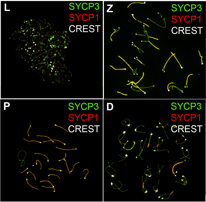
Meiosis () is a special type of cell division of germ cells in sexually-reproducing organisms that produces the gametes, the sperm or egg cells. It involves two rounds of division that ultimately result in four cells, each with only one copy of each chromosome (haploid). Additionally, prior to the division, genetic material from the paternal and maternal copies of each chromosome is crossed over, creating new combinations of code on each chromosome. Later on, during fertilisation, the haploid cells produced by meiosis from a male and a female will fuse to create a zygote, a cell with two copies of each chromosome. Errors in meiosis resulting in aneuploidy (an abnormal number of chromosomes) are the leading known cause of miscarriage and the most frequent genetic cause of developmental disabilities. In meiosis, DNA replication is followed by two rounds of cell division to produce four daughter cells, each with half the number of chromosomes as the original parent cell. The tw ... [...More Info...] [...Related Items...] OR: [Wikipedia] [Google] [Baidu] |
Kinetochore
A kinetochore (, ) is a flared oblique-shaped protein structure associated with duplicated chromatids in eukaryotic cells where the spindle fibers, which can be thought of as the ropes pulling chromosomes apart, attach during cell division to pull sister chromatids apart. The kinetochore assembles on the centromere and links the chromosome to microtubule polymers from the mitotic spindle during mitosis and meiosis. The term kinetochore was first used in a footnote in a 1934 Cytology book by Lester W. Sharp and commonly accepted in 1936. Sharp's footnote reads: "The convenient term ''kinetochore'' (= movement place) has been suggested to the author by J. A. Moore", likely referring to John Alexander Moore who had joined Columbia University as a freshman in 1932. Monocentric organisms, including vertebrates, fungi, and most plants, have a single centromeric region on each chromosome which assembles a single, localized kinetochore. Holocentric organisms, such as nematodes a ... [...More Info...] [...Related Items...] OR: [Wikipedia] [Google] [Baidu] |
Sister Chromatids
A sister chromatid refers to the identical copies ( chromatids) formed by the DNA replication of a chromosome, with both copies joined together by a common centromere. In other words, a sister chromatid may also be said to be 'one-half' of the duplicated chromosome. A pair of sister chromatids is called a dyad. A full set of sister chromatids is created during the synthesis ( S) phase of interphase, when all the chromosomes in a cell are replicated. The two sister chromatids are separated from each other into two different cells during mitosis or during the second division of meiosis. Compare sister chromatids to homologous chromosomes, which are the two ''different'' copies of a chromosome that diploid organisms (like humans) inherit, one from each parent. Sister chromatids are by and large identical (since they carry the same alleles, also called variants or versions, of genes) because they derive from one original chromosome. An exception is towards the end of meiosis, after ... [...More Info...] [...Related Items...] OR: [Wikipedia] [Google] [Baidu] |
Sister Chromatid Exchange
Sister chromatid exchange (SCE) is the exchange of genetic material between two identical sister chromatids. It was first discovered by using the Giemsa staining method on one chromatid belonging to the sister chromatid complex before anaphase in mitosis. The staining revealed that few segments were passed to the sister chromatid which were not dyed. The Giemsa staining was able to stain due to the presence of bromodeoxyuridine analogous base which was introduced to the desired chromatid. The reason for the (SCE) is not known but it is required and used as a mutagenic testing of many products. Four to five sister chromatid exchanges per chromosome pair, per mitosis is in the normal distribution, while 14–100 exchanges is not normal and presents a danger to the organism. SCE is elevated in pathologies including Bloom syndrome, having recombination rates ~10–100 times above normal, depending on cell type. Frequent SCEs may also be related to formation of tumors. Sister chro ... [...More Info...] [...Related Items...] OR: [Wikipedia] [Google] [Baidu] |
Mitosis
Mitosis () is a part of the cell cycle in eukaryote, eukaryotic cells in which replicated chromosomes are separated into two new Cell nucleus, nuclei. Cell division by mitosis is an equational division which gives rise to genetically identical cells in which the total number of chromosomes is maintained. Mitosis is preceded by the S phase of interphase (during which DNA replication occurs) and is followed by telophase and cytokinesis, which divide the cytoplasm, organelles, and cell membrane of one cell into two new cell (biology), cells containing roughly equal shares of these cellular components. The different stages of mitosis altogether define the mitotic phase (M phase) of a cell cycle—the cell division, division of the mother cell into two daughter cells genetically identical to each other. The process of mitosis is divided into stages corresponding to the completion of one set of activities and the start of the next. These stages are preprophase (specific to plant ce ... [...More Info...] [...Related Items...] OR: [Wikipedia] [Google] [Baidu] |
Centromere
The centromere links a pair of sister chromatids together during cell division. This constricted region of chromosome connects the sister chromatids, creating a short arm (p) and a long arm (q) on the chromatids. During mitosis, spindle fibers attach to the centromere via the kinetochore. The physical role of the centromere is to act as the site of assembly of the kinetochores – a highly complex multiprotein structure that is responsible for the actual events of chromosome segregation – i.e. binding microtubules and signaling to the cell cycle machinery when all chromosomes have adopted correct attachments to the spindle, so that it is safe for cell division to proceed to completion and for cells to enter anaphase. There are, broadly speaking, two types of centromeres. "Point centromeres" bind to specific proteins that recognize particular DNA sequences with high efficiency. Any piece of DNA with the point centromere DNA sequence on it will typically form a centr ... [...More Info...] [...Related Items...] OR: [Wikipedia] [Google] [Baidu] |
Homologous Chromosome
Homologous chromosomes or homologs are a set of one maternal and one paternal chromosome that pair up with each other inside a cell during meiosis. Homologs have the same genes in the same locus (genetics), loci, where they provide points along each chromosome that enable a pair of chromosomes to align correctly with each other before separating during meiosis. This is the basis for Mendelian inheritance, which characterizes inheritance patterns of genetic material from an organism to its offspring parent developmental cell at the given time and area. Overview Chromosomes are linear arrangements of condensed DNA, deoxyribonucleic acid (DNA) and histone proteins, which form a complex called chromatin. Homologous chromosomes are made up of chromosome pairs of approximately the same length, centromere position, and staining pattern, for genes with the same corresponding Locus (genetics), loci. One homologous chromosome is inherited from the organism's mother; the other is inherited ... [...More Info...] [...Related Items...] OR: [Wikipedia] [Google] [Baidu] |
Chromosomal Crossover
Chromosomal crossover, or crossing over, is the exchange of genetic material during sexual reproduction between two homologous chromosomes' sister chromatids, non-sister chromatids that results in recombinant chromosomes. It is one of the final phases of genetic recombination, which occurs in the ''pachytene'' stage of prophase I of meiosis during a process called synapsis. Synapsis is usually initiated before the synaptonemal complex develops and is not completed until near the end of prophase I. Crossover usually occurs when matching regions on matching chromosomes break and then reconnect to the other chromosome, resulting in Chiasma (genetics), chiasma which are the visible evidence of crossing over. History of discovery Crossing over was described, in theory, by Thomas Hunt Morgan; the term crossover was coined by Morgan and Eleth Cattell. Hunt relied on the discovery of Frans Alfons Janssens who described the phenomenon in 1909 and had called it "chiasmatypie". Th ... [...More Info...] [...Related Items...] OR: [Wikipedia] [Google] [Baidu] |
Homologous Chromosome
Homologous chromosomes or homologs are a set of one maternal and one paternal chromosome that pair up with each other inside a cell during meiosis. Homologs have the same genes in the same locus (genetics), loci, where they provide points along each chromosome that enable a pair of chromosomes to align correctly with each other before separating during meiosis. This is the basis for Mendelian inheritance, which characterizes inheritance patterns of genetic material from an organism to its offspring parent developmental cell at the given time and area. Overview Chromosomes are linear arrangements of condensed DNA, deoxyribonucleic acid (DNA) and histone proteins, which form a complex called chromatin. Homologous chromosomes are made up of chromosome pairs of approximately the same length, centromere position, and staining pattern, for genes with the same corresponding Locus (genetics), loci. One homologous chromosome is inherited from the organism's mother; the other is inherited ... [...More Info...] [...Related Items...] OR: [Wikipedia] [Google] [Baidu] |


