|
Cerebrospinal Venous System
The cerebrospinal venous system (CSVS) consists of the interconnected venous systems of the brain (the cerebral venous system) and the spine (the vertebral venous system). Introduction The anatomic connections between the cerebral and vertebral venous systems was accurately depicted in 1819 by Gilbert Breschet, a French physician later to become Professor of Anatomy at Faculté de médecine de Paris. However, the significance and physiology of this venous complex remained obscure for more than a century, until the seminal work of Oscar Batson. Batson, a Professor of Anatomy at the University of Pennsylvania, in 1940 detailed the anatomy and physiology of the cerebrospinal venous system and its role in the spread of metastases.Batson, O.V., The Function of the Vertebral Veins and their role in the spread of metastases. Annals of Surgery, 1940. 112: p. 138-149 Batson’s work remains primarily known for its accurate depiction of the vertebral venous system as the route of metastasis ... [...More Info...] [...Related Items...] OR: [Wikipedia] [Google] [Baidu] |
Cerebral Circulation
Cerebral circulation is the movement of blood through a network of cerebral arteries and veins supplying the brain. The rate of cerebral blood flow in an adult human is typically 750 milliliters per minute, or about 15% of cardiac output. Arteries deliver oxygenated blood, glucose and other nutrients to the brain. Veins carry "used or spent" blood back to the heart, to remove carbon dioxide, lactic acid, and other metabolic products. Because the brain would quickly suffer damage from any stoppage in blood supply, the cerebral circulatory system has safeguards including autoregulation of the blood vessels. The failure of these safeguards may result in a stroke. The volume of blood in circulation is called the cerebral blood flow. Sudden intense accelerations change the gravitational forces perceived by bodies and can severely impair cerebral circulation and normal functions to the point of becoming serious life-threatening conditions. The following description is based on ideal ... [...More Info...] [...Related Items...] OR: [Wikipedia] [Google] [Baidu] |
Batson Venous Plexus
The Batson venous plexus (Batson veins) is a network of valveless veins in the human body that connect the deep pelvic veins and thoracic veins (draining the inferior end of the urinary bladder, breast and prostate) to the internal vertebral venous plexuses. Because of their location and lack of valves, they are believed to provide a route for the spread of cancer metastases. These metastases commonly arise from cancer of the pelvic organs such as the rectum and prostate and may spread to the vertebral column or brain. The plexus is named after anatomist Oscar Vivian Batson, who first described it in 1940. Batson's plexus is part of the Cerebrospinal venous system. Batson's venous plexus may also allow the spread of infection in a similar manner. neuroschistomiasis,Carbonell C, Rodríguez-Alonso B, López-Bernús A, Almeida H, Galindo-Pérez I, Velasco-Tirado V, Marcos M, Pardo-Lledías J, Belhassen-García M. Clinical Spectrum of Schistosomiasis: An Update. J Clin Med. 2021 Nov ... [...More Info...] [...Related Items...] OR: [Wikipedia] [Google] [Baidu] |
Gilbert Breschet
Gilbert Breschet (7 July 1784 – 10 May 1845) was a French anatomist born in Clermont-Ferrand. He studied medicine at the University of Paris, and in 1812 was conferred as doctor of medicine in Paris. In 1836 he succeeded Jean Cruveilhier (1791–1874) as professor of anatomy at the Faculté de Médecine de Paris. Breschet made many contributions in comparative anatomy and in his research of zoonotic diseases. In 1813 with François Magendie (1783–1855), he demonstrated that rabies can be transmitted from the saliva of humans to dogs. Also, he was the first to create an accurate figure of the '' rete mirabile'' in whales and dolphins, a vascular network that allows these mammals to survive and adapt in ocean depths; a feature discovered by Edward Tyson. He did extensive anatomical studies of veins of the cranium and spine, and made important investigations of the auditory system in vertebrates. He provided a comprehensive description of the utricle and saccule ... [...More Info...] [...Related Items...] OR: [Wikipedia] [Google] [Baidu] |
Metastasis
Metastasis is a pathogenic agent's spread from an initial or primary site to a different or secondary site within the host's body; the term is typically used when referring to metastasis by a cancerous tumor. The newly pathological sites, then, are metastases (mets). It is generally distinguished from cancer invasion, which is the direct extension and penetration by cancer cells into neighboring tissues. Cancer occurs after cells are genetically altered to proliferate rapidly and indefinitely. This uncontrolled proliferation by mitosis produces a primary heterogeneic tumour. The cells which constitute the tumor eventually undergo metaplasia, followed by dysplasia then anaplasia, resulting in a malignant phenotype. This malignancy allows for invasion into the circulation, followed by invasion to a second site for tumorigenesis. Some cancer cells known as circulating tumor cells acquire the ability to penetrate the walls of lymphatic or blood vessels, after which they a ... [...More Info...] [...Related Items...] OR: [Wikipedia] [Google] [Baidu] |
Magnetic Resonance Imaging
Magnetic resonance imaging (MRI) is a medical imaging technique used in radiology to form pictures of the anatomy and the physiological processes inside the body. MRI scanners use strong magnetic fields, magnetic field gradients, and radio waves to generate images of the organs in the body. MRI does not involve X-rays or the use of ionizing radiation, which distinguishes it from computed tomography (CT) and positron emission tomography (PET) scans. MRI is a medical application of nuclear magnetic resonance (NMR) which can also be used for imaging in other NMR applications, such as NMR spectroscopy. MRI is widely used in hospitals and clinics for medical diagnosis, staging and follow-up of disease. Compared to CT, MRI provides better contrast in images of soft tissues, e.g. in the brain or abdomen. However, it may be perceived as less comfortable by patients, due to the usually longer and louder measurements with the subject in a long, confining tube, although "open ... [...More Info...] [...Related Items...] OR: [Wikipedia] [Google] [Baidu] |
Anastomoses
An anastomosis (, plural anastomoses) is a connection or opening between two things (especially cavities or passages) that are normally diverging or branching, such as between blood vessels, leaf veins, or streams. Such a connection may be normal (such as the foramen ovale in a fetus's heart) or abnormal (such as the patent foramen ovale in an adult's heart); it may be acquired (such as an arteriovenous fistula) or innate (such as the arteriovenous shunt of a metarteriole); and it may be natural (such as the aforementioned examples) or artificial (such as a surgical anastomosis). The reestablishment of an anastomosis that had become blocked is called a reanastomosis. Anastomoses that are abnormal, whether congenital or acquired, are often called fistulas. The term is used in medicine, biology, mycology, geology, and geography. Etymology Anastomosis: medical or Modern Latin, from Greek ἀναστόμωσις, anastomosis, "outlet, opening", Gr ana- "up, on, upon", stoma "mou ... [...More Info...] [...Related Items...] OR: [Wikipedia] [Google] [Baidu] |
Suboccipital Triangle
The suboccipital triangle is a region of the neck bounded by the following three muscles of the suboccipital group of muscles: * Rectus capitis posterior major - above and medially * Obliquus capitis superior - above and laterally * Obliquus capitis inferior - below and laterally (Rectus capitis posterior minor is also in this region but does not form part of the triangle) It is covered by a layer of dense fibro-fatty tissue, situated beneath the semispinalis capitis. The floor is formed by the posterior atlantooccipital membrane, and the posterior arch of the atlas. In the deep groove on the upper surface of the posterior arch of the atlas are the vertebral artery and the first cervical or suboccipital nerve. In the past, the vertebral artery was accessed here in order to conduct angiography of the circle of Willis. Presently, formal angiography of the circle of Willis is performed via catheter angiography, with access usually being acquired at the common femoral artery. Al ... [...More Info...] [...Related Items...] OR: [Wikipedia] [Google] [Baidu] |
Hemostasis
In biology, hemostasis or haemostasis is a process to prevent and stop bleeding, meaning to keep blood within a damaged blood vessel (the opposite of hemostasis is hemorrhage). It is the first stage of wound healing. This involves coagulation, which changes blood from a liquid to a gel. Intact blood vessels are central to moderating blood's tendency to form clots. The endothelial cells of intact vessels prevent blood clotting with a heparin-like molecule and thrombomodulin, and prevent platelet aggregation with nitric oxide and prostacyclin. When endothelium of a blood vessel is damaged, the endothelial cells stop secretion of coagulation and aggregation inhibitors and instead secrete von Willebrand factor, which initiate the maintenance of hemostasis after injury. Hemostasis involves three major steps: * vasoconstriction * temporary blockage of a hole in a damaged blood vessel by a platelet plug * blood coagulation (formation of fibrin clots) These processes seal the injury ... [...More Info...] [...Related Items...] OR: [Wikipedia] [Google] [Baidu] |
Radiocontrast Agent
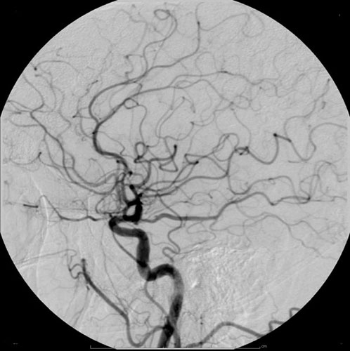
Radiocontrast agents are substances used to enhance the visibility of internal structures in X-ray-based imaging techniques such as computed tomography (contrast CT), projectional radiography, and fluoroscopy. Radiocontrast agents are typically iodine, or more rarely barium sulfate. The contrast agents absorb external X-rays, resulting in decreased exposure on the X-ray detector. This is different from radiopharmaceuticals used in nuclear medicine which emit radiation. Magnetic resonance imaging (MRI) functions through different principles and thus MRI contrast agents have a different mode of action. These compounds work by altering the magnetic properties of nearby hydrogen nuclei. Types and uses Radiocontrast agents used in X-ray examinations can be grouped in positive (iodinated agents, barium sulfate), and negative agents (air, carbon dioxide, methylcellulose). Iodine (circulatory system) Iodinated contrast contains iodine. It is the main type of radiocontrast used for in ... [...More Info...] [...Related Items...] OR: [Wikipedia] [Google] [Baidu] |
Intracranial
The cranial cavity, also known as intracranial space, is the space within the skull that accommodates the brain. The skull minus the mandible is called the ''cranium''. The cavity is formed by eight cranial bones known as the neurocranium that in humans includes the skull cap and forms the protective case around the brain. The remainder of the skull is called the facial skeleton. Meninges are protective membranes that surround the brain to minimize damage of the brain when there is head trauma. Meningitis is the inflammation of meninges caused by bacterial or viral infections. Structure The capacity of an adult human cranial cavity is 1,200–1,700 cm3. The spaces between meninges and the brain are filled with a clear cerebrospinal fluid, increasing the protection of the brain. Facial bones of the skull are not included in the cranial cavity. There are only eight cranial bones: The occipital, sphenoid, frontal, ethmoid, two parietal, and two temporal bones are fused toget ... [...More Info...] [...Related Items...] OR: [Wikipedia] [Google] [Baidu] |
Sun Sentinel
The ''Sun Sentinel'' (also known as the ''South Florida Sun Sentinel'', known until 2008 as the ''Sun-Sentinel'', and stylized on its masthead as ''SunSentinel'') is the main daily newspaper of Fort Lauderdale, Florida, as well as surrounding Broward County and southern Palm Beach County. It circulates all throughout the three counties that comprise South Florida. It is the largest-circulation newspaper in the area. Paul Pham has held the position of general manager since November 2020, and Julie Anderson has held the position of editor-in-chief since February 2018. The newspaper was for many years branded as the ''Sun-Sentinel'', with a hyphen, until a redesign and rebranding on August 17, 2008. The new look also removed the space between "Sun" and "Sentinel" in the newspaper's flag, but its name retained the space. The ''Sun Sentinel'' is owned by parent company, '' Tribune Publishing''. This company was acquired by Alden Global Capital, which operates its media properties th ... [...More Info...] [...Related Items...] OR: [Wikipedia] [Google] [Baidu] |
Glymphatic System
The glymphatic system (or glymphatic clearance pathway, or paravascular system) was described and named in 2013 as a system for waste clearance in the central nervous system (CNS) of vertebrates. According to this model, cerebrospinal fluid (CSF) flows into the paravascular space around cerebral arteries, combining with interstitial fluid (ISF) and parenchymal solutes, and exiting down venous paravascular spaces. The pathway consists of a para-arterial influx route for CSF to enter the brain parenchyma, coupled to a clearance mechanism for the removal of interstitial fluid (ISF) and extracellular solutes from the interstitial compartments of the brain and spinal cord. Exchange of solutes between CSF and ISF is driven primarily by arterial pulsation and regulated during sleep by the expansion and contraction of brain extracellular space. Clearance of soluble proteins, waste products, and excess extracellular fluid is accomplished through convective bulk flow of ISF, facilitated by ... [...More Info...] [...Related Items...] OR: [Wikipedia] [Google] [Baidu] |


