|
Cerebral Polyopia
Cerebral diplopia or polyopia describes seeing two or more images arranged in ordered rows, columns, or diagonals after fixation on a stimulus. The polyopic images occur monocular bilaterally (one eye open on both sides) and binocularly (both eyes open), differentiating it from ocular diplopia or polyopia. The number of duplicated images can range from one to hundreds. Some patients report difficulty in distinguishing the replicated images from the real images, while others report that the false images differ in size, intensity, or color. Cerebral polyopia is sometimes confused with palinopsia ( visual trailing), in which multiple images appear while watching an object. However, in cerebral polyopia, the duplicated images are of a stationary object which are perceived even after the object is removed from the visual field. Movement of the original object causes all of the duplicated images to move, or the polyopic images disappear during motion. In palinoptic polyopia, movement cause ... [...More Info...] [...Related Items...] OR: [Wikipedia] [Google] [Baidu] |
Fixation (visual)
Fixation or visual fixation is the maintaining of the gaze on a single location. An animal can exhibit visual fixation if it possess a fovea in the anatomy of their eye. The fovea is typically located at the center of the retina and is the point of clearest vision. The species in which fixational eye movement has been verified thus far include humans, primates, cats, rabbits, turtles, salamanders, and owls. Regular eye movement alternates between saccades and visual fixations, the notable exception being in smooth pursuit, controlled by a different neural substrate that appears to have developed for hunting prey. The term "fixation" can either be used to refer to the point in time and space of focus or the act of fixating. Fixation, in the act of fixating, is the point between any two saccades, during which the eyes are relatively stationary and virtually all visual input occurs. In the absence of retinal jitter, a laboratory condition known as retinal stabilization, perceptions ... [...More Info...] [...Related Items...] OR: [Wikipedia] [Google] [Baidu] |
Stimulus (psychology)
In psychology Psychology is the scientific study of mind and behavior. Psychology includes the study of conscious and unconscious phenomena, including feelings and thoughts. It is an academic discipline of immense scope, crossing the boundaries betwe ..., a stimulus is any object or event that elicits a sensory or behavioral response in an organism. In this context, a distinction is made between the ''distal stimulus'' (the external, perceived object) and the ''proximal stimulus'' (the stimulation of sensory organs). *In perceptual psychology, a stimulus is an energy change (e.g., light or sound) which is registered by the senses (e.g., vision, hearing, taste, etc.) and constitutes the basis for perception. *In behavioral psychology (i.e., classical conditioning, classical and operant conditioning, operant conditioning), a stimulus constitutes the basis for behavior. The stimulus–response model emphasizes the relation between stimulus and behavior rather than an anim ... [...More Info...] [...Related Items...] OR: [Wikipedia] [Google] [Baidu] |
Palinopsia
Palinopsia (Greek: ''palin'' for "again" and ''opsia'' for "seeing") is the persistent recurrence of a visual image after the stimulus has been removed. Palinopsia is not a diagnosis; it is a diverse group of pathological visual symptoms with a wide variety of causes. Visual perseveration is synonymous with palinopsia. In 2014, Gersztenkorn and Lee comprehensively reviewed all cases of palinopsia in the literature and subdivided it into two clinically relevant groups: illusory palinopsia and hallucinatory palinopsia. Hallucinatory palinopsia, usually due to seizures or posterior cortical lesions, describes afterimages that are formed, long-lasting, and high resolution. Illusory palinopsia, usually due to migraines, head trauma, prescription drugs, visual snow or hallucinogen persisting perception disorder (HPPD), describes afterimages that are affected by ambient light and motion and are unformed, indistinct, or low resolution. Presentation People with palinopsia frequently repor ... [...More Info...] [...Related Items...] OR: [Wikipedia] [Google] [Baidu] |
Illusory Palinopsia
Illusory palinopsia is a subtype of palinopsia, a visual disturbance defined as the persistence or recurrence of a visual image after the stimulus has been removed. Palinopsia is a broad term describing a heterogeneous group of symptoms, which is divided into hallucinatory palinopsia and illusory palinopsia. Illusory palinopsia is likely due to sustained awareness of a stimulus and is similar to a visual illusion: the distorted perception of a real external stimulus. Illusory palinopsia is caused by migraines, hallucinogen persisting perception disorder (HPPD), prescription drugs, and head trauma, but is also sometimes idiopathic. Illusory palinopsia consists of afterimages that are short-lived or unformed, occur at the same location in the visual field as the original stimulus, and are often exposed or exacerbated based on environmental parameters such as stimulus intensity, background contrast, fixation, and movement. Illusory palinopsia symptoms occur continuously or predic ... [...More Info...] [...Related Items...] OR: [Wikipedia] [Google] [Baidu] |
Entomopia
Entomopia (from the Greek roots for "insect" and "eye Eyes are organs of the visual system. They provide living organisms with vision, the ability to receive and process visual detail, as well as enabling several photo response functions that are independent of vision. Eyes detect light and conv ..."), is a form of polyopia in which a grid-like pattern of multiple copies of the same visual image is seen. Entomopia may be due to disease of the occipital lobe, defects in visual integration and fixation or incomplete visual processing due to poor visuospatial localisation in the hemianopic field, although its causes are unknown. Reassurance may be the only treatment. See also * Diplopia References Visual disturbances and blindness {{med-sign-stub ... [...More Info...] [...Related Items...] OR: [Wikipedia] [Google] [Baidu] |
Homonymous Hemianopsia
Hemianopsia, or hemianopia, is a visual field loss on the left or right side of the vertical midline. It can affect one eye but usually affects both eyes. Homonymous hemianopsia (or homonymous hemianopia) is hemianopic visual field loss on the same side of both eyes. Homonymous hemianopsia occurs because the right half of the brain has visual pathways for the left hemifield of both eyes, and the left half of the brain has visual pathways for the right hemifield of both eyes. When one of these pathways is damaged, the corresponding visual field is lost. Signs and symptoms Paris as seen with right homonymous hemianopsia Mobility can be difficult for people with homonymous hemianopsia. "Patients frequently complain of bumping into obstacles on the side of the field loss, thereby bruising their arms and legs." People with homonymous hemianopsia often experience discomfort in crowds. "A patient with this condition may be unaware of what he or she cannot see and frequently bumps ... [...More Info...] [...Related Items...] OR: [Wikipedia] [Google] [Baidu] |
Visual Release Hallucinations
Visual release hallucinations, also known as Charles Bonnet syndrome or CBS, are a type of psychophysical visual disturbance in which a person with partial or severe blindness experiences visual hallucinations. First described by Charles Bonnet in 1760, the term ''Charles Bonnet syndrome'' was first introduced into English-speaking psychiatry in 1982. A related type of hallucination that also occurs with lack of visual input is the closed-eye hallucination. Signs and symptoms People with significant vision loss may have vivid recurrent visual hallucinations (fictive visual percepts). One characteristic of these hallucinations is that they usually are " lilliputian" (hallucinations in which the characters or objects are smaller than normal). Depending on the content, visual hallucinations can be classified as either simple or complex. Simple visual hallucinations are commonly characterized by shapes, photopsias, and grid-like patterns. Complex visual hallucinations consist of highl ... [...More Info...] [...Related Items...] OR: [Wikipedia] [Google] [Baidu] |
Visual Cortex
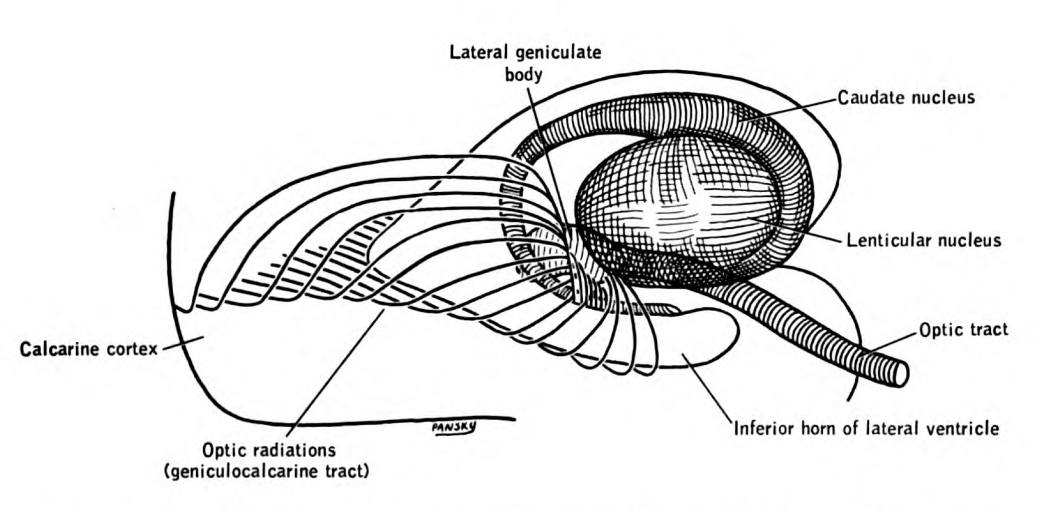
The visual cortex of the brain is the area of the cerebral cortex that processes visual information. It is located in the occipital lobe. Sensory input originating from the eyes travels through the lateral geniculate nucleus in the thalamus and then reaches the visual cortex. The area of the visual cortex that receives the sensory input from the lateral geniculate nucleus is the primary visual cortex, also known as visual area 1 ( V1), Brodmann area 17, or the striate cortex. The extrastriate areas consist of visual areas 2, 3, 4, and 5 (also known as V2, V3, V4, and V5, or Brodmann area 18 and all Brodmann area 19). Both hemispheres of the brain include a visual cortex; the visual cortex in the left hemisphere receives signals from the right visual field, and the visual cortex in the right hemisphere receives signals from the left visual field. Introduction The primary visual cortex (V1) is located in and around the calcarine fissure in the occipital lobe. Each hemisphere's V1 ... [...More Info...] [...Related Items...] OR: [Wikipedia] [Google] [Baidu] |
CT Scan
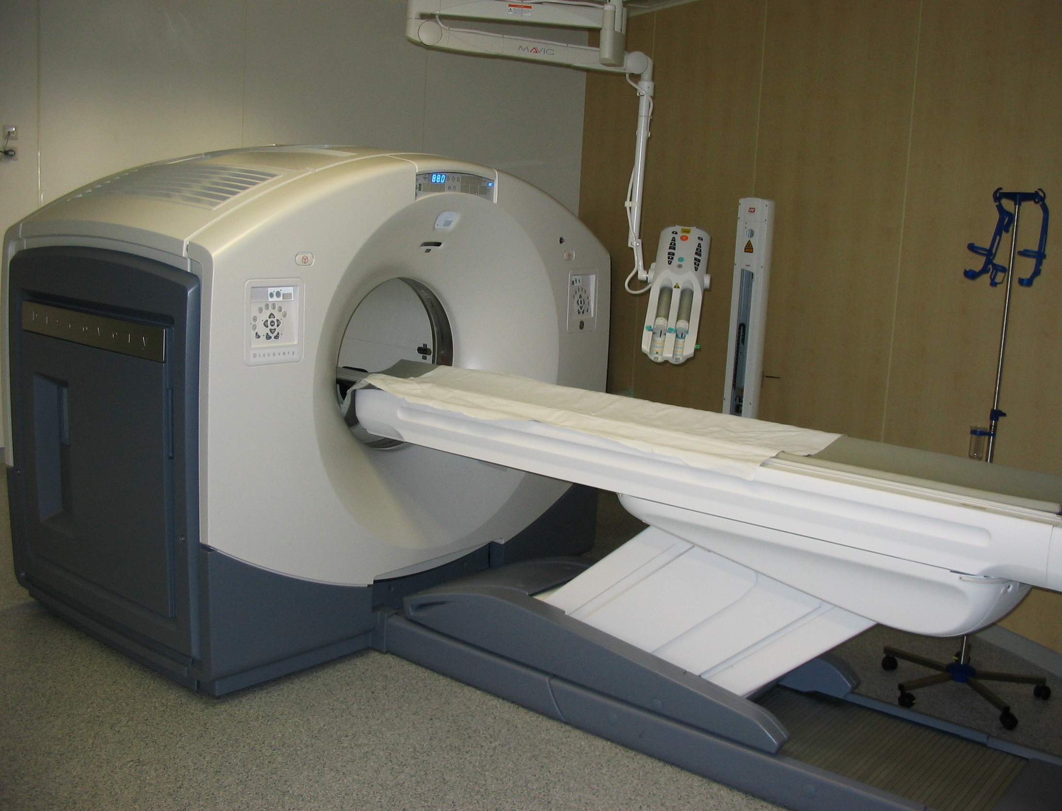
A computed tomography scan (CT scan; formerly called computed axial tomography scan or CAT scan) is a medical imaging technique used to obtain detailed internal images of the body. The personnel that perform CT scans are called radiographers or radiology technologists. CT scanners use a rotating X-ray tube and a row of detectors placed in a gantry (medical), gantry to measure X-ray Attenuation#Radiography, attenuations by different tissues inside the body. The multiple X-ray measurements taken from different angles are then processed on a computer using tomographic reconstruction algorithms to produce Tomography, tomographic (cross-sectional) images (virtual "slices") of a body. CT scans can be used in patients with metallic implants or pacemakers, for whom magnetic resonance imaging (MRI) is Contraindication, contraindicated. Since its development in the 1970s, CT scanning has proven to be a versatile imaging technique. While CT is most prominently used in medical diagnosis, ... [...More Info...] [...Related Items...] OR: [Wikipedia] [Google] [Baidu] |
Positron Emission Tomography
Positron emission tomography (PET) is a functional imaging technique that uses radioactive substances known as radiotracers to visualize and measure changes in Metabolism, metabolic processes, and in other physiological activities including blood flow, regional chemical composition, and absorption. Different tracers are used for various imaging purposes, depending on the target process within the body. For example, 18F-FDG, -FDG is commonly used to detect cancer, Sodium fluoride#Medical imaging, NaF is widely used for detecting bone formation, and Isotopes of oxygen#Oxygen-15, oxygen-15 is sometimes used to measure blood flow. PET is a common medical imaging, imaging technique, a Scintigraphy#Process, medical scintillography technique used in nuclear medicine. A radiopharmaceutical, radiopharmaceutical — a radioisotope attached to a drug — is injected into the body as a radioactive tracer, tracer. When the radiopharmaceutical undergoes beta plus decay, a positron is ... [...More Info...] [...Related Items...] OR: [Wikipedia] [Google] [Baidu] |
Magnetic Resonance Imaging
Magnetic resonance imaging (MRI) is a medical imaging technique used in radiology to form pictures of the anatomy and the physiological processes of the body. MRI scanners use strong magnetic fields, magnetic field gradients, and radio waves to generate images of the organs in the body. MRI does not involve X-rays or the use of ionizing radiation, which distinguishes it from CT and PET scans. MRI is a medical application of nuclear magnetic resonance (NMR) which can also be used for imaging in other NMR applications, such as NMR spectroscopy. MRI is widely used in hospitals and clinics for medical diagnosis, staging and follow-up of disease. Compared to CT, MRI provides better contrast in images of soft-tissues, e.g. in the brain or abdomen. However, it may be perceived as less comfortable by patients, due to the usually longer and louder measurements with the subject in a long, confining tube, though "Open" MRI designs mostly relieve this. Additionally, implants and oth ... [...More Info...] [...Related Items...] OR: [Wikipedia] [Google] [Baidu] |
Electroencephalography
Electroencephalography (EEG) is a method to record an electrogram of the spontaneous electrical activity of the brain. The biosignals detected by EEG have been shown to represent the postsynaptic potentials of pyramidal neurons in the neocortex and allocortex. It is typically non-invasive, with the EEG electrodes placed along the scalp (commonly called "scalp EEG") using the International 10-20 system, or variations of it. Electrocorticography, involving surgical placement of electrodes, is sometimes called " intracranial EEG". Clinical interpretation of EEG recordings is most often performed by visual inspection of the tracing or quantitative EEG analysis. Voltage fluctuations measured by the EEG bioamplifier and electrodes allow the evaluation of normal brain activity. As the electrical activity monitored by EEG originates in neurons in the underlying brain tissue, the recordings made by the electrodes on the surface of the scalp vary in accordance with their orientation and ... [...More Info...] [...Related Items...] OR: [Wikipedia] [Google] [Baidu] |


