|
Cartilage
Cartilage is a resilient and smooth type of connective tissue. Semi-transparent and non-porous, it is usually covered by a tough and fibrous membrane called perichondrium. In tetrapods, it covers and protects the ends of long bones at the joints as articular cartilage, and is a structural component of many body parts including the rib cage, the neck and the bronchial tubes, and the intervertebral discs. In other taxa, such as chondrichthyans and cyclostomes, it constitutes a much greater proportion of the skeleton. It is not as hard and rigid as bone, but it is much stiffer and much less flexible than muscle. The matrix of cartilage is made up of glycosaminoglycans, proteoglycans, collagen fibers and, sometimes, elastin. It usually grows quicker than bone. Because of its rigidity, cartilage often serves the purpose of holding tubes open in the body. Examples include the rings of the trachea, such as the cricoid cartilage and carina. Cartilage is composed of specialized c ... [...More Info...] [...Related Items...] OR: [Wikipedia] [Google] [Baidu] |
Hyaline Cartilage
Hyaline cartilage is the glass-like (hyaline) and translucent cartilage found on many joint surfaces. It is also most commonly found in the ribs, nose, larynx, and trachea. Hyaline cartilage is pearl-gray in color, with a firm consistency and has a considerable amount of collagen. It contains no nerves or blood vessels, and its structure is relatively simple. Structure Hyaline cartilage is the most common kind of cartilage in the human body. It is primarily composed of type II collagen and proteoglycans. Hyaline cartilage is located in the trachea, nose, epiphyseal plate, sternum, and ribs. Hyaline cartilage is covered externally by a fibrous membrane known as the perichondrium. The primary cells of cartilage are chondrocytes, which are in a matrix of fibrous tissue, proteoglycans and glycosaminoglycans. As cartilage does not have lymph glands or blood vessels, the movements of solutes, including nutrients, occur via diffusion within the fluid compartments contiguous with ... [...More Info...] [...Related Items...] OR: [Wikipedia] [Google] [Baidu] |
Cricoid Cartilage
The cricoid cartilage , or simply cricoid (from the Greek ''krikoeides'' meaning "ring-shaped") or cricoid ring, is the only complete ring of cartilage around the trachea. It forms the back part of the voice box and functions as an attachment site for muscles, cartilages, and ligaments involved in opening and closing the airway and in producing speech. Anatomy The cricoid cartilage is the only laryngeal cartilage to form a complete circle around the airway. It is smaller yet thicker and tougher than the thyroid cartilage above. It articulates superiorly with the thyroid cartilage, and the paired arytenoid cartilage. Inferiorly, the trachea attaches onto it. It occurs at the level of the C6 vertebra. Structure The posterior part of the cricoid cartilage (cricoid lamina) is somewhat broader than the anterior and lateral part (cricoid arch). Its shape is said to resemble a signet ring. Cricoid arch The cricoid arch is the curved and vertically narrow anterior portion of ... [...More Info...] [...Related Items...] OR: [Wikipedia] [Google] [Baidu] |
Chondrocyte
Chondrocytes (, ) are the only cells found in healthy cartilage. They produce and maintain the cartilaginous matrix, which consists mainly of collagen and proteoglycans. Although the word '' chondroblast'' is commonly used to describe an immature chondrocyte, the term is imprecise, since the progenitor of chondrocytes (which are mesenchymal stem cells) can differentiate into various cell types, including osteoblasts. Development From least- to terminally-differentiated, the chondrocytic lineage is: # Colony-forming unit-fibroblast # Mesenchymal stem cell / marrow stromal cell # Chondrocyte # Hypertrophic chondrocyte Mesenchymal (mesoderm origin) stem cells are undifferentiated, meaning they can differentiate into a variety of generative cells commonly known as osteochondrogenic (or osteogenic, chondrogenic, osteoprogenitor, etc.) cells. When referring to bone, or in this case cartilage, the originally undifferentiated mesenchymal stem cells lose their pluripotency, proliferate ... [...More Info...] [...Related Items...] OR: [Wikipedia] [Google] [Baidu] |
Knee
In humans and other primates, the knee joins the thigh with the leg and consists of two joints: one between the femur and tibia (tibiofemoral joint), and one between the femur and patella (patellofemoral joint). It is the largest joint in the human body. The knee is a modified hinge joint, which permits flexion and extension (kinesiology), extension as well as slight internal and external rotation. The knee is vulnerable to injury and to the development of osteoarthritis. It is often termed a ''compound joint'' having tibiofemoral and patellofemoral components. (The fibular collateral ligament is often considered with tibiofemoral components.) Structure The knee is a modified hinge joint, a type of synovial joint, which is composed of three functional compartments: the patellofemoral articulation, consisting of the patella, or "kneecap", and the patellar groove on the front of the femur through which it slides; and the medial and lateral tibiofemoral articulations linking the ... [...More Info...] [...Related Items...] OR: [Wikipedia] [Google] [Baidu] |
Elastic Cartilage
Elastic cartilage, fibroelastic cartilage or yellow fibrocartilage is a type of cartilage present in the pinnae (auricles) of the ear giving it shape, provides shape for the lateral region of the external auditory meatus, medial part of the auditory canal Eustachian tube, corniculate and cuneiform laryneal cartilages, and the epiglottis. It contains elastic fiber networks and collagen type II fibers. The principal protein is elastin. Structure Elastic cartilage is histologically similar to hyaline cartilage but contains many yellow elastic fibers lying in a solid matrix. These fibers form bundles that appear dark under a microscope. The elastic fibers require special staining since when it is stained using haematoxylin and eosin (H&E) stain it appears the same as hyaline cartilage. Verhoeff van Geison stains are used (giving the elastic fibers a black color), but aldehyde fuchsin stains, Weigert's elastic stains, and orcein stains also work. These fibers give elastic cartila ... [...More Info...] [...Related Items...] OR: [Wikipedia] [Google] [Baidu] |
Fibrocartilage
Fibrocartilage consists of a mixture of white fibrous tissue and cartilaginous tissue in various proportions. It owes its inflexibility and toughness to the former of these constituents, and its elasticity to the latter. It is the only type of cartilage that contains type I collagen in addition to the normal type II. Structure The extracellular matrix of fibrocartilage is mainly made from type I collagen secreted by chondroblasts. Locations of fibrocartilage in the human body * secondary cartilaginous joints: ** pubic symphysis ** annulus fibrosis of intervertebral discs ** manubriosternal joint * glenoid labrum of shoulder joint * acetabular labrum of hip joint * medial and lateral menisci of the knee joint * location where tendons and ligaments attach to bone * triangular fibrocartilage complex (UTFCC) Function Repair If hyaline cartilage is torn all the way down to the bone, the blood supply from inside the bone is sometimes enough to start some heal ... [...More Info...] [...Related Items...] OR: [Wikipedia] [Google] [Baidu] |
Rib Cage
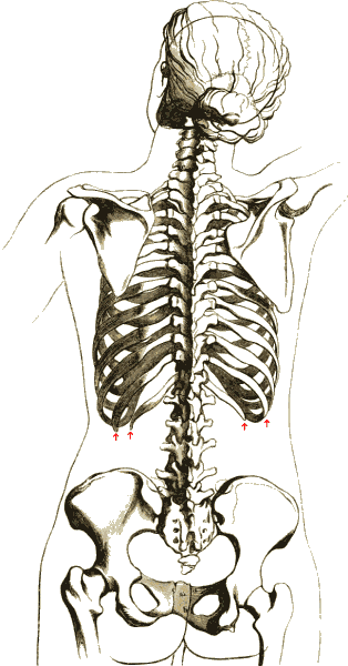
The rib cage or thoracic cage is an endoskeletal enclosure in the thorax of most vertebrates that comprises the ribs, vertebral column and sternum, which protect the vital organs of the thoracic cavity, such as the heart, lungs and great vessels and support the shoulder girdle to form the core part of the axial skeleton. A typical human thoracic cage consists of 12 pairs of ribs and the adjoining costal cartilages, the sternum (along with the manubrium and xiphoid process), and the 12 thoracic vertebrae articulating with the ribs. The thoracic cage also provides attachments for extrinsic skeletal muscles of the neck, upper limbs, upper abdomen and back, and together with the overlying skin and associated fascia and muscles, makes up the thoracic wall. In tetrapods, the rib cage intrinsically holds the muscles of respiration ( diaphragm, intercostal muscles, etc.) that are crucial for active inhalation and forced exhalation, and therefore has a major ventilatory fu ... [...More Info...] [...Related Items...] OR: [Wikipedia] [Google] [Baidu] |
Bone
A bone is a rigid organ that constitutes part of the skeleton in most vertebrate animals. Bones protect the various other organs of the body, produce red and white blood cells, store minerals, provide structure and support for the body, and enable mobility. Bones come in a variety of shapes and sizes and have complex internal and external structures. They are lightweight yet strong and hard and serve multiple functions. Bone tissue (osseous tissue), which is also called bone in the uncountable sense of that word, is hard tissue, a type of specialised connective tissue. It has a honeycomb-like matrix internally, which helps to give the bone rigidity. Bone tissue is made up of different types of bone cells. Osteoblasts and osteocytes are involved in the formation and mineralisation of bone; osteoclasts are involved in the resorption of bone tissue. Modified (flattened) osteoblasts become the lining cells that form a protective layer on the bone surface. The mine ... [...More Info...] [...Related Items...] OR: [Wikipedia] [Google] [Baidu] |
Collagen
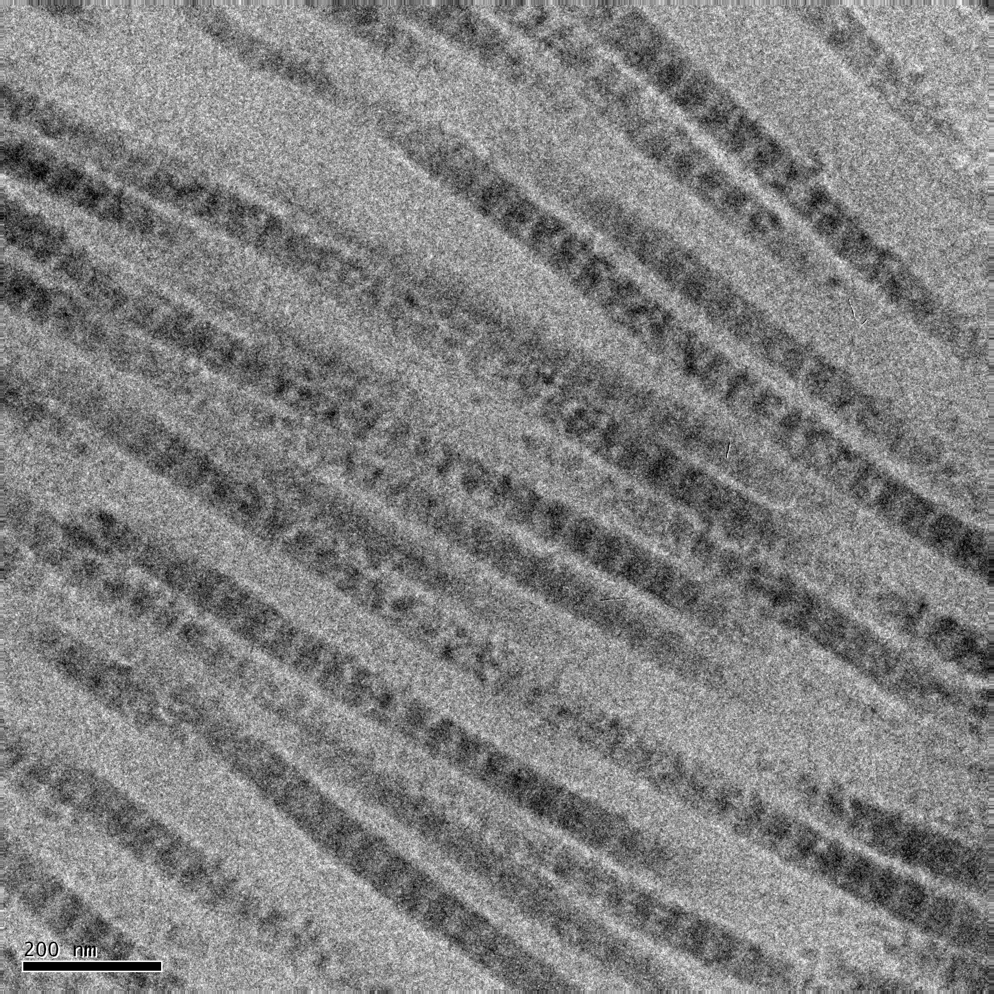
Collagen () is the main structural protein in the extracellular matrix of the connective tissues of many animals. It is the most abundant protein in mammals, making up 25% to 35% of protein content. Amino acids are bound together to form a triple helix of elongated fibril known as a collagen helix. It is mostly found in cartilage, bones, tendons, ligaments, and skin. Vitamin C is vital for collagen synthesis. Depending on the degree of biomineralization, mineralization, collagen tissues may be rigid (bone) or compliant (tendon) or have a gradient from rigid to compliant (cartilage). Collagen is also abundant in corneas, blood vessels, the Gut (anatomy), gut, intervertebral discs, and the dentin in teeth. In muscle tissue, it serves as a major component of the endomysium. Collagen constitutes 1% to 2% of muscle tissue and 6% by weight of skeletal muscle. The fibroblast is the most common cell creating collagen in animals. Gelatin, which is used in food and industry, is collagen t ... [...More Info...] [...Related Items...] OR: [Wikipedia] [Google] [Baidu] |
Joint
A joint or articulation (or articular surface) is the connection made between bones, ossicles, or other hard structures in the body which link an animal's skeletal system into a functional whole.Saladin, Ken. Anatomy & Physiology. 7th ed. McGraw-Hill Connect. Webp.274/ref> They are constructed to allow for different degrees and types of movement. Some joints, such as the knee, elbow, and shoulder, are self-lubricating, almost frictionless, and are able to withstand compression and maintain heavy loads while still executing smooth and precise movements. Other joints such as suture (joint), sutures between the bones of the skull permit very little movement (only during birth) in order to protect the brain and the sense organs. The connection between a tooth and the jawbone is also called a joint, and is described as a fibrous joint known as a gomphosis. Joints are classified both structurally and functionally. Joints play a vital role in the human body, contributing to movement, sta ... [...More Info...] [...Related Items...] OR: [Wikipedia] [Google] [Baidu] |
Perichondrium
The perichondrium (from Greek and ) is a layer of dense irregular connective tissue that surrounds the cartilage of developing bone. It consists of two separate layers: an outer fibrous layer and inner chondrogenic layer. The fibrous layer contains fibroblasts, which produce collagenous fibres. The chondrogenic layer remains undifferentiated and can form chondroblasts. Perichondrium can be found around the perimeter of elastic cartilage and hyaline cartilage. Perichondrium is a type of irregular collagenous ordinary connective tissue, and also functions in the growth and repair of cartilage. Perichondrium contains type I collagen Type I collagen is the most abundant collagen of the human body, consisting of around 90% of the body's total collagen in vertebrates. Due to this, it is also the most abundant protein type found in all vertebrates. Type I forms large, eosinop ... and type XII collagen. References External links * Iowa Histology Index 4/iv-05' - "Slide 12, ... [...More Info...] [...Related Items...] OR: [Wikipedia] [Google] [Baidu] |


