|
Carotid Sheath
The carotid sheath is an anatomical term for the fibrous connective tissue that surrounds the vascular compartment of the neck. It is part of the deep cervical fascia of the neck, below the superficial cervical fascia meaning the subcutaneous adipose tissue immediately beneath the skin. The deep cervical fascia of the neck includes four parts: * The investing layer (encloses the SCM and Trapezius) * The carotid sheath (encloses the vascular region of the neck) * The pretracheal fascia (encloses the visceral region of the neck) * The prevertebral fascia (encloses the vertebral region of the neck) Structure The carotid sheath is located at the lateral boundary of the retropharyngeal space at the level of the oropharynx on each side of the neck and deep to the sternocleidomastoid muscle. It extends from the base of the skull to the first rib and sternum, varying between C7 and T4. It merges with the axillary sheath when it reaches the subclavian vein. Contents The four majo ... [...More Info...] [...Related Items...] OR: [Wikipedia] [Google] [Baidu] |
Connective Tissue
Connective tissue is one of the four primary types of animal tissue, along with epithelial tissue, muscle tissue, and nervous tissue. It develops from the mesenchyme derived from the mesoderm the middle embryonic germ layer. Connective tissue is found in between other tissues everywhere in the body, including the nervous system. The three meninges, membranes that envelop the brain and spinal cord are composed of connective tissue. Most types of connective tissue consists of three main components: elastic and collagen fibers, ground substance, and cells. Blood, and lymph are classed as specialized fluid connective tissues that do not contain fiber. All are immersed in the body water. The cells of connective tissue include fibroblasts, adipocytes, macrophages, mast cells and leucocytes. The term "connective tissue" (in German, ''Bindegewebe'') was introduced in 1830 by Johannes Peter Müller. The tissue was already recognized as a distinct class in the 18th century. ... [...More Info...] [...Related Items...] OR: [Wikipedia] [Google] [Baidu] |
Vagus Nerve
The vagus nerve, also known as the tenth cranial nerve, cranial nerve X, or simply CN X, is a cranial nerve that interfaces with the parasympathetic control of the heart, lungs, and digestive tract. It comprises two nerves—the left and right vagus nerves—but they are typically referred to collectively as a single subsystem. The vagus is the longest nerve of the autonomic nervous system in the human body and comprises both sensory and motor fibers. The sensory fibers originate from neurons of the nodose ganglion, whereas the motor fibers come from neurons of the dorsal motor nucleus of the vagus and the nucleus ambiguus. The vagus was also historically called the pneumogastric nerve. Structure Upon leaving the medulla oblongata between the olive and the inferior cerebellar peduncle, the vagus nerve extends through the jugular foramen, then passes into the carotid sheath between the internal carotid artery and the internal jugular vein down to the neck, chest, ... [...More Info...] [...Related Items...] OR: [Wikipedia] [Google] [Baidu] |
Cervical Spinal Nerve 3
The cervical spinal nerve 3 (C3) is a spinal nerve of the cervical segment. Nervous System -- Groups of Nerves It originates from the spinal column from above the cervical vertebra 3
In tetrapods, cervical vertebrae (singular: vertebra) are the vertebrae of the neck, immediately below the skull. Truncal vertebrae (divided into thoracic and lumbar vertebrae in mammals) lie caudal (toward the tail) of cervical vertebrae. In sa ... (C3).
References Spinal nerves {{neuroanatomy-stub ...[...More Info...] [...Related Items...] OR: [Wikipedia] [Google] [Baidu] |
Cervical Spinal Nerve 2
The cervical spinal nerve 2 (C2) is a spinal nerve of the cervical segment. Nervous System -- Groups of Nerves It is a part of the ansa cervicalis along with the C1 and C3 nerves sometimes forming part of . it also connects into the inferior root of the ansa cervicalis
The ansa cervicalis (or an ...
[...More Info...] [...Related Items...] OR: [Wikipedia] [Google] [Baidu] |
Cervical Spinal Nerve 1
The cervical spinal nerve 1 (C1) is a spinal nerve of the cervical segment. from spinalcordinjuryzone.com. Published February 23, 2004 Archived Dec 23, 2011. Retrieved June 12, 2018. C1 carries predominantly motor fibres, but also a small meningeal branch that supplies sensation to parts of the dura around the foramen magnum (via dorsal rami). It originates from the spinal column from above the cervical vertebra 1 (C1). The dorsal root and ganglion of the first cervical ner ... [...More Info...] [...Related Items...] OR: [Wikipedia] [Google] [Baidu] |
Ansa Cervicalis
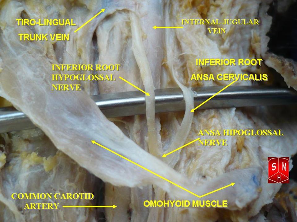
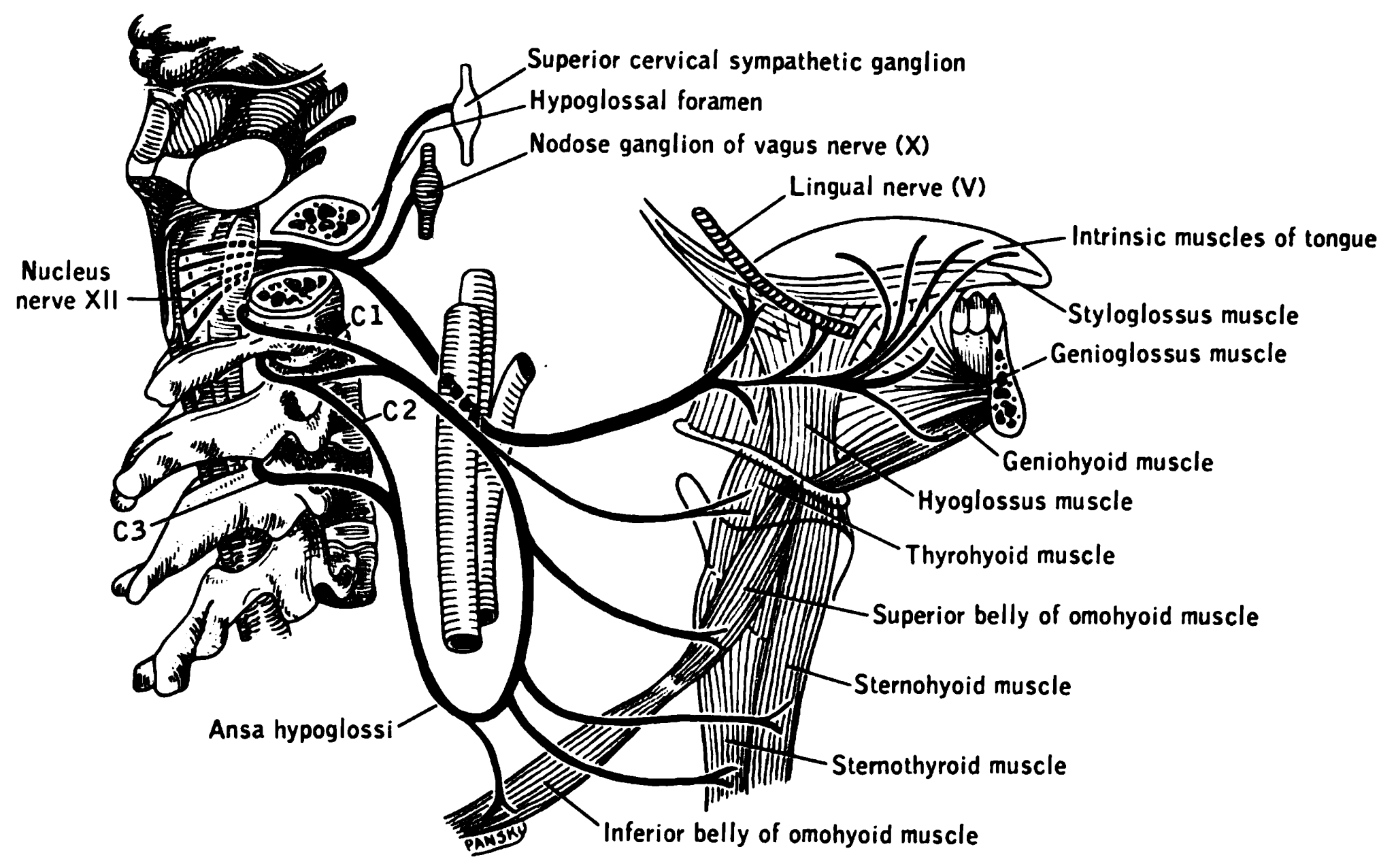
The ansa cervicalis (or ansa hypoglossi in older literature) is a loop of nerves that are part of the cervical plexus. It lies superficial to the internal jugular vein in the carotid triangle. Its name means "handle of the neck" in Latin. Branches from the ansa cervicalis innervate most of the infrahyoid muscles, including the sternothyroid muscle, sternohyoid muscle and the omohyoid muscle. Note that the thyrohyoid muscle, which is also an infrahyoid muscle and the geniohyoid muscle which is a suprahyoid muscle are innervated by cervical spinal nerve 1 via the hypoglossal nerve. Roots Two roots make up the ansa cervicalis, a superior root, and an inferior root. The superior root of the ansa cervicalis is formed from cervical spinal nerve 1 of the cervical plexus. These nerve fibers travel in the hypoglossal nerve before separating in the carotid triangle to form the superior root. The superior root goes around the occipital artery and then descends on the caroti ... [...More Info...] [...Related Items...] OR: [Wikipedia] [Google] [Baidu] |
Jugular Foramen
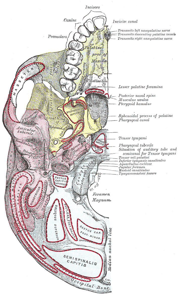
A jugular foramen is one of the two (left and right) large foramina (openings) in the base of the skull, located behind the carotid canal. It is formed by the temporal bone and the occipital bone. It allows many structures to pass, including the inferior petrosal sinus, three cranial nerves, the sigmoid sinus, and meningeal arteries. Structure The jugular foramen is formed in front by the petrous portion of the temporal bone, and behind by the occipital bone. It is generally slightly larger on the right side than on the left side. Contents The jugular foramen may be subdivided into three compartments, each with their own contents. * The ''anterior'' compartment transmits the inferior petrosal sinus. * The ''intermediate'' compartment transmits the glossopharyngeal nerve, the vagus nerve, and the accessory nerve. * The ''posterior'' compartment transmits the sigmoid sinus (becoming the internal jugular vein The internal jugular vein is a paired jugular vein that ... [...More Info...] [...Related Items...] OR: [Wikipedia] [Google] [Baidu] |
Carotid Canal
The carotid canal is a passageway in the temporal bone of the skull through which the internal carotid artery enters the middle cranial fossa from the neck. Structure The carotid canal is located within the middle cranial fossa, at the petrous part of the temporal bone. Anteriorly, it is limited by posterior margin of the greater wing of sphenoid bone. Posteromedially, it is limited by basilar part of occipital bone. It is divided in three parts, namely, ascending petrous, transverse petrous, and ascending cavernous parts. The carotid canal has two openings, namely internal and external openings. The internal opening is situated laterally to foramen lacerum. The external opening of the carotid canal is located posterolaterally to the foramen lacerum. Both internal and external openings of the carotid canal lies anterior to the jugular foramen, where the latter is located inside the posterior cranial fossa. The carotid canal is separated from middle ear and inner ear by a thi ... [...More Info...] [...Related Items...] OR: [Wikipedia] [Google] [Baidu] |
Base Of Skull
The base of skull, also known as the cranial base or the cranial floor, is the most inferior area of the skull. It is composed of the endocranium and the lower parts of the calvaria. Structure Structures found at the base of the skull are for example: Bones There are five bones that make up the base of the skull: * Ethmoid bone * Sphenoid bone *Occipital bone *Frontal bone *Temporal bone Sinuses * Occipital sinus * Superior sagittal sinus * Superior petrosal sinus Foramina of the skull * Foramen cecum * Optic foramen * Foramen lacerum * Foramen rotundum * Foramen magnum *Foramen ovale *Jugular foramen * Internal auditory meatus * Mastoid foramen * Sphenoidal emissary foramen * Foramen spinosum Sutures * Frontoethmoidal suture * Sphenofrontal suture *Sphenopetrosal suture The sphenopetrosal fissure (or sphenopetrosal suture) is the cranial suture between the sphenoid bone The sphenoid bone is an unpaired bone of the neurocranium. It is situated in the middle of the ... [...More Info...] [...Related Items...] OR: [Wikipedia] [Google] [Baidu] |
Sympathetic Trunk
The sympathetic trunks (sympathetic chain, gangliated cord) are a paired bundle of nerve fibers that run from the base of the skull to the coccyx. They are a major component of the sympathetic nervous system. Structure The sympathetic trunk lies just lateral to the vertebral bodies for the entire length of the vertebral column. It interacts with the anterior rami of spinal nerves by way of rami communicantes. The sympathetic trunk permits preganglionic fibers of the sympathetic nervous system to ascend to spinal levels superior to T1 and descend to spinal levels inferior to L2/3.Greenstein B., Greenstein A. (2002): Color atlas of neuroscience – Neuroanatomy and neurophysiology. Thieme, Stuttgart – New York, . The superior end of it is continued upward through the carotid canal into the skull, and forms a plexus on the internal carotid artery; the inferior part travels in front of the coccyx, where it converges with the other trunk at a structure known as the ganglion ... [...More Info...] [...Related Items...] OR: [Wikipedia] [Google] [Baidu] |
Hypoglossal Nerve
The hypoglossal nerve, also known as the twelfth cranial nerve, cranial nerve XII, or simply CN XII, is a cranial nerve that innervates all the extrinsic and intrinsic muscles of the tongue except for the palatoglossus, which is innervated by the vagus nerve. CN XII is a nerve with a solely motor function. The nerve arises from the hypoglossal nucleus in the medulla as a number of small rootlets, passes through the hypoglossal canal and down through the neck, and eventually passes up again over the tongue muscles it supplies into the tongue. The nerve is involved in controlling tongue movements required for speech and swallowing, including sticking out the tongue and moving it from side to side. Damage to the nerve or the neural pathways which control it can affect the ability of the tongue to move and its appearance, with the most common sources of damage being injury from trauma or surgery, and motor neuron disease. The first recorded description of the nerve is by Her ... [...More Info...] [...Related Items...] OR: [Wikipedia] [Google] [Baidu] |
Accessory Nerve
The accessory nerve, also known as the eleventh cranial nerve, cranial nerve XI, or simply CN XI, is a cranial nerve that supplies the sternocleidomastoid and trapezius muscles. It is classified as the eleventh of twelve pairs of cranial nerves because part of it was formerly believed to originate in the brain. The sternocleidomastoid muscle tilts and rotates the head, whereas the trapezius muscle, connecting to the scapula, acts to shrug the shoulder. Traditional descriptions of the accessory nerve divide it into a spinal part and a cranial part. The cranial component rapidly joins the vagus nerve, and there is ongoing debate about whether the cranial part should be considered part of the accessory nerve proper. Consequently, the term "accessory nerve" usually refers only to nerve supplying the sternocleidomastoid and trapezius muscles, also called the spinal accessory nerve. Strength testing of these muscles can be measured during a neurological examination to assess funct ... [...More Info...] [...Related Items...] OR: [Wikipedia] [Google] [Baidu] |


