|
Cantlie's Line
In human anatomy, the Cantlie line or Cantlie's line is an imaginary division of the liver. The division divides the liver into two planes, extending from the middle hepatic vein to the middle of the gallbladder. It is useful for performing hepatectomies. Structure The division divides the liver into two planes. It extends from the middle hepatic vein (or the inferior vena cava) to the middle of the gallbladder. Using Couinaud's classification system, segments two, three, and both parts of four are on the left side of the division, while segments five, six, seven, and eight are on the right. Clinical significance Cantlie's line is useful when performing hepatectomies. History It was first described by Scottish surgeon James Cantlie in 1887 when he noticed a difference in the amount of atrophy on both sides of this line of the liver while performing an autopsy. He concluded that the line dividing the atrophied segment from the hypertrophied segment must be the true mid ... [...More Info...] [...Related Items...] OR: [Wikipedia] [Google] [Baidu] |
Liver
The liver is a major organ only found in vertebrates which performs many essential biological functions such as detoxification of the organism, and the synthesis of proteins and biochemicals necessary for digestion and growth. In humans, it is located in the right upper quadrant of the abdomen, below the diaphragm. Its other roles in metabolism include the regulation of glycogen storage, decomposition of red blood cells, and the production of hormones. The liver is an accessory digestive organ that produces bile, an alkaline fluid containing cholesterol and bile acids, which helps the breakdown of fat. The gallbladder, a small pouch that sits just under the liver, stores bile produced by the liver which is later moved to the small intestine to complete digestion. The liver's highly specialized tissue, consisting mostly of hepatocytes, regulates a wide variety of high-volume biochemical reactions, including the synthesis and breakdown of small and complex molecule ... [...More Info...] [...Related Items...] OR: [Wikipedia] [Google] [Baidu] |
Human Anatomy
The human body is the structure of a human being. It is composed of many different types of cells that together create tissues and subsequently organ systems. They ensure homeostasis and the viability of the human body. It comprises a head, hair, neck, trunk (which includes the thorax and abdomen), arms and hands, legs and feet. The study of the human body involves anatomy, physiology, histology and embryology. The body varies anatomically in known ways. Physiology focuses on the systems and organs of the human body and their functions. Many systems and mechanisms interact in order to maintain homeostasis, with safe levels of substances such as sugar and oxygen in the blood. The body is studied by health professionals, physiologists, anatomists, and by artists to assist them in their work. Composition The human body is composed of elements including hydrogen, oxygen, carbon, calcium and phosphorus. These elements reside in trillions of cells and non-cellular c ... [...More Info...] [...Related Items...] OR: [Wikipedia] [Google] [Baidu] |
Liver
The liver is a major organ only found in vertebrates which performs many essential biological functions such as detoxification of the organism, and the synthesis of proteins and biochemicals necessary for digestion and growth. In humans, it is located in the right upper quadrant of the abdomen, below the diaphragm. Its other roles in metabolism include the regulation of glycogen storage, decomposition of red blood cells, and the production of hormones. The liver is an accessory digestive organ that produces bile, an alkaline fluid containing cholesterol and bile acids, which helps the breakdown of fat. The gallbladder, a small pouch that sits just under the liver, stores bile produced by the liver which is later moved to the small intestine to complete digestion. The liver's highly specialized tissue, consisting mostly of hepatocytes, regulates a wide variety of high-volume biochemical reactions, including the synthesis and breakdown of small and complex molecule ... [...More Info...] [...Related Items...] OR: [Wikipedia] [Google] [Baidu] |
Hepatic Veins
In human anatomy, the hepatic veins are the veins that drain venous blood from the liver into the inferior vena cava (as opposed to the hepatic portal vein which conveys blood from the gastrointestinal organs to the liver). There are usually three large upper hepatic veins draining from the left, middle, and right parts of the liver, as well as a number (6-20) of lower hepatic veins. All hepatic veins are valveless. Structure All the hepatic veins drain into the inferior vena cava. The hepatic veins are divided into an upper and a lower group. Upper group The upper group consists of three hepatic veins - the right, middle, and left hepatic veins - draining the central veins from the right, middle, and left regions of the liver and are larger than the lower group of veins. The veins of the upper group drain into the suprahepatic part of the inferior vena cava (i.e. part superior to the liver). Right hepatic vein The right hepatic vein is the longest and largest of all the he ... [...More Info...] [...Related Items...] OR: [Wikipedia] [Google] [Baidu] |
Gallbladder
In vertebrates, the gallbladder, also known as the cholecyst, is a small hollow organ where bile is stored and concentrated before it is released into the small intestine. In humans, the pear-shaped gallbladder lies beneath the liver, although the structure and position of the gallbladder can vary significantly among animal species. It receives and stores bile, produced by the liver, via the common hepatic duct, and releases it via the common bile duct into the duodenum, where the bile helps in the digestion of fats. The gallbladder can be affected by gallstones, formed by material that cannot be dissolved – usually cholesterol or bilirubin, a product of haemoglobin breakdown. These may cause significant pain, particularly in the upper-right corner of the abdomen, and are often treated with removal of the gallbladder (called a cholecystectomy). Cholecystitis, inflammation of the gallbladder, has a wide range of causes, including result from the impaction of gallstones, inf ... [...More Info...] [...Related Items...] OR: [Wikipedia] [Google] [Baidu] |
Hepatectomy
Hepatectomy is the surgical resection (removal of all or part) of the liver. While the term is often employed for the removal of the liver from a liver transplant donor, this article will focus on partial resections of hepatic tissue and hepatoportoenterostomy. History The first hepatectomies were reported by Dr. Ichio Honjo (1913–1987) of ( Kyoto University) in 1949, and Dr. Jean-Louis Lortat-Jacob (1908–1992) of France in 1952. In the latter case, the patient was a 58-year-old woman diagnosed with colorectal cancer which had metastasized to the liver. Indications Most hepatectomies are performed for the treatment of hepatic neoplasms, both benign or malign. Benign neoplasms include hepatocellular adenoma, hepatic hemangioma and focal nodular hyperplasia. The most common malignant neoplasms (cancers) of the liver are metastases; those arising from colorectal cancer are among the most common, and the most amenable to surgical resection. The most common primary malignant tum ... [...More Info...] [...Related Items...] OR: [Wikipedia] [Google] [Baidu] |
Inferior Vena Cava
The inferior vena cava is a large vein that carries the deoxygenated blood from the lower and middle body into the right atrium of the heart. It is formed by the joining of the right and the left common iliac veins, usually at the level of the fifth lumbar vertebra. The inferior vena cava is the lower (" inferior") of the two venae cavae, the two large veins that carry deoxygenated blood from the body to the right atrium of the heart: the inferior vena cava carries blood from the lower half of the body whilst the superior vena cava carries blood from the upper half of the body. Together, the venae cavae (in addition to the coronary sinus, which carries blood from the muscle of the heart itself) form the venous counterparts of the aorta. It is a large retroperitoneal vein that lies posterior to the abdominal cavity and runs along the right side of the vertebral column. It enters the right auricle at the lower right, back side of the heart. The name derives from la, ve ... [...More Info...] [...Related Items...] OR: [Wikipedia] [Google] [Baidu] |
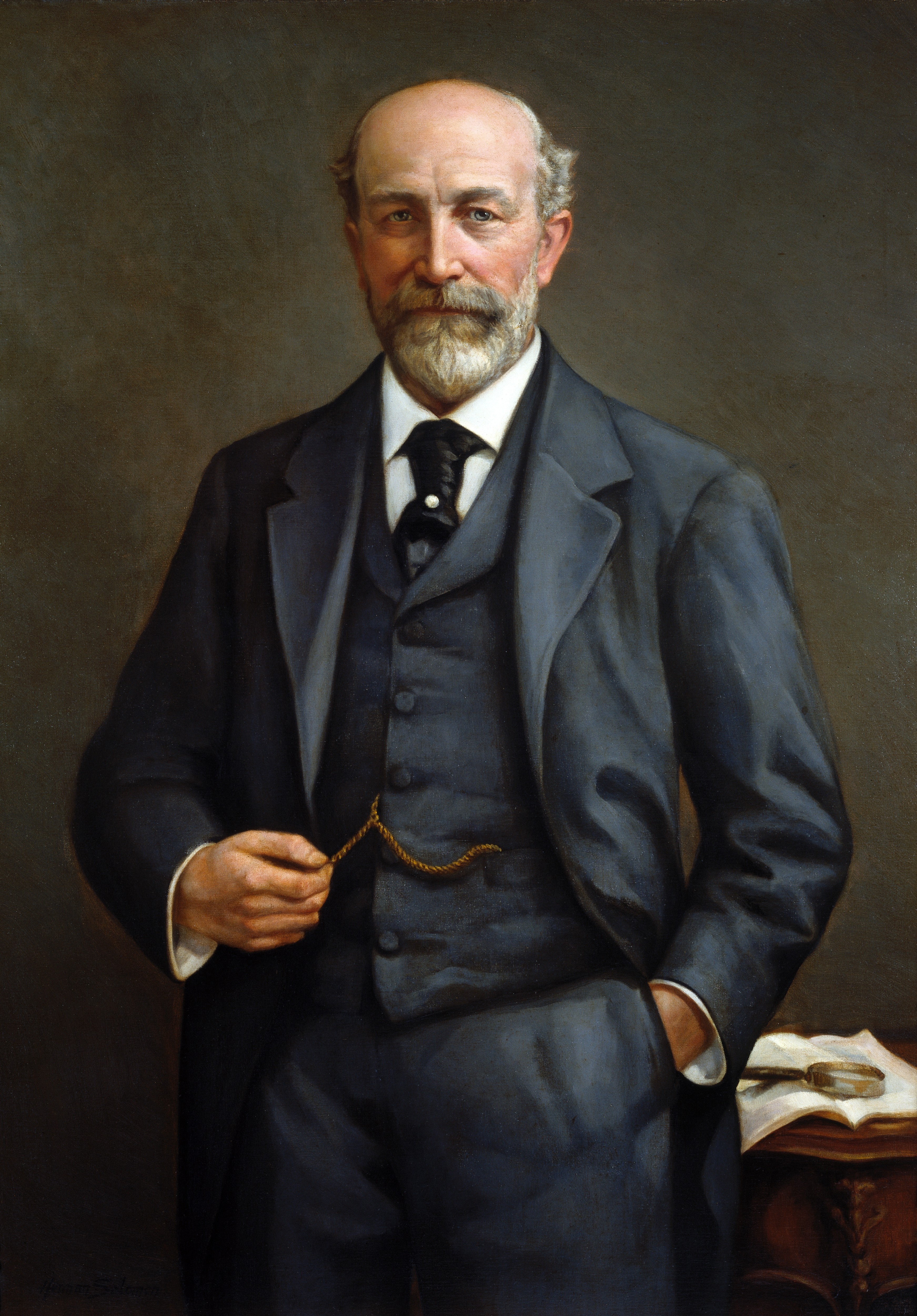
James Cantlie
Sir James Cantlie (17 January 1851 – 28 May 1926) was a British physician. He was a pioneer of first aid, which in 1875 was unknown: even the police had no knowledge of basic techniques such as how to stop serious bleeding and applying splints. He was also influential in the study of tropical diseases and in the debates concerning degeneration theory. Cantlie was born in Banffshire and took his first degree at Aberdeen University, carrying out his clinical training at Charing Cross Hospital, London. In 1877, Cantlie became a Fellow of the Royal College of Surgeons and Assistant Surgeon to Charing Cross Hospital; in 1886 he became Surgeon at Charing Cross. In 1888 he resigned to take up a position in Hong Kong. While in the crown colony, he co-founded the Hong Kong College of Medicine for Chinese, which later grew into the University of Hong Kong. One of his first pupils at the College was the future Chinese leader Sun Yat-sen. Cantlie's work in Hong Kong included investiga ... [...More Info...] [...Related Items...] OR: [Wikipedia] [Google] [Baidu] |
Atrophy
Atrophy is the partial or complete wasting away of a part of the body. Causes of atrophy include mutations (which can destroy the gene to build up the organ), poor nourishment, poor circulation, loss of hormonal support, loss of nerve supply to the target organ, excessive amount of apoptosis of cells, and disuse or lack of exercise or disease intrinsic to the tissue itself. In medical practice, hormonal and nerve inputs that maintain an organ or body part are said to have ''trophic'' effects. A diminished muscular trophic condition is designated as ''atrophy''. Atrophy is reduction in size of cell, organ or tissue, after attaining its normal mature growth. In contrast, hypoplasia is the reduction in the cellular numbers of an organ, or tissue that has not attained normal maturity. Atrophy is the general physiological process of reabsorption and breakdown of tissues, involving apoptosis. When it occurs as a result of disease or loss of trophic support because of other diseases ... [...More Info...] [...Related Items...] OR: [Wikipedia] [Google] [Baidu] |
Autopsy
An autopsy (post-mortem examination, obduction, necropsy, or autopsia cadaverum) is a surgical procedure that consists of a thorough examination of a corpse by dissection to determine the cause, mode, and manner of death or to evaluate any disease or injury that may be present for research or educational purposes. (The term " necropsy" is generally reserved for non-human animals). Autopsies are usually performed by a specialized medical doctor called a pathologist. In most cases, a medical examiner or coroner can determine the cause of death. However, only a small portion of deaths require an autopsy to be performed, under certain circumstances. Purposes of performance Autopsies are performed for either legal or medical purposes. Autopsies can be performed when any of the following information is desired: * Determine if death was natural or unnatural * Injury source and extent on the corpse * Manner of death must be determined * Post mortem interval * Determining the dece ... [...More Info...] [...Related Items...] OR: [Wikipedia] [Google] [Baidu] |
Hepatic Portal Vein
The portal vein or hepatic portal vein (HPV) is a blood vessel that carries blood from the gastrointestinal tract, gallbladder, pancreas and spleen to the liver. This blood contains nutrients and toxins extracted from digested contents. Approximately 75% of total liver blood flow is through the portal vein, with the remainder coming from the hepatic artery proper. The blood leaves the liver to the heart in the hepatic veins. The portal vein is not a true vein, because it conducts blood to capillary beds in the liver and not directly to the heart. It is a major component of the hepatic portal system, one of only two portal venous systems in the body – with the hypophyseal portal system being the other. The portal vein is usually formed by the confluence of the superior mesenteric, splenic veins, inferior mesenteric, left, right gastric veins and the pancreatic vein. Conditions involving the portal vein cause considerable illness and death. An important example of such ... [...More Info...] [...Related Items...] OR: [Wikipedia] [Google] [Baidu] |
Porta Hepatis
The porta hepatis or transverse fissure of the liver is a short but deep fissure, about 5 cm long, extending transversely beneath the left portion of the right lobe of the liver, nearer its posterior surface than its anterior border. It joins nearly at right angles with the left sagittal fossa, and separates the quadrate lobe in front from the caudate lobe and process behind. Function It transmits the following (in anterior to posterior order): * common hepatic duct (leaving) * proper hepatic artery (entering) * hepatic portal vein (entering) The hepatic duct lies in front and to the right, the hepatic artery to the left, and the portal vein behind and between the duct and artery. It also transmits nerves and lymphatics. * Sympathetic nerves - these provide afferent pain impulses from the liver and gall bladder to the brain. Pain may be referred to the lower pole of the right scapula (T7). * Hepatic branch of the vagus nerve (CN X). Location The porta hepatis runs in th ... [...More Info...] [...Related Items...] OR: [Wikipedia] [Google] [Baidu] |


