|
Calcaneal Apophysis
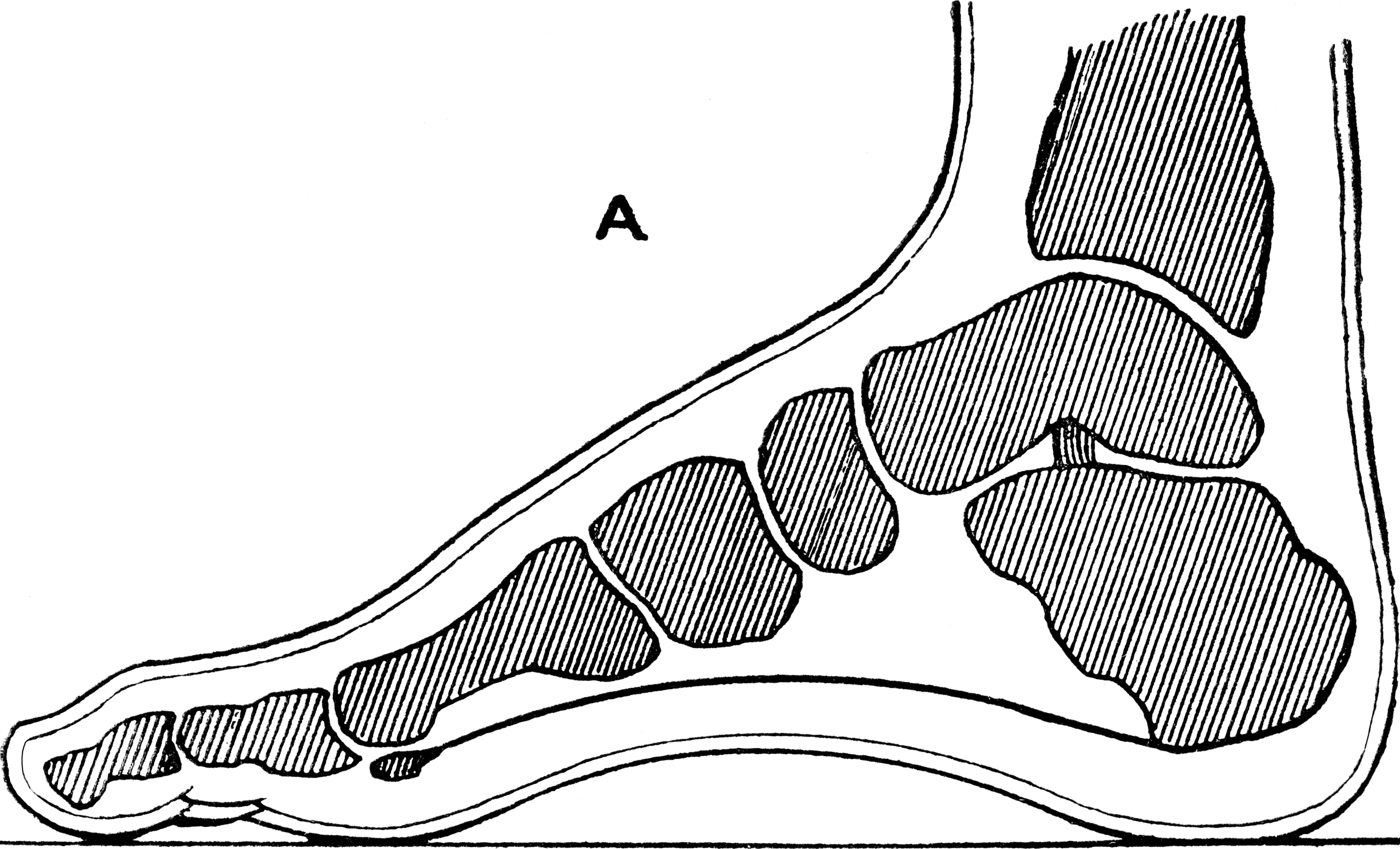
In humans and many other primates, the calcaneus (; from the Latin ''calcaneus'' or ''calcaneum'', meaning heel) or heel bone is a bone of the tarsus of the foot which constitutes the heel. In some other animals, it is the point of the hock. Structure In humans, the calcaneus is the largest of the tarsal bones and the largest bone of the foot. Its long axis is pointed forwards and laterally. The talus bone, calcaneus, and navicular bone are considered the proximal row of tarsal bones. In the calcaneus, several important structures can be distinguished:Platzer (2004), p 216 There is a large calcaneal tuberosity located posteriorly on plantar surface with medial and lateral tubercles on its surface. Besides, there is another peroneal tubecle on its lateral surface. On its lower edge on either side are its lateral and medial processes (serving as the origins of the abductor hallucis and abductor digiti minimi). The Achilles tendon is inserted into a roughened area on its superior ... [...More Info...] [...Related Items...] OR: [Wikipedia] [Google] [Baidu] |
Heel
The heel is the prominence at the posterior end of the foot. It is based on the projection of one bone, the calcaneus or heel bone, behind the articulation of the bones of the lower Human leg, leg. Structure To distribute the compressive forces exerted on the heel during gait, and especially the stance phase when the heel contacts the ground, the sole (foot), sole of the foot is covered by a layer of subcutaneous connective tissue up to 2 cm thick (under the heel). This tissue has a system of pressure chambers that both acts as a shock absorber and stabilises the sole. Each of these chambers contains fibrofatty tissue covered by a layer of tough connective tissue made of collagen fibers. These septum, septa ("walls") are firmly attached both to the plantar aponeurosis above and the sole's Dermis, skin below. The sole of the foot is one of the most highly vascularized regions of the body surface, and the dense system of blood vessels further stabilize the septa. The Ach ... [...More Info...] [...Related Items...] OR: [Wikipedia] [Google] [Baidu] |
Deltoid Ligament
The deltoid ligament (or medial ligament of talocrural joint) is a strong, flat, triangular band, attached, above, to the apex and anterior and posterior borders of the medial malleolus. The deltoid ligament is composed of 4 fibers: 1. Anterior tibiotalar ligament 2. Tibiocalcaneal ligament 3. Posterior tibiotalar ligament 4. Tibionavicular ligament. It consists of two sets of fibers, superficial and deep. Superficial fibres Of the superficial fibres, * ''tibionavicular'' pass forward to be inserted into the tuberosity of the navicular bone, and immediately behind this they blend with the medial margin of the plantar calcaneonavicular ligament; * ''tibiocalcaneal'' descend almost perpendicularly to be inserted into the whole length of the sustentaculum tali of the calcaneus; * ''posterior tibiotalar'' from the posterior colliculus of the medial malleolus to the posteromedial surface of the talus Deep fibres The deep fibres (''anterior tibiotalar'') are attached from the anterio ... [...More Info...] [...Related Items...] OR: [Wikipedia] [Google] [Baidu] |
Gray268
Grey (more common in British English) or gray (more common in American English) is an intermediate color between black and white. It is a neutral or achromatic color, meaning literally that it is "without color", because it can be composed of black and white. It is the color of a cloud-covered sky, of ash and of lead. The first recorded use of ''grey'' as a color name in the English language was in 700 CE.Maerz and Paul ''A Dictionary of Color'' New York:1930 McGraw-Hill Page 196 ''Grey'' is the dominant spelling in European and Commonwealth English, while ''gray'' has been the preferred spelling in American English; both spellings are valid in both varieties of English. In Europe and North America, surveys show that grey is the color most commonly associated with neutrality, conformity, boredom, uncertainty, old age, indifference, and modesty. Only one percent of respondents chose it as their favorite color. Etymology ''Grey'' comes from the Middle English or ... [...More Info...] [...Related Items...] OR: [Wikipedia] [Google] [Baidu] |
Anatomical Terms Of Motion
Motion, the process of movement, is described using specific anatomical terms. Motion includes movement of organs, joints, limbs, and specific sections of the body. The terminology used describes this motion according to its direction relative to the anatomical position of the body parts involved. Anatomists and others use a unified set of terms to describe most of the movements, although other, more specialized terms are necessary for describing unique movements such as those of the hands, feet, and eyes. In general, motion is classified according to the anatomical plane it occurs in. ''Flexion'' and ''extension'' are examples of ''angular'' motions, in which two axes of a joint are brought closer together or moved further apart. ''Rotational'' motion may occur at other joints, for example the shoulder, and are described as ''internal'' or ''external''. Other terms, such as ''elevation'' and ''depression'', describe movement above or below the horizontal plane. Many anatomica ... [...More Info...] [...Related Items...] OR: [Wikipedia] [Google] [Baidu] |
Posterior Compartment Of The Leg
The posterior compartment of the leg is one of the fascial compartments of the leg and is divided further into deep and superficial compartments. Structure Muscles Superficial posterior compartment Deep posterior compartment Blood supply Posterior tibial artery Innervation The posterior compartment of the leg is supplied by the tibial nerve The tibial nerve is a branch of the sciatic nerve. The tibial nerve passes through the popliteal fossa to pass below the arch of soleus. Structure Popliteal fossa The tibial nerve is the larger terminal branch of the sciatic nerve with root val .... Function * It contains the plantar flexors: Additional images File:From below - Superficial posterior compartment of leg - animation.gif, Superficial posterior compartment. Animation. File:From below - Deep posterior compartment of leg - animation.gif, Deep posterior compartment. Animation. References External links Diagram at patientcareonline.com {{DEFAULTSORT ... [...More Info...] [...Related Items...] OR: [Wikipedia] [Google] [Baidu] |
Plantaris Muscle
The plantaris is one of the superficial muscles of the superficial posterior compartment of the leg, one of the fascial compartments of the leg. It is composed of a thin muscle belly and a long thin tendon. While not as thick as the achilles tendon, the plantaris tendon (which tends to be between in length) is the longest tendon in the human body. Not including the tendon, the plantaris muscle is approximately long and is absent in 8-12% of the population. It is one of the plantar flexors in the posterior compartment of the leg, along with the gastrocnemius and soleus muscles. The plantaris is considered to have become an unimportant muscle when human ancestors switched from climbing trees to bipedalism and in anatomically modern humans it mainly acts with the gastrocnemius. Structure The plantaris muscle arises from the inferior part of the lateral supracondylar ridge of the femur at a position slightly superior to the origin of the lateral head of gastrocnemius. It passes ... [...More Info...] [...Related Items...] OR: [Wikipedia] [Google] [Baidu] |
Soleus Muscle
In humans and some other mammals, the soleus is a powerful muscle in the back part of the lower leg (the calf). It runs from just below the knee to the heel, and is involved in standing and walking. It is closely connected to the gastrocnemius muscle and some anatomists consider them to be a single muscle, the triceps surae. Its name is derived from the Latin word "solea", meaning "sandal". Structure The soleus is located in the superficial posterior compartment of the leg. The soleus exhibits significant morphological differences across species. It is unipennate in many species. In some animals, such as the rabbit, it is fused for much of its length with the gastrocnemius muscle. In humans, the soleus is a complex, multi-pennate muscle, usually having a separate (posterior) aponeurosis from the gastrocnemius muscle. A majority of soleus muscle fibers originate from each side of the anterior aponeurosis, attached to the tibia and fibula. Other fibers originate from the posteri ... [...More Info...] [...Related Items...] OR: [Wikipedia] [Google] [Baidu] |
Gastrocnemius Muscle
The gastrocnemius muscle (plural ''gastrocnemii'') is a superficial two-headed muscle that is in the back part of the lower leg of humans. It runs from its two heads just above the knee to the heel, a three joint muscle (knee, ankle and subtalar joints). The muscle is named via Latin, from Greek γαστήρ (''gaster'') 'belly' or 'stomach' and κνήμη (''knḗmē'') 'leg', meaning 'stomach of the leg' (referring to the bulging shape of the calf). Structure The gastrocnemius is located with the soleus in the posterior (back) compartment of the leg. The lateral head originates from the lateral condyle of the femur, while the medial head originates from the medial condyle of the femur. Its other end forms a common tendon with the soleus muscle; this tendon is known as the calcaneal tendon or Achilles tendon and inserts onto the posterior surface of the calcaneus, or heel bone. It is considered a superficial muscle as it is located directly under skin, and its shape may often ... [...More Info...] [...Related Items...] OR: [Wikipedia] [Google] [Baidu] |
Prenatal Development
Prenatal development () includes the development of the embryo and of the fetus during a viviparous animal's gestation. Prenatal development starts with fertilization, in the germinal stage of embryonic development, and continues in fetal development until birth. In human pregnancy, prenatal development is also called antenatal development. The development of the human embryo follows fertilization, and continues as fetal development. By the end of the tenth week of gestational age the embryo has acquired its basic form and is referred to as a fetus. The next period is that of fetal development where many organs become fully developed. This fetal period is described both topically (by organ) and chronologically (by time) with major occurrences being listed by gestational age. The very early stages of embryonic development are the same in all mammals, but later stages of development, and the length of gestation varies. Terminology In the human: Different terms are used to ... [...More Info...] [...Related Items...] OR: [Wikipedia] [Google] [Baidu] |
Ossification Center
An ossification center is a point where ossification of the cartilage begins. The first step in ossification is that the cartilage cells at this point enlarge and arrange themselves in rows.Gray and Spitzka (1910), page 44. The matrix in which they are imbedded increases in quantity, so that the cells become further separated from each other. A deposit of calcareous material now takes place in this matrix, between the rows of cells, so that they become separated from each other by longitudinal columns of calcified matrix, presenting a granular and opaque appearance. Here and there the matrix between two cells of the same row also becomes calcified, and transverse bars of calcified substance stretch across from one calcareous column to another. Thus there are longitudinal groups of the cartilage cells enclosed in oblong cavities, the walls of which are formed of calcified matrix which cuts off all nutrition from the cells; the cells, in consequence, atrophy, leaving spaces calle ... [...More Info...] [...Related Items...] OR: [Wikipedia] [Google] [Baidu] |
Bursa (anatomy)
( grc-gre, Προῦσα, Proûsa, Latin: Prusa, ota, بورسه, Arabic:بورصة) is a city in northwestern Turkey and the administrative center of Bursa Province. The fourth-most populous city in Turkey and second-most populous in the Marmara Region, Bursa is one of the industrial centers of the country. Most of Turkey's automotive production takes place in Bursa. As of 2019, the Metropolitan Province was home to 3,056,120 inhabitants, 2,161,990 of whom lived in the 3 city urban districts ( Osmangazi, Yildirim and Nilufer) plus Gursu and Kestel, largely conurbated. Bursa was the first major and second overall capital of the Ottoman State between 1335 and 1363. The city was referred to as (, meaning "God's Gift" in Ottoman Turkish, a name of Persian origin) during the Ottoman period, while a more recent nickname is ("") in reference to the parks and gardens located across its urban fabric, as well as to the vast and richly varied forests of the surrounding re ... [...More Info...] [...Related Items...] OR: [Wikipedia] [Google] [Baidu] |
Talocalcaneal Joint
In human anatomy, the subtalar joint, also known as the talocalcaneal joint, is a joint of the foot. It occurs at the meeting point of the talus and the calcaneus. The joint is classed structurally as a synovial joint, and functionally as a plane joint. Structure The talus is oriented slightly obliquely on the anterior surface of the calcaneus. There are three points of articulation between the two bones: two anteriorly and one posteriorly. The three articulations are known as facets, and they are the posterior, middle and anterior facets. * At the ''anterior and middle talocalcaneal articulation'', convex areas of the talus fits on to concave surfaces of the calcaneus. * The ''posterior talocalcaneal articulation'' is formed by a concave surface of the talus and a convex surface of the calcaneus. The sustentaculum tali forms the floor of middle facet, and the anterior facet articulates with the head of the talus, and sits lateral and congruent to the middle facet. In som ... [...More Info...] [...Related Items...] OR: [Wikipedia] [Google] [Baidu] |


