|
CYSLTR1
Cysteinyl leukotriene receptor 1, also termed CYSLTR1, is a receptor for cysteinyl leukotrienes (LT) (see leukotrienes#Cysteinyl leukotrienes). CYSLTR1, by binding these cysteinyl LTs (CysLTs; viz, LTC4, LTD4, and to a much lesser extent, LTE4) contributes to mediating various allergic and hypersensitivity reactions in humans as well as models of the reactions in other animals. Gene The human gene maps to the X chromosome at position Xq13-Xq21, contains three exons with the entire open reading frame located in exon 3, and codes for a protein composed of 337 amino acids. The ''CYSLTR1'' gene promoter region is distanced from 665 to 30 bp upstream of its transcription start site. Expression CYSLTR1 mRNA is expressed in lung smooth muscle, lung macrophages, monocytes, eosinophils, basophils, neutrophils, platelets, T cells, B lymphocytes, pluripotent hematopoietic stem cells (CD34+), mast cells, pancreas, small intestine, prostate, interstitial cells of the nasal mucosa, airway ... [...More Info...] [...Related Items...] OR: [Wikipedia] [Google] [Baidu] |
Leukotrienes
Leukotrienes are a family of eicosanoid inflammatory mediators produced in leukocytes by the oxidation of arachidonic acid (AA) and the essential fatty acid eicosapentaenoic acid (EPA) by the enzyme arachidonate 5-lipoxygenase. Leukotrienes use lipid signaling to convey information to either the cell producing them (autocrine signaling) or neighboring cells (paracrine signaling) in order to regulate immune responses. The production of leukotrienes is usually accompanied by the production of histamine and prostaglandins, which also act as inflammatory mediators. One of their roles (specifically, leukotriene D4) is to trigger contractions in the smooth muscles lining the bronchioles; their overproduction is a major cause of inflammation in asthma and allergic rhinitis. Leukotriene antagonists are used to treat these disorders by inhibiting the production or activity of leukotrienes. History and name The name ''leukotriene'', introduced by Swedish biochemist Bengt Samuelss ... [...More Info...] [...Related Items...] OR: [Wikipedia] [Google] [Baidu] |
LTC4
Leukotriene C4 (LTC4) is a leukotriene. LTC4 has been extensively studied in the context of allergy and asthma. In cells of myeloid origin such as mast cells, its biosynthesis is orchestrated by translocation to the nuclear envelope along with co-localization of cytosolic phospholipase A2 (cPLA2), Arachidonate 5-lipoxygenase (5-LO), 5-lipoxygenase-activating protein (FLAP) and LTC4 synthase (LTC4S), which couples glutathione to an LTA4 intermediate. The MRP1 transporter then secretes cytosolic LTC4 and cell surface proteases further metabolize it by sequential cleavage of the γ-glutamyl and glycine residues off its glutathione segment, generating the more stable products LTD4 and LTE4. All three leukotrienes then bind at different affinities to two G-protein coupled receptors: CYSLTR1 and CYSLTR2, triggering pulmonary vasoconstriction and bronchoconstriction. In cells of non-haematopoietic lineage, endoplasmic reticulum (ER) stress and chemotherapy induce LTC4 biosynthesi ... [...More Info...] [...Related Items...] OR: [Wikipedia] [Google] [Baidu] |
Gq Alpha Subunit
Gq protein alpha subunit is a family of heterotrimeric G protein alpha subunits. This family is also commonly called the Gq/11 (Gq/G11) family or Gq/11/14/15 family to include closely related family members. G alpha subunits may be referred to as Gq alpha, Gαq, or Gqα. Gq proteins couple to G protein-coupled receptors to activate beta-type phospholipase C (PLC-β) enzymes. PLC-β in turn hydrolyzes phosphatidylinositol 4,5-bisphosphate (PIP2) to diacyl glycerol (DAG) and inositol trisphosphate (IP3). IP3 acts as a second messenger to release stored calcium into the cytoplasm, while DAG acts as a second messenger that activates protein kinase C (PKC). Family members In humans, there are four distinct proteins in the Gq alpha subunit family: * Gαq is encoded by the gene GNAQ. * Gα11 is encoded by the gene GNA11. * Gα14 is encoded by the gene GNA14. * Gα15 is encoded by the gene GNA15. Function The general function of Gq is to activate intracellular signaling p ... [...More Info...] [...Related Items...] OR: [Wikipedia] [Google] [Baidu] |
Ligand
In coordination chemistry, a ligand is an ion or molecule (functional group) that binds to a central metal atom to form a coordination complex. The bonding with the metal generally involves formal donation of one or more of the ligand's electron pairs, often through Lewis bases. The nature of metal–ligand bonding can range from covalent to ionic. Furthermore, the metal–ligand bond order can range from one to three. Ligands are viewed as Lewis bases, although rare cases are known to involve Lewis acidic "ligands". Metals and metalloids are bound to ligands in almost all circumstances, although gaseous "naked" metal ions can be generated in a high vacuum. Ligands in a complex dictate the reactivity of the central atom, including ligand substitution rates, the reactivity of the ligands themselves, and redox. Ligand selection requires critical consideration in many practical areas, including bioinorganic and medicinal chemistry, homogeneous catalysis, and environmental chemi ... [...More Info...] [...Related Items...] OR: [Wikipedia] [Google] [Baidu] |
G Protein–coupled Receptor
G protein-coupled receptors (GPCRs), also known as seven-(pass)-transmembrane domain receptors, 7TM receptors, heptahelical receptors, serpentine receptors, and G protein-linked receptors (GPLR), form a large group of evolutionarily-related proteins that are cell surface receptors that detect molecules outside the cell and activate cellular responses. Coupling with G proteins, they are called seven-transmembrane receptors because they pass through the cell membrane seven times. Text was copied from this source, which is available under Attribution 2.5 Generic (CC BY 2.5) license. Ligands can bind either to extracellular N-terminus and loops (e.g. glutamate receptors) or to the binding site within transmembrane helices (Rhodopsin-like family). They are all activated by agonists although a spontaneous auto-activation of an empty receptor can also be observed. G protein-coupled receptors are found only in eukaryotes, including yeast, choanoflagellates, and ... [...More Info...] [...Related Items...] OR: [Wikipedia] [Google] [Baidu] |
Endothelial Cells
The endothelium is a single layer of squamous endothelial cells that line the interior surface of blood vessels and lymphatic vessels. The endothelium forms an interface between circulating blood or lymph in the lumen and the rest of the vessel wall. Endothelial cells form the barrier between vessels and tissue and control the flow of substances and fluid into and out of a tissue. Endothelial cells in direct contact with blood are called vascular endothelial cells whereas those in direct contact with lymph are known as lymphatic endothelial cells. Vascular endothelial cells line the entire circulatory system, from the heart to the smallest capillaries. These cells have unique functions that include fluid filtration, such as in the glomerulus of the kidney, blood vessel tone, hemostasis, neutrophil recruitment, and hormone trafficking. Endothelium of the interior surfaces of the heart chambers is called endocardium. An impaired function can lead to serious health issues throug ... [...More Info...] [...Related Items...] OR: [Wikipedia] [Google] [Baidu] |
Fibroblast
A fibroblast is a type of cell (biology), biological cell that synthesizes the extracellular matrix and collagen, produces the structural framework (Stroma (tissue), stroma) for animal Tissue (biology), tissues, and plays a critical role in wound healing. Fibroblasts are the most common cells of connective tissue in animals. Structure Fibroblasts have a branched cytoplasm surrounding an elliptical, speckled cell nucleus, nucleus having two or more nucleoli. Active fibroblasts can be recognized by their abundant Endoplasmic reticulum#Rough endoplasmic reticulum, rough endoplasmic reticulum. Inactive fibroblasts (called fibrocytes) are smaller, spindle-shaped, and have a reduced amount of rough endoplasmic reticulum. Although disjointed and scattered when they have to cover a large space, fibroblasts, when crowded, often locally align in parallel clusters. Unlike the epithelial cells lining the body structures, fibroblasts do not form flat monolayers and are not restricted by a ... [...More Info...] [...Related Items...] OR: [Wikipedia] [Google] [Baidu] |
Bronchial
A bronchus is a passage or airway in the lower respiratory tract that conducts air into the lungs. The first or primary bronchi pronounced (BRAN-KAI) to branch from the trachea at the carina are the right main bronchus and the left main bronchus. These are the widest bronchi, and enter the right lung, and the left lung at each hilum. The main bronchi branch into narrower secondary bronchi or lobar bronchi, and these branch into narrower tertiary bronchi or segmental bronchi. Further divisions of the segmental bronchi are known as 4th order, 5th order, and 6th order segmental bronchi, or grouped together as subsegmental bronchi. The bronchi, when too narrow to be supported by cartilage, are known as bronchioles. No gas exchange takes place in the bronchi. Structure The trachea (windpipe) divides at the carina into two main or primary bronchi, the left bronchus and the right bronchus. The carina of the trachea is located at the level of the sternal angle and the fifth thoracic verte ... [...More Info...] [...Related Items...] OR: [Wikipedia] [Google] [Baidu] |
Nasal Mucosa
The nasal mucosa lines the nasal cavity. It is part of the respiratory mucosa, the mucous membrane lining the respiratory tract. The nasal mucosa is intimately adherent to the periosteum or perichondrium of the nasal conchae. It is continuous with the skin through the nostrils, and with the mucous membrane of the nasal part of the pharynx through the choanae. From the nasal cavity its continuity with the conjunctiva may be traced, through the nasolacrimal and lacrimal ducts; and with the frontal, ethmoidal, sphenoidal, and maxillary sinuses, through the several openings in the nasal meatuses. The mucous membrane is thickest, and most vascular, over the nasal conchae. It is also thick over the nasal septum where increased numbers of goblet cells produce a greater amount of nasal mucus. It is very thin in the meatuses on the floor of the nasal cavities, and in the various sinuses. It is one of the most commonly infected tissues in adults and children. Inflammation of this tissue ... [...More Info...] [...Related Items...] OR: [Wikipedia] [Google] [Baidu] |
Interstitial Cell
Interstitial cell refers to any cell that lies in the spaces between the functional cells of a tissue. Examples include: * Interstitial cell of Cajal (ICC) * Leydig cells, cells present in the male testes responsible for the production of androgen (male sex hormone) * A portion of the stroma of ovary * Certain cells in the pineal gland * Renal interstitial cells * neuroglial cells See also *List of human cell types derived from the germ layers This is a list of cells in humans derived from the three embryonic germ layers – ectoderm, mesoderm, and endoderm. Cells derived from ectoderm Surface ectoderm Skin * Trichocyte * Keratinocyte Anterior pituitary * Gonadotrope * Corticotro ... References *Sybil B Parker (ed). "Interstitial cell". McGraw Hill Dictionary of Scientific and Technical Terms. Fifth Edition. International Edition. 1994. Page 1041. {{DEFAULTSORT:Interstitial cells Cell biology Biology-related lists ... [...More Info...] [...Related Items...] OR: [Wikipedia] [Google] [Baidu] |
Prostate
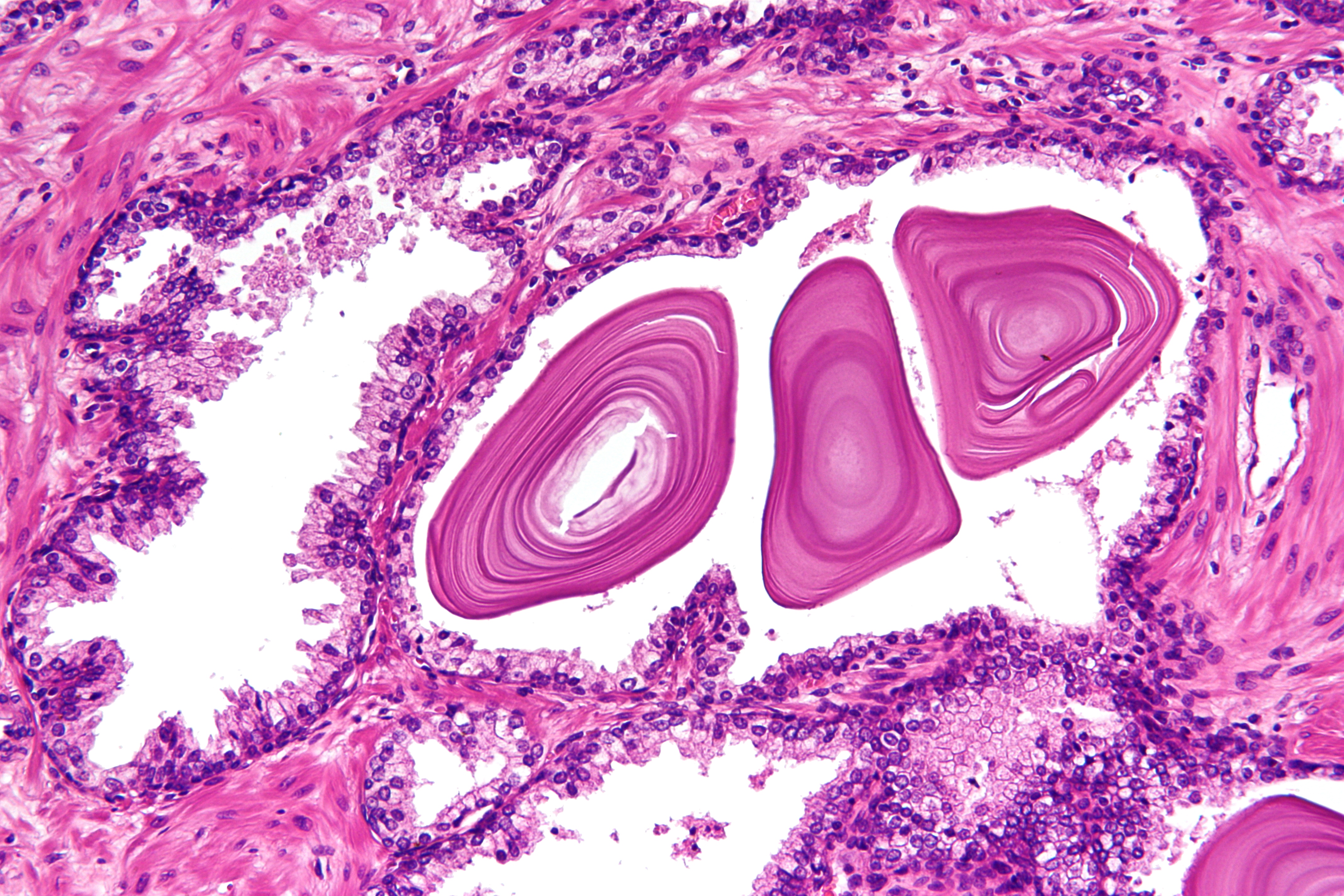
The prostate is both an Male accessory gland, accessory gland of the male reproductive system and a muscle-driven mechanical switch between urination and ejaculation. It is found only in some mammals. It differs between species anatomically, chemically, and physiologically. Anatomically, the prostate is found below the Urinary bladder, bladder, with the urethra passing through it. It is described in gross anatomy as consisting of lobes and in microanatomy by zone. It is surrounded by an elastic, fibromuscular capsule and contains glandular tissue as well as connective tissue. The prostate glands produce and contain fluid that forms part of semen, the substance emitted during ejaculation as part of the male Human sexual response cycle, sexual response. This prostatic fluid is slightly alkaline, milky or white in appearance. The alkalinity of semen helps neutralize the acidity of the vagina, vaginal tract, prolonging the lifespan of sperm. The prostatic fluid is expelled in the ... [...More Info...] [...Related Items...] OR: [Wikipedia] [Google] [Baidu] |
4-3D-balls.png)


.jpg)

