|
CD34⁺
CD34 is a transmembrane phosphoglycoprotein protein encoded by the CD34 gene in humans, mice, rats and other species. CD34 derives its name from the cluster of differentiation protocol that identifies cell surface antigens. CD34 was first described on hematopoietic stem cells independently by Civin et al. and Tindle et al. as a cell surface glycoprotein and functions as a cell-cell adhesion factor. It may also mediate the attachment of hematopoietic stem cells to bone marrow extracellular matrix or directly to stromal cells. Clinically, it is associated with the selection and enrichment of hematopoietic stem cells for bone marrow transplants. Due to these historical and clinical associations, CD34 expression is almost ubiquitously related to hematopoietic cells; however, it is actually found on many other cell types as well. Function The CD34 protein is a member of a family of single-pass transmembrane sialomucin proteins that show expression on early haematopoietic and ... [...More Info...] [...Related Items...] OR: [Wikipedia] [Google] [Baidu] |
Protein
Proteins are large biomolecules and macromolecules that comprise one or more long chains of amino acid residues. Proteins perform a vast array of functions within organisms, including catalysing metabolic reactions, DNA replication, responding to stimuli, providing structure to cells and organisms, and transporting molecules from one location to another. Proteins differ from one another primarily in their sequence of amino acids, which is dictated by the nucleotide sequence of their genes, and which usually results in protein folding into a specific 3D structure that determines its activity. A linear chain of amino acid residues is called a polypeptide. A protein contains at least one long polypeptide. Short polypeptides, containing less than 20–30 residues, are rarely considered to be proteins and are commonly called peptides. The individual amino acid residues are bonded together by peptide bonds and adjacent amino acid residues. The sequence of amino acid residue ... [...More Info...] [...Related Items...] OR: [Wikipedia] [Google] [Baidu] |
Lymphatics
The lymphatic vessels (or lymph vessels or lymphatics) are thin-walled vessels (tubes), structured like blood vessels, that carry lymph. As part of the lymphatic system, lymph vessels are complementary to the cardiovascular system. Lymph vessels are lined by endothelial cells, and have a thin layer of smooth muscle, and adventitia that binds the lymph vessels to the surrounding tissue. Lymph vessels are devoted to the propulsion of the lymph from the lymph capillaries, which are mainly concerned with the absorption of interstitial fluid from the tissues. Lymph capillaries are slightly bigger than their counterpart capillaries of the vascular system. Lymph vessels that carry lymph to a lymph node are called afferent lymph vessels, and those that carry it from a lymph node are called efferent lymph vessels, from where the lymph may travel to another lymph node, may be returned to a vein, or may travel to a larger lymph duct. Lymph ducts drain the lymph into one of the subclavian ve ... [...More Info...] [...Related Items...] OR: [Wikipedia] [Google] [Baidu] |
Graft-versus-host Disease
Graft-versus-host disease (GvHD) is a syndrome, characterized by inflammation in different organs. GvHD is commonly associated with bone marrow transplants and stem cell transplants. White blood cells of the donor's immune system which remain within the donated tissue (the graft) recognize the recipient (the host) as foreign (non-self). The white blood cells present within the transplanted tissue then attack the recipient's body's cells, which leads to GvHD. This should not be confused with a transplant rejection, which occurs when the immune system of the transplant recipient rejects the transplanted tissue; GvHD occurs when the donor's immune system's white blood cells reject the recipient. The underlying principle (alloimmunity) is the same, but the details and course may differ. GvHD can also occur after a blood transfusion if the blood products used have not been irradiated or treated with an approved pathogen reduction system. Types In the clinical setting, graft-versus-hos ... [...More Info...] [...Related Items...] OR: [Wikipedia] [Google] [Baidu] |
Magnetic-activated Cell Sorting
Magnetic-activated cell sorting (MACS) is a method for separation of various cell populations depending on their surface antigens ( CD molecules) invented by Miltenyi Biotec. The name MACS is a registered trademark of the company. The method was developed with Miltenyi Biotec's MACS system, which uses superparamagnetic nanoparticles and columns. The superparamagnetic nanoparticles are of the order of 100 nm. They are used to tag the targeted cells in order to capture them inside the column. The column is placed between permanent magnets so that when the magnetic particle-cell complex passes through it, the tagged cells can be captured. The column consists of steel wool which increases the magnetic field gradient to maximize separation efficiency when the column is placed between the permanent magnets. Magnetic-activated cell sorting is a commonly used method in areas like immunology, cancer research, neuroscience, and stem cell research. Miltenyi sells microbeads which are magn ... [...More Info...] [...Related Items...] OR: [Wikipedia] [Google] [Baidu] |
Blood
Blood is a body fluid in the circulatory system of humans and other vertebrates that delivers necessary substances such as nutrients and oxygen to the cells, and transports metabolic waste products away from those same cells. Blood in the circulatory system is also known as ''peripheral blood'', and the blood cells it carries, ''peripheral blood cells''. Blood is composed of blood cells suspended in blood plasma. Plasma, which constitutes 55% of blood fluid, is mostly water (92% by volume), and contains proteins, glucose, mineral ions, hormones, carbon dioxide (plasma being the main medium for excretory product transportation), and blood cells themselves. Albumin is the main protein in plasma, and it functions to regulate the colloidal osmotic pressure of blood. The blood cells are mainly red blood cells (also called RBCs or erythrocytes), white blood cells (also called WBCs or leukocytes) and platelets (also called thrombocytes). The most abundant cells in vertebrate blo ... [...More Info...] [...Related Items...] OR: [Wikipedia] [Google] [Baidu] |
Malignant Peripheral Nerve Sheath Tumor
A malignant peripheral nerve sheath tumor (MPNST) is a form of cancer of the connective tissue surrounding nerves. Given its origin and behavior it is classified as a sarcoma. About half the cases are diagnosed in people with neurofibromatosis; the lifetime risk for an MPNST in patients with neurofibromatosis type 1 is 8–13%. MPNST with rhabdomyoblastomatous component are called malignant triton tumors. The first-line treatment is surgical resection with wide margins. Chemotherapy (e.g. high-dose doxorubicin) and often radiotherapy are done as adjuvant and/or neoadjuvant treatment. Signs and symptoms Symptoms may include: * Swelling in the extremities (arms or legs), also called peripheral edema; the swelling often is painless. * Difficulty in moving the extremity that has the tumor, including a limp. * Soreness localized to the area of the tumor or in the extremity. * Neurological symptoms. * Pain or discomfort: numbness, burning, or "pins and needles." * Dizziness and/or lo ... [...More Info...] [...Related Items...] OR: [Wikipedia] [Google] [Baidu] |
Hemangiopericytoma
A hemangiopericytoma is a type of soft-tissue sarcoma that originates in the pericytes in the walls of capillaries. When inside the nervous system, although not strictly a meningioma tumor, it is a meningeal tumor with a special aggressive behavior. It was first characterized in 1942. Signs and symptoms Symptoms of hemangiopericytoma vary greatly depending on both tumor stage and affected organs. Most patients report pain and mass-related symptoms, while others also report vascular disease-related symptoms, and some have no symptoms until late in the disease process. Hemangiopericytomas are most commonly found in the meninges, lower extremities, retroperitoneum, pelvis, lungs, and pleura. Histopathology Hemangiopericytomas are tumors that are derived from specialized spindle shaped cells called pericytes, which line capillaries. Hemangiopericytoma located in the cerebral cavity is an aggressive tumor of the mesenchyme with oval nuclei with scant cytoplasm. "There is dense inter ... [...More Info...] [...Related Items...] OR: [Wikipedia] [Google] [Baidu] |
Solitary Fibrous Tumour
Solitary fibrous tumor (SFT), also known as fibrous tumor of the pleura, is a rare mesenchymal tumor originating in the pleuraTravis WD, Brambilla E, Muller-Hermelink HK, Harris CC (Eds.): World Health Organization Classification of Tumours. Pathology and Genetics of Tumours of the Lung, Pleura, Thymus and Heart. IARC Press: Lyon 2004. or at virtually any site in the soft tissue including seminal vesicle. Approximately 78% to 88% of SFT's are benign and 12% to 22% are malignant.Robinson LA. Solitary fibrous tumor of the pleura. Cancer Control 2006;13:264-9. The World Health Organization (2020) classified SET as specific type of tumor in the category of malignant fibroblastic and myofibroblastic tumors. Signs and symptoms About 80% of pleural SFTs originate in the visceral pleura, while 20% arise from parietal pleura.Briselli M, Mark EJ, Dickersin GR. Solitary fibrous tumors of the pleura: eight new cases and review of 360 cases in the literature" ''Cancer'' 1981;47:2678-89. Althou ... [...More Info...] [...Related Items...] OR: [Wikipedia] [Google] [Baidu] |
Gastrointestinal Stromal Tumor
Gastrointestinal stromal tumors (GISTs) are the most common mesenchymal neoplasms of the gastrointestinal tract. GISTs arise in the smooth muscle pacemaker interstitial cell of Cajal, or similar cells. They are defined as tumors whose behavior is driven by mutations in the KIT gene (85%), PDGFRA gene (10%), or BRAF kinase (rare). 95% of GISTs stain positively for KIT (CD117). Most (66%) occur in the stomach and gastric GISTs have a lower malignant potential than tumors found elsewhere in the GI tract. Classification GIST was introduced as a diagnostic term in 1983. Until the late 1990s, many non-epithelial tumors of the gastrointestinal tract were called "gastrointestinal stromal tumors". Histopathologists were unable to specifically distinguish among types we now know to be dissimilar molecularly. Subsequently, CD34, and later CD117 were identified as markers that could distinguish the various types. Additionally, in the absence of specific therapy, the diagnostic categori ... [...More Info...] [...Related Items...] OR: [Wikipedia] [Google] [Baidu] |
Dermatofibrosarcoma Protuberans
Dermatofibrosarcoma protuberans (DFSP) is a rare locally aggressive malignant cutaneous soft-tissue sarcoma. DFSP develops in the connective tissue cells in the middle layer of the skin (dermis). Estimates of the overall occurrence of DFSP in the United States are 0.8 to 4.5 cases per million persons per year. In the United States, DFSP accounts for between 1 and 6 percent of all soft tissue sarcomas and 18 percent of all cutaneous soft tissue sarcomas. In the Surveillance, Epidemiology and End Results (SEER) tumor registry from 1992 through 2004, DFSP was second only to Kaposi sarcoma. Presentation Dermatofibrosarcoma protuberans begins as a minor firm area of skin most commonly about to 1 to 5 cm in diameter. It can resemble a bruise, birthmark, or pimple. It is a slow-growing tumor and is usually found on the torso but can occur anywhere on the body. About 90% of DFSPs are low-grade sarcomas. About 10% are mixed, containing a high-grade sarcomatous component (DFSP-FS) ... [...More Info...] [...Related Items...] OR: [Wikipedia] [Google] [Baidu] |
Dermis
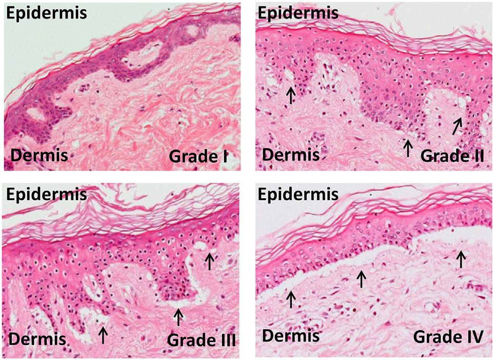
The dermis or corium is a layer of skin between the epidermis (with which it makes up the cutis) and subcutaneous tissues, that primarily consists of dense irregular connective tissue and cushions the body from stress and strain. It is divided into two layers, the superficial area adjacent to the epidermis called the papillary region and a deep thicker area known as the reticular dermis.James, William; Berger, Timothy; Elston, Dirk (2005). ''Andrews' Diseases of the Skin: Clinical Dermatology'' (10th ed.). Saunders. Pages 1, 11–12. . The dermis is tightly connected to the epidermis through a basement membrane. Structural components of the dermis are collagen, elastic fibers, and extrafibrillar matrix.Marks, James G; Miller, Jeffery (2006). ''Lookingbill and Marks' Principles of Dermatology'' (4th ed.). Elsevier Inc. Page 8–9. . It also contains mechanoreceptors that provide the sense of touch and thermoreceptors that provide the sense of heat. In addition, hair follicles, ... [...More Info...] [...Related Items...] OR: [Wikipedia] [Google] [Baidu] |
Skin Appendage
Skin appendages (or adnexa of skin) are anatomical skin-associated structures that serve a particular function including sensation, contractility, lubrication and heat loss in animals. In humans, some of the more common skin appendages are hairs (sensation, heat loss, filter for breathing, protection), arrector pilli (smooth muscles that pull hairs straight), sebaceous glands (secrete sebum onto hair follicle, which oils the hair), sweat glands (can secrete sweat with strong odour (apocrine) or with a faint odour (merocrine or eccrine)), and nails (protection). Skin appendages are derived from the skin, and are usually adjacent to it. Types of appendages include hair, glands, and nails. Glands * Sweat glands are distributed all over the body except nipples and outer genitals. Although the nipples do have the mammary glands, these are known as ''modified sweat glands''. * Sebaceous gland A sebaceous gland is a microscopic exocrine gland in the skin that opens into a hai ... [...More Info...] [...Related Items...] OR: [Wikipedia] [Google] [Baidu] |


