|
CBED
Convergent beam electron diffraction (CBED) is a diffraction technique where a convergent or divergent beam (conical electron beam) of electrons is used to study materials. History This technique was first introduced in 1939 by Kossel and Möllenstedt, who worked with large (~40 μm) probes and small convergence angles. The development of the Field Emission Gun (FEG) in the 1970s, the Scanning Transmission Electron Microscopy (STEM), energy filtering devices and so on, made possible smaller probe diameters and larger convergence angles, and all this made CBED more popular. In the seventies, CBED was being used for the determination of the point group and space group symmetries by Goodman and Lehmpfuh, Steeds, Buxton and starting on 1985, by Tanaka et al. by using different techniques of CBED which covered different applications. Applications By using CBED, different types of information can be obtained: *crystal structural parameters such as lattice parameters, sample thickne ... [...More Info...] [...Related Items...] OR: [Wikipedia] [Google] [Baidu] |
CBED Sketch
Convergent beam electron diffraction (CBED) is a diffraction technique where a convergent or divergent beam (conical electron beam) of electrons is used to study materials. History This technique was first introduced in 1939 by Kossel and Möllenstedt, who worked with large (~40 μm) probes and small convergence angles. The development of the Field Emission Gun (FEG) in the 1970s, the Scanning Transmission Electron Microscopy (STEM), energy filtering devices and so on, made possible smaller probe diameters and larger convergence angles, and all this made CBED more popular. In the seventies, CBED was being used for the determination of the point group and space group symmetries by Goodman and Lehmpfuh, Steeds, Buxton and starting on 1985, by Tanaka et al. by using different techniques of CBED which covered different applications. Applications By using CBED, different types of information can be obtained: *crystal structural parameters such as lattice parameters, sample thickne ... [...More Info...] [...Related Items...] OR: [Wikipedia] [Google] [Baidu] |
Scanning Transmission Electron Microscopy
A scanning transmission electron microscope (STEM) is a type of transmission electron microscope (TEM). Pronunciation is tɛmor �sti:i:ɛm As with a conventional transmission electron microscope (CTEM), images are formed by electrons passing through a sufficiently thin specimen. However, unlike CTEM, in STEM the electron beam is focused to a fine spot (with the typical spot size 0.05 – 0.2 nm) which is then scanned over the sample in a raster illumination system constructed so that the sample is illuminated at each point with the beam parallel to the optical axis. The rastering of the beam across the sample makes STEM suitable for analytical techniques such as Z-contrast annular dark-field imaging, and spectroscopic mapping by energy dispersive X-ray (EDX) spectroscopy, or electron energy loss spectroscopy (EELS). These signals can be obtained simultaneously, allowing direct correlation of images and spectroscopic data. A typical STEM is a conventional transmission el ... [...More Info...] [...Related Items...] OR: [Wikipedia] [Google] [Baidu] |
Electron Diffraction
Electron diffraction refers to the bending of electron beams around atomic structures. This behaviour, typical for waves, is applicable to electrons due to the wave–particle duality stating that electrons behave as both particles and waves. Since the diffracted beams interfere, they generate diffraction patterns widely used for analysis of the objects which caused the diffraction. Therefore, electron diffraction can also refer to derived experimental techniques used for material characterization. This technique is similar to X-ray and neutron diffraction. Electron diffraction is most frequently used in solid state physics and chemistry to study crystalline, quasi-crystalline and amorphous materials using electron microscopes. In these instruments, electrons are accelerated by an electrostatic potential in order to gain energy and shorten their wavelength. With the wavelength sufficiently short, the atomic structure acts as a diffraction grating generating diffraction patte ... [...More Info...] [...Related Items...] OR: [Wikipedia] [Google] [Baidu] |
Selected Area Diffraction
Selected area (electron) diffraction (abbreviated as SAD or SAED), is a crystallographic experimental technique typically performed using a transmission electron microscope (TEM). It is a specific case of electron diffraction used primarily in material science and solid state physics as one of the most common experimental techniques. Especially with appropriate analytical software, SAD patterns (SADP) can be used to determine crystal orientation, measure lattice constants or examine its defects. Principle In transmission electron microscope, a thin crystalline sample is illuminated by parallel beam of electrons accelerated to energy of hundreds of kiloelectron volts. At these energies, even metallic samples are transparent for the electrons if the sample is thinned enough (typically less than 100 nm). Due to the wave–particle duality, the high-energetic electrons behave as waves with wavelength of a few thousandths of a nanometer. The relativistic wavelength is ... [...More Info...] [...Related Items...] OR: [Wikipedia] [Google] [Baidu] |
Transmission Electron Microscopy
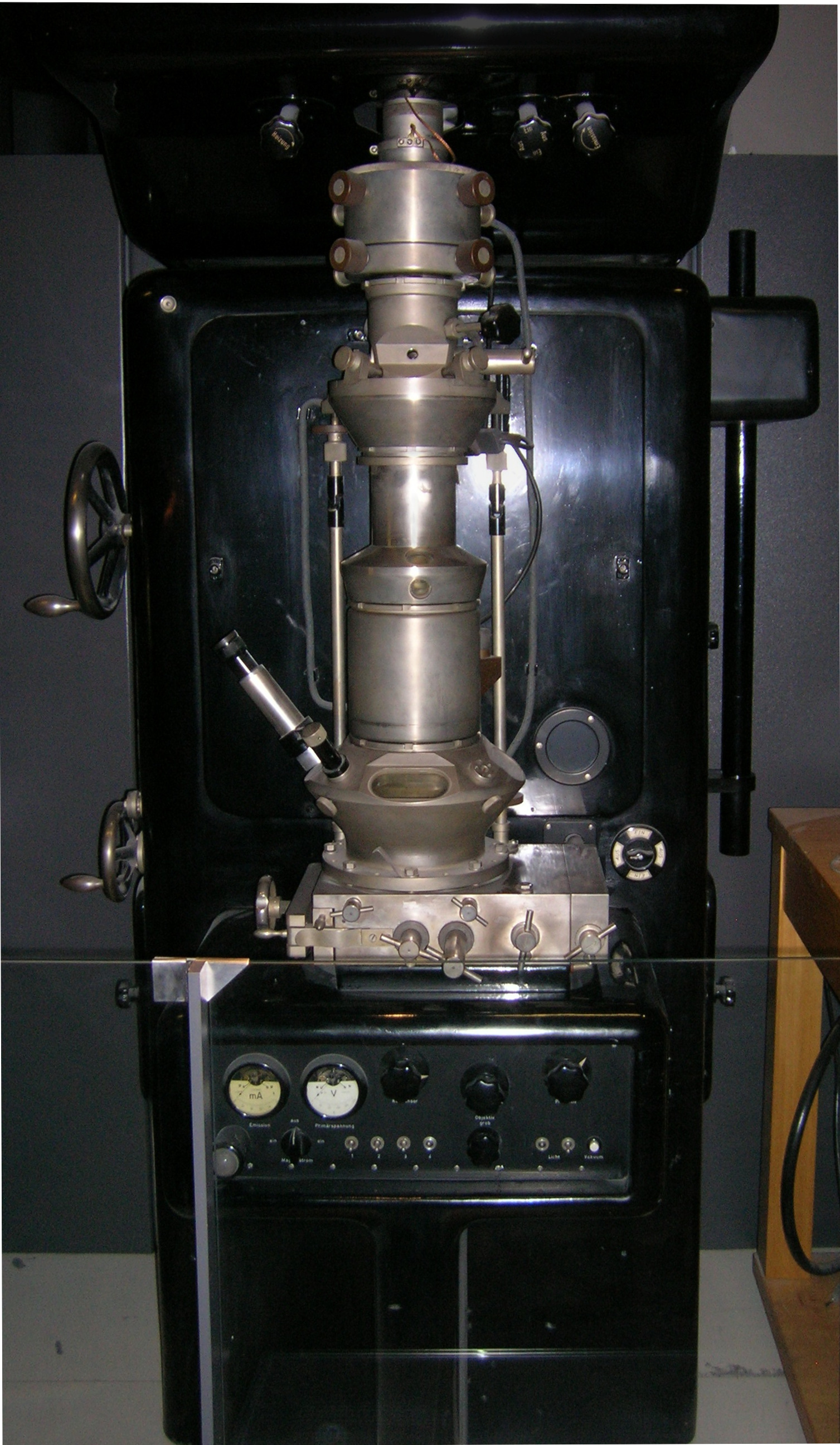
Transmission electron microscopy (TEM) is a microscopy technique in which a beam of electrons is transmitted through a specimen to form an image. The specimen is most often an ultrathin section less than 100 nm thick or a suspension on a grid. An image is formed from the interaction of the electrons with the sample as the beam is transmitted through the specimen. The image is then magnified and focused onto an imaging device, such as a fluorescent screen, a layer of photographic film, or a sensor such as a scintillator attached to a charge-coupled device. Transmission electron microscopes are capable of imaging at a significantly higher resolution than light microscopes, owing to the smaller de Broglie wavelength of electrons. This enables the instrument to capture fine detail—even as small as a single column of atoms, which is thousands of times smaller than a resolvable object seen in a light microscope. Transmission electron microscopy is a major analytical method i ... [...More Info...] [...Related Items...] OR: [Wikipedia] [Google] [Baidu] |
Band-pass Filter
A band-pass filter or bandpass filter (BPF) is a device that passes frequencies within a certain range and rejects (attenuates) frequencies outside that range. Description In electronics and signal processing, a filter is usually a two-port circuit or device which removes frequency components of a signal (an alternating voltage or current). A band-pass filter allows through components in a specified band of frequencies, called its ''passband'' but blocks components with frequencies above or below this band. This contrasts with a high-pass filter, which allows through components with frequencies above a specific frequency, and a low-pass filter, which allows through components with frequencies below a specific frequency. In digital signal processing, in which signals represented by digital numbers are processed by computer programs, a band-pass filter is a computer algorithm that performs the same function. The term band-pass filter is also used for optical filters, sh ... [...More Info...] [...Related Items...] OR: [Wikipedia] [Google] [Baidu] |
Laboratory Techniques In Condensed Matter Physics
A laboratory (; ; colloquially lab) is a facility that provides controlled conditions in which scientific or technological research, experiments, and measurement may be performed. Laboratory services are provided in a variety of settings: physicians' offices, clinics, hospitals, and regional and national referral centers. Overview The organisation and contents of laboratories are determined by the differing requirements of the specialists working within. A physics laboratory might contain a particle accelerator or vacuum chamber, while a metallurgy laboratory could have apparatus for casting or refining metals or for testing their strength. A chemist or biologist might use a wet laboratory, while a psychologist's laboratory might be a room with one-way mirrors and hidden cameras in which to observe behavior. In some laboratories, such as those commonly used by computer scientists, computers (sometimes supercomputers) are used for either simulations or the analysis of data. Scienti ... [...More Info...] [...Related Items...] OR: [Wikipedia] [Google] [Baidu] |
Measurement
Measurement is the quantification of attributes of an object or event, which can be used to compare with other objects or events. In other words, measurement is a process of determining how large or small a physical quantity is as compared to a basic reference quantity of the same kind. The scope and application of measurement are dependent on the context and discipline. In natural sciences and engineering, measurements do not apply to nominal properties of objects or events, which is consistent with the guidelines of the ''International vocabulary of metrology'' published by the International Bureau of Weights and Measures. However, in other fields such as statistics as well as the social and behavioural sciences, measurements can have multiple levels, which would include nominal, ordinal, interval and ratio scales. Measurement is a cornerstone of trade, science, technology and quantitative research in many disciplines. Historically, many measurement systems existed fo ... [...More Info...] [...Related Items...] OR: [Wikipedia] [Google] [Baidu] |
Bilayer
A bilayer is a double layer of closely packed atoms or molecules. The properties of bilayers are often studied in condensed matter physics, particularly in the context of semiconductor devices, where two distinct materials are united to form junctions (such as p–n junctions, Schottky junctions, etc.). Layered materials, such as graphene, boron nitride, or transition metal dichalchogenides, have unique electronic properties as a bilayer system and are an active area of current research. In biology a common example is the Lipid bilayer, which describes the structure of multiple organic structures, such as the membrane of a cell. See also * Monolayer * Non-carbon nanotube A non-carbon nanotube is a cylindrical molecule often composed of metal oxides, or group III-Nitrides and morphologically similar to a carbon nanotube. Non-carbon nanotubes have been observed to occur naturally in some mineral deposits. A few year ... * Semiconductor * Thin film References Phases of ... [...More Info...] [...Related Items...] OR: [Wikipedia] [Google] [Baidu] |
Monolayer
A monolayer is a single, closely packed layer of atoms, molecules, or cells. In some cases it is referred to as a self-assembled monolayer. Monolayers of layered crystals like graphene and molybdenum disulfide are generally called 2D materials. Chemistry A Langmuir monolayer or insoluble monolayer is a one-molecule thick layer of an insoluble organic material spread onto an aqueous subphase in a Langmuir-Blodgett trough. Traditional compounds used to prepare Langmuir monolayers are amphiphilic materials that possess a hydrophilic headgroup and a hydrophobic tail. Since the 1980s a large number of other materials have been employed to produce Langmuir monolayers, some of which are semi-amphiphilic, including polymeric, ceramic or metallic nanoparticles and macromolecules such as polymers. Langmuir monolayers are extensively studied for the fabrication of Langmuir-Blodgett film (LB films), which are formed by transferred monolayers on a solid substrate. A Gibbs monolayer or ... [...More Info...] [...Related Items...] OR: [Wikipedia] [Google] [Baidu] |
Angstrom
The angstromEntry "angstrom" in the Oxford online dictionary. Retrieved on 2019-03-02 from https://en.oxforddictionaries.com/definition/angstrom.Entry "angstrom" in the Merriam-Webster online dictionary. Retrieved on 2019-03-02 from https://www.merriam-webster.com/dictionary/angstrom. (, ; , ) or ångström is a metric unit of length equal to m; that is, one ten-billionth ( US) of a metre, a hundred-millionth of a centimetre,Entry "angstrom" in the Oxford English Dictionary, 2nd edition (1986). Retrieved on 2021-11-22 from https://www.oed.com/oed2/00008552. 0.1 nanometre, or 100 picometres. Its symbol is Å, a letter of the Swedish alphabet. The unit is named after the Swedish physicist Anders Jonas Ångström (1814–1874). The angstrom is often used in the natural sciences and technology to express sizes of atoms, molecules, microscopic biological structures, and lengths of chemical bonds, arrangement of atoms in crystals,Arturas Vailionis (2015):Geometry of Crystals Lect ... [...More Info...] [...Related Items...] OR: [Wikipedia] [Google] [Baidu] |
Phase Retrieval
Phase retrieval is the process of algorithmically finding solutions to the phase problem. Given a complex signal F(k), of amplitude , F (k), , and phase \psi(k): ::F(k) = , F(k), e^ =\int_^ f(x)\ e^\,dx where ''x'' is an ''M''-dimensional spatial coordinate and ''k'' is an ''M''-dimensional spatial frequency coordinate. Phase retrieval consists of finding the phase that satisfies a set of constraints for a measured amplitude. Important applications of phase retrieval include X-ray crystallography, transmission electron microscopy and coherent diffractive imaging, for which M = 2. Uniqueness theorems for both 1-D and 2-D cases of the phase retrieval problem, including the phaseless 1-D inverse scattering problem, were proven by Klibanov and his collaborators (see References). Problem formulation Here we consider 1-D discrete Fourier transform (DFT) phase retrieval problem. The DFT of a complex signal f /math> is given by F \sum_^ f e^=, F \cdot e^ \quad k=0,1, \ldots, N-1, ... [...More Info...] [...Related Items...] OR: [Wikipedia] [Google] [Baidu] |


