|
Bronchopulmonary Segment
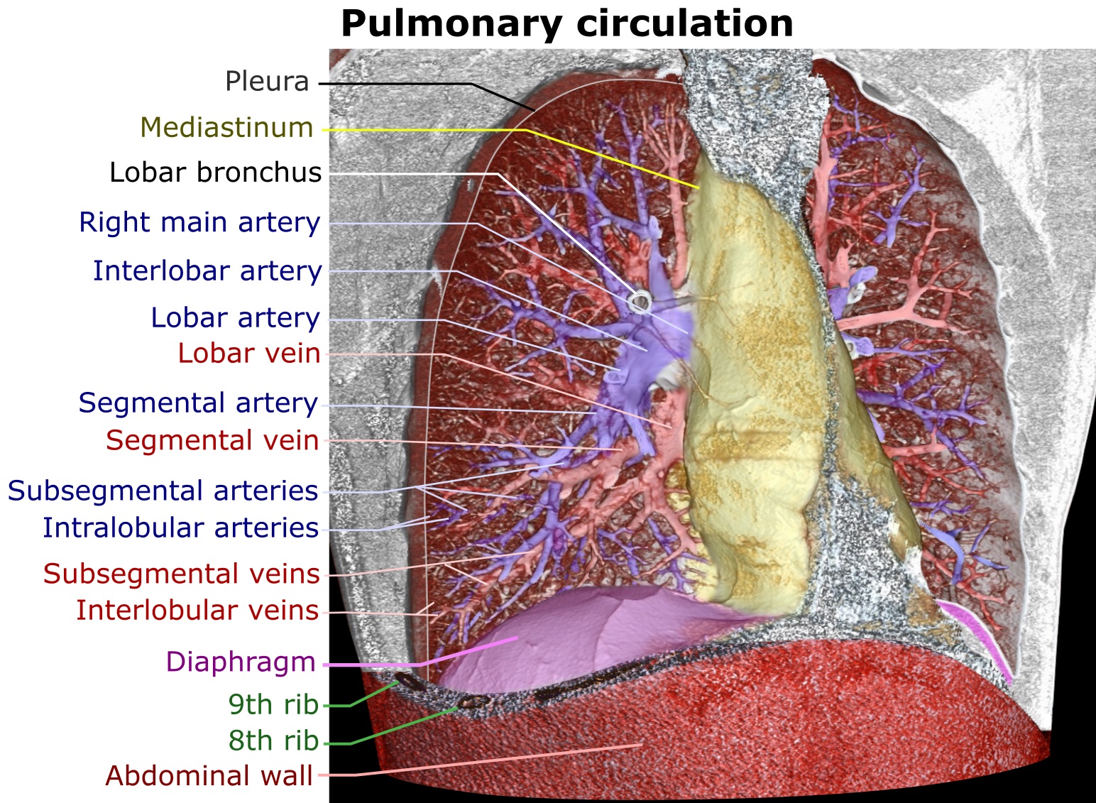
A bronchopulmonary segment is a portion of lung supplied by a specific segmental bronchus and its vessels. These arteries branch from the pulmonary and bronchial arteries, and run together through the center of the segment. Veins and lymphatic vessels drain along the edges of the segment. The segments are separated from each other by layers of connective tissue that forms them into discrete anatomical and functional units. This separation means that a bronchopulmonary segment can be surgically removed without affecting the function of the others. There are ten bronchopulmonary segments in the right lung: three in the superior lobe, two in the middle lobe, and five in the inferior lobe. Some of the segments may fuse in the left lung to form usually eight to nine segments (four to five in the upper lobe and four to five in the lower lobe. The delineation of the bronchopulmonary segments was made by Chevalier Jackson and John Franklin Huber at Temple University Hospital. Right l ... [...More Info...] [...Related Items...] OR: [Wikipedia] [Google] [Baidu] |
Lung
The lungs are the primary organs of the respiratory system in humans and most other animals, including some snails and a small number of fish. In mammals and most other vertebrates, two lungs are located near the backbone on either side of the heart. Their function in the respiratory system is to extract oxygen from the air and transfer it into the bloodstream, and to release carbon dioxide from the bloodstream into the atmosphere, in a process of gas exchange. Respiration is driven by different muscular systems in different species. Mammals, reptiles and birds use their different muscles to support and foster breathing. In earlier tetrapods, air was driven into the lungs by the pharyngeal muscles via buccal pumping, a mechanism still seen in amphibians. In humans, the main muscle of respiration that drives breathing is the diaphragm. The lungs also provide airflow that makes vocal sounds including human speech possible. Humans have two lungs, one on the left an ... [...More Info...] [...Related Items...] OR: [Wikipedia] [Google] [Baidu] |
Segmental Bronchus
A bronchus is a passage or airway in the lower respiratory tract that conducts air into the lungs. The first or primary bronchi pronounced (BRAN-KAI) to branch from the trachea at the carina are the right main bronchus and the left main bronchus. These are the widest bronchi, and enter the right lung, and the left lung at each hilum. The main bronchi branch into narrower secondary bronchi or lobar bronchi, and these branch into narrower tertiary bronchi or segmental bronchi. Further divisions of the segmental bronchi are known as 4th order, 5th order, and 6th order segmental bronchi, or grouped together as subsegmental bronchi. The bronchi, when too narrow to be supported by cartilage, are known as bronchioles. No gas exchange takes place in the bronchi. Structure The trachea (windpipe) divides at the carina into two main or primary bronchi, the left bronchus and the right bronchus. The carina of the trachea is located at the level of the sternal angle and the fifth thoracic ver ... [...More Info...] [...Related Items...] OR: [Wikipedia] [Google] [Baidu] |
Pulmonary Artery
A pulmonary artery is an artery in the pulmonary circulation that carries deoxygenated blood from the right side of the heart to the lungs. The largest pulmonary artery is the ''main pulmonary artery'' or ''pulmonary trunk'' from the heart, and the smallest ones are the arterioles, which lead to the capillaries that surround the pulmonary alveoli. Structure The pulmonary arteries are blood vessels that carry systemic venous blood from the right ventricle of the heart to the microcirculation of the lungs. Unlike in other organs where arteries supply oxygenated blood, the blood carried by the pulmonary arteries is deoxygenated, as it is venous blood returning to the heart. The main pulmonary arteries emerge from the right side of the heart, and then split into smaller arteries that progressively divide and become arterioles, eventually narrowing into the capillary microcirculation of the lungs where gas exchange occurs. Pulmonary trunk In order of blood flow, the pulmonary ... [...More Info...] [...Related Items...] OR: [Wikipedia] [Google] [Baidu] |
Bronchial Artery
In human anatomy, the bronchial arteries supply the lungs with nutrition and oxygenated blood. Although there is much variation, there are usually two bronchial arteries that run to the left lung, and one to the right lung and are a vital part of the respiratory system. Structure There are typically two left and one right bronchial arteries. The ''left bronchial arteries'' (superior and inferior) usually arise directly from the thoracic aorta. The single ''right bronchial artery'' usually arises from one of the following: * 1) the thoracic aorta at a common trunk with the right 3rd posterior intercostal artery * 2) the superior bronchial artery on the left side * 3) any number of the right intercostal arteries mostly the third right posterior. Function The bronchial arteries supply blood to the bronchi and connective tissue of the lungs. They travel with and branch with the bronchi, ending about at the level of the respiratory bronchioles. They anastomose with the bran ... [...More Info...] [...Related Items...] OR: [Wikipedia] [Google] [Baidu] |
Connective Tissue
Connective tissue is one of the four primary types of animal tissue, along with epithelial tissue, muscle tissue, and nervous tissue. It develops from the mesenchyme derived from the mesoderm the middle embryonic germ layer. Connective tissue is found in between other tissues everywhere in the body, including the nervous system. The three meninges, membranes that envelop the brain and spinal cord are composed of connective tissue. Most types of connective tissue consists of three main components: elastic and collagen fibers, ground substance, and cells. Blood, and lymph are classed as specialized fluid connective tissues that do not contain fiber. All are immersed in the body water. The cells of connective tissue include fibroblasts, adipocytes, macrophages, mast cells and leucocytes. The term "connective tissue" (in German, ''Bindegewebe'') was introduced in 1830 by Johannes Peter Müller. The tissue was already recognized as a distinct class in the 18th century. ... [...More Info...] [...Related Items...] OR: [Wikipedia] [Google] [Baidu] |
Segmental Resection
Segmental resection (or segmentectomy) is a surgical procedure to remove part of an organ or gland, as a sub-type of a resection, which might involve removing the whole body part. It may also be used to remove a tumor and normal tissue around it. In lung cancer surgery, segmental resection refers to removing a section of a lobe of the lung. The resection margin is the edge of the removed tissue; it is important that this shows free of cancerous cells on examination by a pathologist Pathology is the study of the causes and effects of disease or injury. The word ''pathology'' also refers to the study of disease in general, incorporating a wide range of biology research fields and medical practices. However, when used in th .... References * External links Segmental resectionentry in the public domain NCI Dictionary of Cancer Terms Surgical procedures and techniques Surgical removal procedures {{oncology-stub ... [...More Info...] [...Related Items...] OR: [Wikipedia] [Google] [Baidu] |
Chevalier Jackson
Chevalier Quixote Jackson (November 4, 1865 – August 16, 1958) was an American pioneer in laryngology. He is sometimes known as the "father of endoscopy", although Philipp Bozzini (1773–1809) is also often given this sobriquet. Chevalier Q. Jackson extracted over 2000 swallowed foreign bodies from patients. The collection is currently on display at the Mütter Museum in Philadelphia. Biography Jackson was born in Pittsburgh, Pennsylvania. He went to school at the Western University of Pennsylvania (now the University of Pittsburgh) from 1879 to 1883, and received his MD from Jefferson Medical College in Philadelphia. He also studied laryngology in England. His work reduced the risks involved in a tracheotomy. He essentially invented the modern science of endoscopy of the upper airway and esophagus, using hollow tubes with illumination (esophagoscopes and bronchoscopes). He developed methods for removing foreign bodies from the esophagus and the airway with great safety — a ... [...More Info...] [...Related Items...] OR: [Wikipedia] [Google] [Baidu] |
Temple University School Of Medicine
The Lewis Katz School of Medicine at Temple University (LKSOM), located on the Health Science Campus of Temple University in Philadelphia, PA, is one of 7 schools of medicine in Pennsylvania conferring the M.D. (Doctor of Medicine) degree. It also confers the Ph.D. (Doctor of Philosophy) and M.S. (Master of Science) degrees in biomedical sciences. In addition, LKSOM offers a Narrative Medicine Program. In July 2014, Lewis Katz School of Medicine's scientists became the first to remove HIV from human cells. Temple University's Fox Chase Cancer Center is ranked 9th best Hospital for Adult Cancer by '' U.S. News & World Report''. LKSOM reported 15,624 applications in 2020 (class of 2024) for a class size of 210 students; 340 of the total 9,624 applications received acceptance, translating to a 1.3% acceptance rate. History Founded in 1901 as Pennsylvania's first co-educational medical school, the institution has attained a national reputation for training humanistic and dedicated ... [...More Info...] [...Related Items...] OR: [Wikipedia] [Google] [Baidu] |
Carina Of Trachea
In anatomy, the carina or tracheal bifurcation is a ridge of cartilage in the trachea that occurs between the division of the two main bronchi. Structure The carina occurs at the lower end of the trachea (usually at the level of the 4th to 5th thoracic vertebra). This is in line with the sternal angle, but the carina may raise or descend up to two vertebrae higher or lower with breathing. The carina lies to the left of the midline, and runs antero-posteriorly (front to back). The bronchial arteries supply the carina and the rest of the lower trachea. The carina is around the area posterior to where the aortic arch crosses to the left of the trachea. The azygos vein crosses right to the trachea above the carina. Clinical significance Foreign bodies that fall down the trachea are more likely to enter the right bronchus. The mucous membrane of the carina is the most sensitive area of the trachea and larynx for triggering a cough reflex. Widening and distortion of the carin ... [...More Info...] [...Related Items...] OR: [Wikipedia] [Google] [Baidu] |
Trachea
The trachea, also known as the windpipe, is a cartilaginous tube that connects the larynx to the bronchi of the lungs, allowing the passage of air, and so is present in almost all air- breathing animals with lungs. The trachea extends from the larynx and branches into the two primary bronchi. At the top of the trachea the cricoid cartilage attaches it to the larynx. The trachea is formed by a number of horseshoe-shaped rings, joined together vertically by overlying ligaments, and by the trachealis muscle at their ends. The epiglottis closes the opening to the larynx during swallowing. The trachea begins to form in the second month of embryo development, becoming longer and more fixed in its position over time. It is epithelium lined with column-shaped cells that have hair-like extensions called cilia, with scattered goblet cells that produce protective mucins. The trachea can be affected by inflammation or infection, usually as a result of a viral illness affecting othe ... [...More Info...] [...Related Items...] OR: [Wikipedia] [Google] [Baidu] |


