|
Bronchogenic Cyst
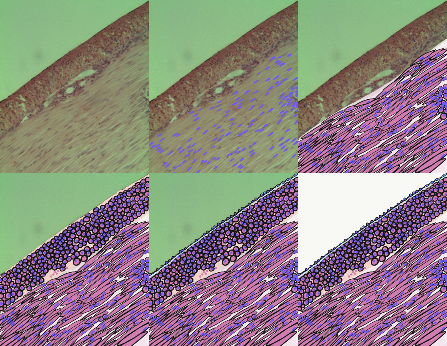
Bronchogenic cysts are small, solitary cysts or sinuses, most typically located in the region of the suprasternal notch or behind the manubrium. Clinical features These cysts are found most often in young adults and are rare in infancy. The usual symptoms are the result of compression by the cyst, e.g., difficulty breathing or swallowing, cough, and chest pain. Malignant degeneration has been reported in these cysts on rare occasions. Bronchogenic cysts are usually found in the middle mediastinum. Chest x-rays show a smooth density just in front of the trachea or main stem bronchi at the carinal level. When the cyst communicates with the tracheobronchial tree, the air-fluid level may be seen within the cyst. CT scanning is useful in localizing these cysts. Pathology Bronchogenic cysts are formed in the 6th week of gestation from an abnormal budding of the tracheal diverticulum. They are lined by respiratory type (ciliated) epithelium, which is characterized by cilia. Histo ... [...More Info...] [...Related Items...] OR: [Wikipedia] [Google] [Baidu] |
Micrograph
A micrograph or photomicrograph is a photograph or digital image taken through a microscope or similar device to show a magnified image of an object. This is opposed to a macrograph or photomacrograph, an image which is also taken on a microscope but is only slightly magnified, usually less than 10 times. Micrography is the practice or art of using microscopes to make photographs. A micrograph contains extensive details of microstructure. A wealth of information can be obtained from a simple micrograph like behavior of the material under different conditions, the phases found in the system, failure analysis, grain size estimation, elemental analysis and so on. Micrographs are widely used in all fields of microscopy. Types Photomicrograph A light micrograph or photomicrograph is a micrograph prepared using an optical microscope, a process referred to as ''photomicroscopy''. At a basic level, photomicroscopy may be performed simply by connecting a camera to a microscope ... [...More Info...] [...Related Items...] OR: [Wikipedia] [Google] [Baidu] |
Cilia
The cilium, plural cilia (), is a membrane-bound organelle found on most types of eukaryotic cell, and certain microorganisms known as ciliates. Cilia are absent in bacteria and archaea. The cilium has the shape of a slender threadlike projection that extends from the surface of the much larger cell body. Eukaryotic flagella found on sperm cells and many protozoans have a similar structure to motile cilia that enables swimming through liquids; they are longer than cilia and have a different undulating motion. There are two major classes of cilia: ''motile'' and ''non-motile'' cilia, each with a subtype, giving four types in all. A cell will typically have one primary cilium or many motile cilia. The structure of the cilium core called the axoneme determines the cilium class. Most motile cilia have a central pair of single microtubules surrounded by nine pairs of double microtubules called a 9+2 axoneme. Most non-motile cilia have a 9+0 axoneme that lacks the central pai ... [...More Info...] [...Related Items...] OR: [Wikipedia] [Google] [Baidu] |
Cutaneous Columnar Cyst
Cutaneous columnar cysts are a cutaneous condition, a group of different cysts lined by columnar epithelium. Types of cysts included in this group are: :* Bronchogenic cyst :* Branchial cyst :* Thyroglossal duct cyst :* Cutaneous ciliated cyst :* Median raphe cyst See also * Pseudocyst of the auricle * List of cutaneous conditions References Epidermal nevi, neoplasms, and cysts {{Epidermal-growth-stub ... [...More Info...] [...Related Items...] OR: [Wikipedia] [Google] [Baidu] |
Observation
Observation is the active acquisition of information from a primary source. In living beings, observation employs the senses. In science, observation can also involve the perception and recording of data via the use of scientific instruments. The term may also refer to any data collected during the scientific activity. Observations can be qualitative, that is, only the absence or presence of a property is noted, or quantitative if a numerical value is attached to the observed phenomenon by counting or measuring. Science The scientific method requires observations of natural phenomena to formulate and test hypotheses. It consists of the following steps: # Ask a question about a natural phenomenon # Make observations of the phenomenon # Formulate a hypothesis that tentatively answers the question # Predict logical, observable consequences of the hypothesis that have not yet been investigated # Test the hypothesis' predictions by an experiment, observational study, field study ... [...More Info...] [...Related Items...] OR: [Wikipedia] [Google] [Baidu] |
Biopsy
A biopsy is a medical test commonly performed by a surgeon, interventional radiologist, or an interventional cardiologist. The process involves extraction of sample cells or tissues for examination to determine the presence or extent of a disease. The tissue is then fixed, dehydrated, embedded, sectioned, stained and mounted before it is generally examined under a microscope by a pathologist; it may also be analyzed chemically. When an entire lump or suspicious area is removed, the procedure is called an excisional biopsy. An incisional biopsy or core biopsy samples a portion of the abnormal tissue without attempting to remove the entire lesion or tumor. When a sample of tissue or fluid is removed with a needle in such a way that cells are removed without preserving the histological architecture of the tissue cells, the procedure is called a needle aspiration biopsy. Biopsies are most commonly performed for insight into possible cancerous or inflammatory conditions. Histor ... [...More Info...] [...Related Items...] OR: [Wikipedia] [Google] [Baidu] |
Bronchi
A bronchus is a passage or airway in the lower respiratory tract that conducts air into the lungs. The first or primary bronchi pronounced (BRAN-KAI) to branch from the trachea at the carina are the right main bronchus and the left main bronchus. These are the widest bronchi, and enter the right lung, and the left lung at each hilum. The main bronchi branch into narrower secondary bronchi or lobar bronchi, and these branch into narrower tertiary bronchi or segmental bronchi. Further divisions of the segmental bronchi are known as 4th order, 5th order, and 6th order segmental bronchi, or grouped together as subsegmental bronchi. The bronchi, when too narrow to be supported by cartilage, are known as bronchioles. No gas exchange takes place in the bronchi. Structure The trachea (windpipe) divides at the carina into two main or primary bronchi, the left bronchus and the right bronchus. The carina of the trachea is located at the level of the sternal angle and the fifth thoracic vert ... [...More Info...] [...Related Items...] OR: [Wikipedia] [Google] [Baidu] |
Smooth Muscle
Smooth muscle is an involuntary non- striated muscle, so-called because it has no sarcomeres and therefore no striations (''bands'' or ''stripes''). It is divided into two subgroups, single-unit and multiunit smooth muscle. Within single-unit muscle, the whole bundle or sheet of smooth muscle cells contracts as a syncytium. Smooth muscle is found in the walls of hollow organs, including the stomach, intestines, bladder and uterus; in the walls of passageways, such as blood, and lymph vessels, and in the tracts of the respiratory, urinary, and reproductive systems. In the eyes, the ciliary muscles, a type of smooth muscle, dilate and contract the iris and alter the shape of the lens. In the skin, smooth muscle cells such as those of the arrector pili cause hair to stand erect in response to cold temperature or fear. Structure Gross anatomy Smooth muscle is grouped into two types: single-unit smooth muscle, also known as visceral smooth muscle, and multiunit ... [...More Info...] [...Related Items...] OR: [Wikipedia] [Google] [Baidu] |
Cartilage
Cartilage is a resilient and smooth type of connective tissue. In tetrapods, it covers and protects the ends of long bones at the joints as articular cartilage, and is a structural component of many body parts including the rib cage, the neck and the bronchial tubes, and the intervertebral discs. In other taxa, such as chondrichthyans, but also in cyclostomes, it may constitute a much greater proportion of the skeleton. It is not as hard and rigid as bone, but it is much stiffer and much less flexible than muscle. The matrix of cartilage is made up of glycosaminoglycans, proteoglycans, collagen fibers and, sometimes, elastin. Because of its rigidity, cartilage often serves the purpose of holding tubes open in the body. Examples include the rings of the trachea, such as the cricoid cartilage and carina. Cartilage is composed of specialized cells called chondrocytes that produce a large amount of collagenous extracellular matrix, abundant ground substance that is rich in ... [...More Info...] [...Related Items...] OR: [Wikipedia] [Google] [Baidu] |
Diverticulum
In medicine or biology, a diverticulum is an outpouching of a hollow (or a fluid-filled) structure in the body. Depending upon which layers of the structure are involved, diverticula are described as being either true or false. In medicine, the term usually implies the structure is not normally present, but in embryology, the term is used for some normal structures arising from others, as for instance the thyroid diverticulum, which arises from the tongue. The word comes from Latin ''dīverticulum'', "bypath" or "byway". Classification Diverticula are described as being true or false depending upon the layers involved: *False diverticula (also known as "pseudodiverticula") do not involve muscular layers or adventitia. False diverticula, in the gastrointestinal tract for instance, involve only the submucosa and mucosa. *True diverticula involve all layers of the structure, including muscularis propria and adventitia, such as Meckel's diverticulum. Embryology *The kidne ... [...More Info...] [...Related Items...] OR: [Wikipedia] [Google] [Baidu] |
H&E Stain
Hematoxylin and eosin stain ( or haematoxylin and eosin stain or hematoxylin-eosin stain; often abbreviated as H&E stain or HE stain) is one of the principal tissue stains used in histology. It is the most widely used stain in medical diagnosis and is often the gold standard. For example, when a pathologist looks at a biopsy of a suspected cancer, the histological section is likely to be stained with H&E. H&E is the combination of two histological stains: hematoxylin and eosin. The hematoxylin stains cell nuclei a purplish blue, and eosin stains the extracellular matrix and cytoplasm pink, with other structures taking on different shades, hues, and combinations of these colors. Hence a pathologist can easily differentiate between the nuclear and cytoplasmic parts of a cell, and additionally, the overall patterns of coloration from the stain show the general layout and distribution of cells and provides a general overview of a tissue sample's structure. Thus, pattern recognit ... [...More Info...] [...Related Items...] OR: [Wikipedia] [Google] [Baidu] |
Pregnancy
Pregnancy is the time during which one or more offspring develops (gestation, gestates) inside a woman, woman's uterus (womb). A multiple birth, multiple pregnancy involves more than one offspring, such as with twins. Pregnancy usually occurs by sexual intercourse, but can also occur through assisted reproductive technology procedures. A pregnancy may end in a Live birth (human), live birth, a miscarriage, an Abortion#Induced, induced abortion, or a stillbirth. Childbirth typically occurs around 40 weeks from the start of the Menstruation#Onset and frequency, last menstrual period (LMP), a span known as the Gestational age (obstetrics), gestational age. This is just over nine months. Counting by Human fertilization#Fertilization age, fertilization age, the length is about 38 weeks. Pregnancy is "the presence of an implanted human embryo or fetus in the uterus"; Implantation (embryology), implantation occurs on average 8–9 days after fertilization. An ''embryo'' ... [...More Info...] [...Related Items...] OR: [Wikipedia] [Google] [Baidu] |


