|
Brodmann Area 38
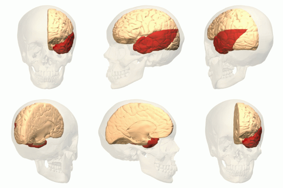
Brodmann area 38, also BA38 or temporopolar area 38 (H), is part of the temporal cortex in the human brain. BA 38 is at the anterior end of the temporal lobe, known as the temporal pole. BA38 is a subdivision of the cytoarchitecturally defined temporal region of cerebral cortex. It is located primarily in the most rostral portions of the superior temporal gyrus and the middle temporal gyrus. Cytoarchitecturally it is bounded caudally by the inferior temporal area 20, the middle temporal area 21, the superior temporal area 22 and the ectorhinal area 36 (Brodmann-1909). The temporal pole is a paralimbic region involved in high level semantic representation and socio-emotional processing. The uncinate fasciculus provides a direct bidirectional path to the orbitofrontal cortex, allowing mnemonic representations stored in the temporal pole to bias decision making in the frontal lobe. The temporal pole appears to be a convergence zone where concepts (also known as semantic memori ... [...More Info...] [...Related Items...] OR: [Wikipedia] [Google] [Baidu] |
Temporal Lobe
The temporal lobe is one of the four Lobes of the brain, major lobes of the cerebral cortex in the brain of mammals. The temporal lobe is located beneath the lateral fissure on both cerebral hemispheres of the mammalian brain. The temporal lobe is involved in processing sensory input into derived meanings for the appropriate retention of visual memory, language comprehension, and emotion association. ''Temporal'' refers to the head's Temple (anatomy), temples. Structure The Temple (anatomy)#Etymology, temporal Lobe (anatomy), lobe consists of structures that are vital for declarative or long-term memory. Declarative memory, Declarative (denotative) or Explicit memory, explicit memory is conscious memory divided into semantic memory (facts) and episodic memory (events). Medial temporal lobe structures that are critical for long-term memory include the hippocampus, along with the surrounding Hippocampal formation, hippocampal region consisting of the Perirhinal cortex, perirhinal, ... [...More Info...] [...Related Items...] OR: [Wikipedia] [Google] [Baidu] |
Semantic Memories
Semantic memory refers to general world knowledge that humans have accumulated throughout their lives. This general knowledge (word meanings, concepts, facts, and ideas) is intertwined in experience and dependent on culture. We can learn about new concepts by applying our knowledge learned from things in the past. Semantic memory is distinct from episodic memory, which is our memory of experiences and specific events that occur during our lives, from which we can recreate at any given point. For instance, semantic memory might contain information about what a cat is, whereas episodic memory might contain a specific memory of petting a particular cat. Semantic memory and episodic memory are both types of explicit memory (or declarative memory), that is, memory of facts or events that can be consciously recalled and "declared". The counterpart to declarative or explicit memory is nondeclarative memory or implicit memory. History The idea of semantic memory was first intro ... [...More Info...] [...Related Items...] OR: [Wikipedia] [Google] [Baidu] |
List Of Regions In The Human Brain
The human brain anatomical regions are ordered following standard neuroanatomy hierarchies. Functional, connective, and developmental regions are listed in parentheses where appropriate. Hindbrain (rhombencephalon) Myelencephalon * Medulla oblongata **Medullary pyramids **Arcuate nucleus **Olivary body ***Inferior olivary nucleus **Rostral ventrolateral medulla **Caudal ventrolateral medulla **Solitary nucleus (Nucleus of the solitary tract) **Respiratory center- Respiratory groups ***Dorsal respiratory group ***Ventral respiratory group or Apneustic centre ****Pre-Bötzinger complex ****Botzinger complex ****Retrotrapezoid nucleus ****Nucleus retrofacialis ****Nucleus retroambiguus ****Nucleus para-ambiguus **Paramedian reticular nucleus **Gigantocellular reticular nucleus **Parafacial zone **Cuneate nucleus ** Gracile nucleus ** Perihypoglossal nuclei *** Intercalated nucleus *** Prepositus nucleus *** Sublingual nucleus **Area postrema **Medullary cranial nerve nucl ... [...More Info...] [...Related Items...] OR: [Wikipedia] [Google] [Baidu] |
Brodmann Area
A Brodmann area is a region of the cerebral cortex, in the human or other primate brain, defined by its cytoarchitecture, or histological structure and organization of cells. History Brodmann areas were originally defined and numbered by the German anatomist Korbinian Brodmann based on the cytoarchitectural organization of neurons he observed in the cerebral cortex using the Nissl method of cell staining. Brodmann published his maps of cortical areas in humans, monkeys, and other species in 1909, along with many other findings and observations regarding the general cell types and laminar organization of the mammalian cortex. The same Brodmann area number in different species does not necessarily indicate homologous areas. A similar, but more detailed cortical map was published by Constantin von Economo and Georg N. Koskinas in 1925. Present importance Brodmann areas have been discussed, debated, refined, and renamed exhaustively for nearly a century and remain the most wid ... [...More Info...] [...Related Items...] OR: [Wikipedia] [Google] [Baidu] |
Temporal Lobe Epilepsy
Temporal lobe epilepsy (TLE) is a chronic disorder of the nervous system which is characterized by recurrent, unprovoked focal seizures that originate in the temporal lobe of the brain and last about one or two minutes. TLE is the most common form of epilepsy with focal seizures. A focal seizure in the temporal lobe may spread to other areas in the brain when it may become a ''focal to bilateral seizure''. TLE is diagnosed by taking a medical history, blood tests, and brain imaging. It can have a number of causes such as head injury, stroke, brain infections, structural lesions in the brain, brain tumors, or it can be of ''unknown onset''. The first line of treatment is through anticonvulsants. Surgery may be an option, especially when there is an observable abnormality in the brain. Another treatment option is electrical stimulation of the brain through an implanted device called the vagus nerve stimulator (VNS). Types Over forty types of epilepsy are recognized and these ... [...More Info...] [...Related Items...] OR: [Wikipedia] [Google] [Baidu] |
Frontotemporal Lobar Degeneration
Frontotemporal lobar degeneration (FTLD) is a pathological process that occurs in frontotemporal dementia. It is characterized by atrophy in the frontal lobe and temporal lobe of the brain, with sparing of the parietal and occipital lobes. Common proteinopathies that are found in FTLD include the accumulation of tau proteins and TAR DNA-binding protein 43 (TDP-43). Mutations in the ''C9orf72'' gene have been established as a major genetic contribution of FTLD, although defects in the granulin (GRN) and microtubule-associated proteins (MAPs) are also associated with it. Classification There are 3 main histological subtypes found at post-mortem: * FTLD-tau is characterised by tau positive inclusion bodies often referred to as Pick-bodies. Examples of FTLD-tau include; Pick's disease, corticobasal degeneration, progressive supranuclear palsy. * FTLD-TDP (or FTLD-U ) is characterised by ubiquitin and TDP-43 positive, tau negative, FUS negative inclusion bodies. The pathological h ... [...More Info...] [...Related Items...] OR: [Wikipedia] [Google] [Baidu] |
Frontotemporal Dementia
Frontotemporal dementia (FTD), or frontotemporal degeneration disease, or frontotemporal neurocognitive disorder, encompasses several types of dementia involving the progressive degeneration of frontal and temporal lobes. FTDs broadly present as behavioral or language disorders with gradual onsets. The three main subtypes or variant syndromes are a behavioral variant (bvFTD) previously known as ''Pick's disease'', and two variants of primary progressive aphasia – semantic variant (svPPA), and nonfluent variant (nfvPPA). Two rare distinct subtypes of FTD are neuronal intermediate filament inclusion disease (NIFID), and basophilic inclusion body disease. Other related disorders include corticobasal syndrome and FTD with amyotrophic lateral sclerosis (ALS) ''FTD-ALS'' also called ''FTD- MND''. Frontotemporal dementias are mostly early-onset syndromes that are linked to frontotemporal lobar degeneration (FTLD), which is characterized by progressive neuronal loss predominantly i ... [...More Info...] [...Related Items...] OR: [Wikipedia] [Google] [Baidu] |
Alzheimer's Disease
Alzheimer's disease (AD) is a neurodegeneration, neurodegenerative disease that usually starts slowly and progressively worsens. It is the cause of 60–70% of cases of dementia. The most common early symptom is difficulty in short-term memory, remembering recent events. As the disease advances, symptoms can include primary progressive aphasia, problems with language, Orientation (mental), disorientation (including easily getting lost), mood swings, loss of motivation, self-neglect, and challenging behaviour, behavioral issues. As a person's condition declines, they often withdraw from family and society. Gradually, bodily functions are lost, ultimately leading to death. Although the speed of progression can vary, the typical life expectancy following diagnosis is three to nine years. The cause of Alzheimer's disease is poorly understood. There are many environmental and genetic risk factors associated with its development. The strongest genetic risk factor is from an alle ... [...More Info...] [...Related Items...] OR: [Wikipedia] [Google] [Baidu] |
Amygdala
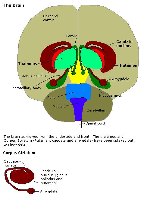
The amygdala (; plural: amygdalae or amygdalas; also '; Latin from Greek, , ', 'almond', 'tonsil') is one of two almond-shaped clusters of nuclei located deep and medially within the temporal lobes of the brain's cerebrum in complex vertebrates, including humans. Shown to perform a primary role in the processing of memory, decision making, and emotional responses (including fear, anxiety, and aggression), the amygdalae are considered part of the limbic system. The term "amygdala" was first introduced by Karl Friedrich Burdach in 1822. Structure The regions described as amygdala nuclei encompass several structures of the cerebrum with distinct connectional and functional characteristics in humans and other animals. Among these nuclei are the basolateral complex, the cortical nucleus, the medial nucleus, the central nucleus, and the intercalated cell clusters. The basolateral complex can be further subdivided into the lateral, the basal, and the accessory basal nucle ... [...More Info...] [...Related Items...] OR: [Wikipedia] [Google] [Baidu] |
Prosopagnosia
Prosopagnosia (from Greek ''prósōpon'', meaning "face", and ''agnōsía'', meaning "non-knowledge"), also called face blindness, ("illChoisser had even begun tpopularizea name for the condition: face blindness.") is a cognitive disorder of face perception in which the ability to recognize familiar faces, including one's own face (self-recognition), is impaired, while other aspects of visual processing (e.g., object discrimination) and intellectual functioning (e.g., decision-making) remain intact. The term originally referred to a condition following acute brain damage (acquired prosopagnosia), but a congenital or developmental form of the disorder also exists, with a prevalence of 2.5%. The brain area usually associated with prosopagnosia is the fusiform gyrus, which activates specifically in response to faces. The functionality of the fusiform gyrus allows most people to recognize faces in more detail than they do similarly complex inanimate objects. For those with prosopagno ... [...More Info...] [...Related Items...] OR: [Wikipedia] [Google] [Baidu] |
Uncinate Fasciculus
The uncinate fasciculus is a white matter association tract in the human brain that connects parts of the limbic system such as the temporal pole, anterior parahippocampus, and amygdala in the temporal lobe with inferior portions of the frontal lobe such as the orbitofrontal cortex. Its function is unknown though it is affected in several psychiatric conditions. It is one of the last white matter tracts to mature in the human brain. Anatomy The uncinate fasciculus is a hook-shaped bundle of axons that links anterior portions of the temporal lobe with the inferior frontal gyrus and the lower surfaces of the frontal lobe. It arises in the anterior temporal lobe and amygdala, in the temporal lobe curving in an upward pathway behind the external capsule inward of the insular cortex and continuing up into the posterior part of the orbital gyrus. It does not appear to have cell bodies in the hippocampus proper. The average length of the uncinate fasciculus is 45 mm with a ra ... [...More Info...] [...Related Items...] OR: [Wikipedia] [Google] [Baidu] |


