|
Blumensaat's Line
Blumensaat's line is a line which corresponds to the roof of the intercondylar fossa of femur as seen on a lateral radiograph of the knee joint. The angle at which this line appears on the radiograph can be used to determine the position of the patella or diagnose an ACL injury. Clinical Use On a normal radiograph, the line intersects the inferior pole of the patella, and so can be useful in diagnosing a broken femur as well as a patellar tendon rupture Patellar tendon rupture is a tear of the tendon that connects the knee cap (patella) to the tibia. Often there is sudden onset of pain and walking is difficult. In a complete rupture, the ability to extend that knee is decreased. A pop may be felt .... It also helps to define "Schottle point" intra-operatively for reconstruction of MPFL(Medial patello-femoral ligament.) It may also be used to describe the course of an ACL graft. References Musculoskeletal radiographic signs {{Orthopedics-stub ... [...More Info...] [...Related Items...] OR: [Wikipedia] [Google] [Baidu] |
Intercondylar Fossa Of Femur
The intercondylar fossa of femur (intercondyloid fossa of femur, intercondylar notch of femur) is a deep notch between the rear surfaces of the medial and lateral epicondyle of the femur, two protrusions on the distal end of the femur (thigh bone) that joins the knee. On the front of the femur, the condyles are but much less prominent and are separated from one another by a smooth shallow articular depression called the patellar surface because it articulates with the posterior surface of the patella (kneecap). The intercondylar fossa of femur and/or the patellar surface may also be referred to as the patellar groove, patellar sulcus, patellofemoral groove, femoropatellar groove, femoral groove, femoral sulcus, trochlear groove of femur, trochlear sulcus of femur, trochlear surface of femur, or trochlea of femur. On a lateral radiograph, it is evident as Blumensaat's line Blumensaat's line is a line which corresponds to the roof of the intercondylar fossa of femur as seen on a la ... [...More Info...] [...Related Items...] OR: [Wikipedia] [Google] [Baidu] |
Lateral (anatomy)
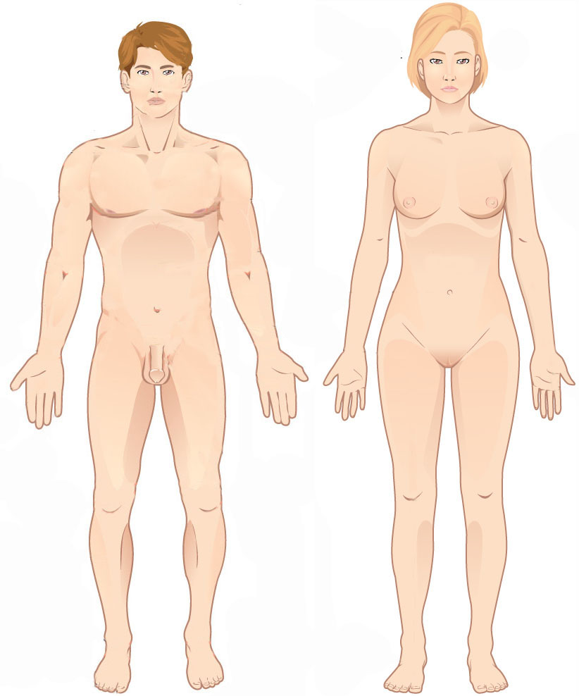
Standard anatomical terms of location are used to unambiguously describe the anatomy of animals, including humans. The terms, typically derived from Latin or Greek roots, describe something in its standard anatomical position. This position provides a definition of what is at the front ("anterior"), behind ("posterior") and so on. As part of defining and describing terms, the body is described through the use of anatomical planes and anatomical axes. The meaning of terms that are used can change depending on whether an organism is bipedal or quadrupedal. Additionally, for some animals such as invertebrates, some terms may not have any meaning at all; for example, an animal that is radially symmetrical will have no anterior surface, but can still have a description that a part is close to the middle ("proximal") or further from the middle ("distal"). International organisations have determined vocabularies that are often used as standard vocabularies for subdisciplines of anatom ... [...More Info...] [...Related Items...] OR: [Wikipedia] [Google] [Baidu] |
Radiography
Radiography is an imaging technique using X-rays, gamma rays, or similar ionizing radiation and non-ionizing radiation to view the internal form of an object. Applications of radiography include medical radiography ("diagnostic" and "therapeutic") and industrial radiography. Similar techniques are used in airport security (where "body scanners" generally use backscatter X-ray). To create an image in conventional radiography, a beam of X-rays is produced by an X-ray generator and is projected toward the object. A certain amount of the X-rays or other radiation is absorbed by the object, dependent on the object's density and structural composition. The X-rays that pass through the object are captured behind the object by a detector (either photographic film or a digital detector). The generation of flat two dimensional images by this technique is called projectional radiography. In computed tomography (CT scanning) an X-ray source and its associated detectors rotate around the su ... [...More Info...] [...Related Items...] OR: [Wikipedia] [Google] [Baidu] |
Knee Joint
In humans and other primates, the knee joins the thigh with the leg and consists of two joints: one between the femur and tibia (tibiofemoral joint), and one between the femur and patella (patellofemoral joint). It is the largest joint in the human body. The knee is a modified hinge joint, which permits flexion and extension as well as slight internal and external rotation. The knee is vulnerable to injury and to the development of osteoarthritis. It is often termed a ''compound joint'' having tibiofemoral and patellofemoral components. (The fibular collateral ligament is often considered with tibiofemoral components.) Structure The knee is a modified hinge joint, a type of synovial joint, which is composed of three functional compartments: the patellofemoral articulation, consisting of the patella, or "kneecap", and the patellar groove on the front of the femur through which it slides; and the medial and lateral tibiofemoral articulations linking the femur, or thigh bone, ... [...More Info...] [...Related Items...] OR: [Wikipedia] [Google] [Baidu] |
Anatomical Terms Of Location
Standard anatomical terms of location are used to unambiguously describe the anatomy of animals, including humans. The terms, typically derived from Latin or Greek roots, describe something in its standard anatomical position. This position provides a definition of what is at the front ("anterior"), behind ("posterior") and so on. As part of defining and describing terms, the body is described through the use of anatomical planes and anatomical axes. The meaning of terms that are used can change depending on whether an organism is bipedal or quadrupedal. Additionally, for some animals such as invertebrates, some terms may not have any meaning at all; for example, an animal that is radially symmetrical will have no anterior surface, but can still have a description that a part is close to the middle ("proximal") or further from the middle ("distal"). International organisations have determined vocabularies that are often used as standard vocabularies for subdisciplines of anatom ... [...More Info...] [...Related Items...] OR: [Wikipedia] [Google] [Baidu] |
Patella
The patella, also known as the kneecap, is a flat, rounded triangular bone which articulates with the femur (thigh bone) and covers and protects the anterior articular surface of the knee joint. The patella is found in many tetrapods, such as mice, cats, birds and dogs, but not in whales, or most reptiles. In humans, the patella is the largest sesamoid bone (i.e., embedded within a tendon or a muscle) in the body. Babies are born with a patella of soft cartilage which begins to ossify into bone at about four years of age. Structure The patella is a sesamoid bone roughly triangular in shape, with the apex of the patella facing downwards. The apex is the most inferior (lowest) part of the patella. It is pointed in shape, and gives attachment to the patellar ligament. The front and back surfaces are joined by a thin margin and towards centre by a thicker margin. The tendon of the quadriceps femoris muscle attaches to the base of the patella., with the vastus intermedius muscle ... [...More Info...] [...Related Items...] OR: [Wikipedia] [Google] [Baidu] |
Patellar Tendon Rupture
Patellar tendon rupture is a tear of the tendon that connects the knee cap (patella) to the tibia. Often there is sudden onset of pain and walking is difficult. In a complete rupture, the ability to extend that knee is decreased. A pop may be felt when it occurs. Injury to the patellar tendon generally requires a significant force such as falling directly on the knee or jumping from a height. Risk factors include patellar tendinitis, kidney failure, diabetes, and corticosteroid, steroid or fluoroquinolone use. There are two main types of ruptures: partial and complete. In most cases, the patellar tendon tears at the point where it attaches to the knee cap. Diagnosis is based on symptoms, physical examination, examination, and medical imaging. Small tears may be treated with rest and orthopedic splinting, splinting, followed by physiotherapy. Larger tears typically require surgery within a couple of weeks. Outcomes are generally good. Rates in the general population are not clear ... [...More Info...] [...Related Items...] OR: [Wikipedia] [Google] [Baidu] |


