|
Bile Canaliculi
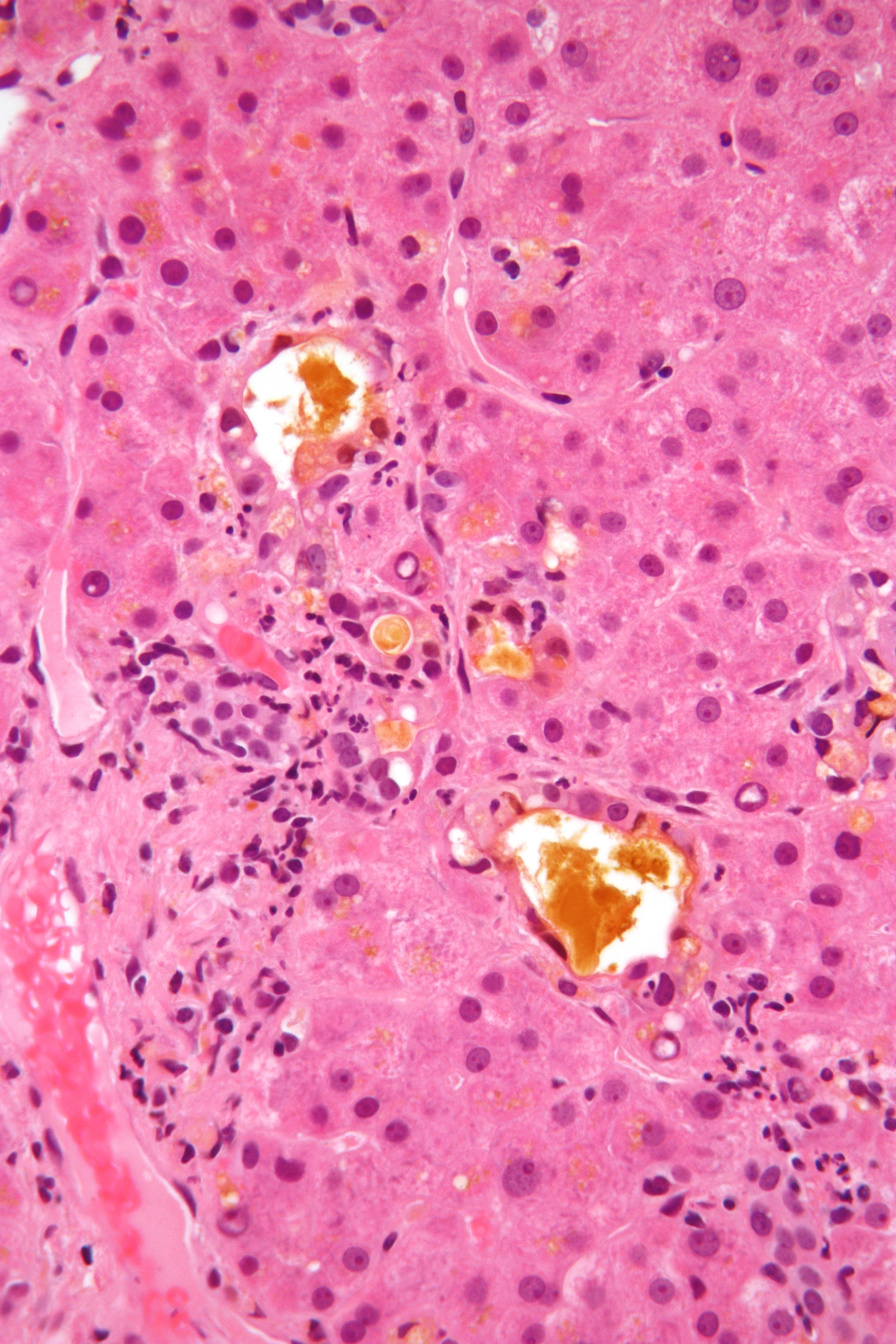
Bile canaliculus (plural:bile canaliculi; also called bile capillaries) is a thin tube that collects bile secreted by hepatocytes. The bile canaliculi empty into a series of progressively larger bile ductules and ducts, which eventually become common hepatic duct. The bile canaliculi empty directly into the Canals of Hering. Hepatocytes are polyhedral in shape, therefore having no set shape or design, although they are made of cuboidal epithelial cells. They have surfaces facing the sinusoids, (called sinusoidal faces) and surfaces which contact other hepatocytes, (called lateral faces). Bile canaliculi are formed by grooves on some of the lateral faces of these hepatocytes. Microvilli Microvilli (singular: microvillus) are microscopic cellular membrane protrusions that increase the surface area for diffusion and minimize any increase in volume, and are involved in a wide variety of functions, including absorption, secretion, ... are present in the canaliculi. External links ... [...More Info...] [...Related Items...] OR: [Wikipedia] [Google] [Baidu] |
Golgi's Method
Golgi's method is a silver staining technique that is used to visualize nervous tissue under light microscopy. The method was discovered by Camillo Golgi, an Italian physician and scientist, who published the first picture made with the technique in 1873. It was initially named the black reaction (''la reazione nera'') by Golgi, but it became better known as the Golgi stain or later, Golgi method. Golgi staining was used by Spanish neuroanatomist Santiago Ramón y Cajal (1852–1934) to discover a number of novel facts about the organization of the nervous system, inspiring the birth of the neuron doctrine. Ultimately, Ramón y Cajal improved the technique by using a method he termed "double impregnation". Ramón y Cajal's staining technique, still in use, is called Cajal's Stain. Mechanism The cells in nervous tissue are densely packed and little information on their structures and interconnections can be obtained if all the cells are stained. Furthermore, the thin filamentar ... [...More Info...] [...Related Items...] OR: [Wikipedia] [Google] [Baidu] |
Bile
Bile (from Latin ''bilis''), or gall, is a dark-green-to-yellowish-brown fluid produced by the liver of most vertebrates that aids the digestion of lipids in the small intestine. In humans, bile is produced continuously by the liver (liver bile) and stored and concentrated in the gallbladder. After eating, this stored bile is discharged into the duodenum. The composition of hepatic bile is (97–98)% water, 0.7% bile salts, 0.2% bilirubin, 0.51% fats (cholesterol, fatty acids, and lecithin), and 200 meq/L inorganic salts. The two main pigments of bile are bilirubin, which is yellow, and its oxidised form biliverdin, which is green. When mixed, they are responsible for the brown color of feces. About 400 to 800 millilitres of bile is produced per day in adult human beings. Function Bile or gall acts to some extent as a surfactant, helping to emulsify the lipids in food. Bile salt anions are hydrophilic on one side and hydrophobic on the other side; consequently, they tend t ... [...More Info...] [...Related Items...] OR: [Wikipedia] [Google] [Baidu] |
Hepatocytes
A hepatocyte is a cell of the main parenchymal tissue of the liver. Hepatocytes make up 80% of the liver's mass. These cells are involved in: * Protein synthesis * Protein storage * Transformation of carbohydrates * Synthesis of cholesterol, bile salts and phospholipids * Detoxification, modification, and excretion of exogenous and endogenous substances * Initiation of formation and secretion of bile Structure The typical hepatocyte is cubical with sides of 20-30 μm, (in comparison, a human hair has a diameter of 17 to 180 μm).The diameter of human hair ranges from 17 to 181 μm. The typical volume of a hepatocyte is 3.4 x 10⁻⁹ cm3. Smooth endoplasmic reticulum is abundant in hepatocytes, in contrast to most other cell types. Microanatomy Hepatocytes display an eosinophilic cytoplasm, reflecting numerous mitochondria, and basophilic stippling due to large amounts of smooth endoplasmic reticulum and free ribosomes. Brown lipofuscin granules are also observed (wi ... [...More Info...] [...Related Items...] OR: [Wikipedia] [Google] [Baidu] |
Common Hepatic Duct
The common hepatic duct is the first part of the biliary tract. It joins the cystic duct coming from the gallbladder to form the common bile duct. Structure The common hepatic duct is the first part of the biliary tract. It is formed by the convergence of the right hepatic duct (which drains bile from the right functional lobe of the liver) and the left hepatic duct (which drains bile from the left functional lobe of the liver). It then joins the cystic duct coming from the gallbladder to form the common bile duct. The duct is usually 6–8 cm long. The common hepatic duct is about 6 mm in diameter in adults, with some variation.Gray's Anatomy, 39th ed, p. 1228 The inner surface is covered in a simple columnar epithelium. Variation Around 1.7% of people have additional accessory hepatic ducts that join onto the common hepatic duct. Rarely, the common hepatic duct joins onto the gallbladder directly, leading to illness. Function The hepatic duct is part of the bi ... [...More Info...] [...Related Items...] OR: [Wikipedia] [Google] [Baidu] |
Canals Of Hering
The canals of Hering, or intrahepatic bile ductules, are part of the outflow system of exocrine bile product from the liver. Liver stem cells are located in the canals of Hering. Structure They are found between the bile canaliculi and interlobular bile ducts near the outer edge of a classic liver lobule. Histology Histologically, the cells of the ductule are described as simple cuboidal epithelium, lined partially by cholangiocytes and hepatocytes. They may not be readily visible but can be differentially stained by cytokeratins CK19 and CK7. Clinical relevance The canals of Hering are destroyed early in primary biliary cholangitis and may be primary sites of scarring in methotrexate toxicity. Research has indicated the presence of intraorgan stem cells of the liver that can proliferate in disease states, so-called oval cells. History They are named for Ewald Hering Karl Ewald Konstantin Hering (5 August 1834 – 26 January 1918) was a German physiologist who did m ... [...More Info...] [...Related Items...] OR: [Wikipedia] [Google] [Baidu] |
Polyhedra
In geometry, a polyhedron (plural polyhedra or polyhedrons; ) is a three-dimensional shape with flat polygonal faces, straight edges and sharp corners or vertices. A convex polyhedron is the convex hull of finitely many points, not all on the same plane. Cubes and pyramids are examples of convex polyhedra. A polyhedron is a 3-dimensional example of a polytope, a more general concept in any number of dimensions. Definition Convex polyhedra are well-defined, with several equivalent standard definitions. However, the formal mathematical definition of polyhedra that are not required to be convex has been problematic. Many definitions of "polyhedron" have been given within particular contexts,. some more rigorous than others, and there is not universal agreement over which of these to choose. Some of these definitions exclude shapes that have often been counted as polyhedra (such as the self-crossing polyhedra) or include shapes that are often not considered as valid pol ... [...More Info...] [...Related Items...] OR: [Wikipedia] [Google] [Baidu] |
Liver Sinusoid
A liver sinusoid is a type of capillary known as a sinusoidal capillary, discontinuous capillary or sinusoid, that is similar to a fenestrated capillary, having discontinuous endothelium that serves as a location for mixing of the oxygen-rich blood from the hepatic artery and the nutrient-rich blood from the portal vein. The liver sinusoid has a larger caliber than other types of capillaries and has a lining of specialised endothelial cells known as the liver sinusoidal endothelial cells (LSECs), and Kupffer cells. The cells are porous and have a scavenging function. The LSECs make up around half of the non-parenchymal cells in the liver and are flattened and fenestrated. LSECs have many fenestrae that gives easy communication between the sinusoidal lumen and the space of Disse. They play a part in filtration, endocytosis, and in the regulation of blood flow in the sinusoids. The Kupffer cells can take up and destroy foreign material such as bacteria. Hepatocytes are separate ... [...More Info...] [...Related Items...] OR: [Wikipedia] [Google] [Baidu] |
Microvilli
Microvilli (singular: microvillus) are microscopic cellular membrane protrusions that increase the surface area for diffusion and minimize any increase in volume, and are involved in a wide variety of functions, including absorption, secretion, cellular adhesion, and mechanotransduction. Structure Microvilli are covered in plasma membrane, which encloses cytoplasm and microfilaments. Though these are cellular extensions, there are little or no cellular organelles present in the microvilli. Each microvillus has a dense bundle of cross-linked actin filaments, which serves as its structural core. 20 to 30 tightly bundled actin filaments are cross-linked by bundling proteins fimbrin (or plastin-1), villin and espin to form the core of the microvilli. In the enterocyte microvillus, the structural core is attached to the plasma membrane along its length by lateral arms made of myosin 1a and Ca2+ binding protein calmodulin. Myosin 1a functions through a binding site for filamento ... [...More Info...] [...Related Items...] OR: [Wikipedia] [Google] [Baidu] |

