|
Biceps Brachii
The biceps or biceps brachii ( la, musculus biceps brachii, "two-headed muscle of the arm") is a large muscle that lies on the front of the upper arm between the shoulder and the elbow. Both heads of the muscle arise on the scapula and join to form a single muscle belly which is attached to the upper forearm. While the biceps crosses both the shoulder and elbow joints, its main function is at the elbow where it flexes the forearm and supinates the forearm. Both these movements are used when opening a bottle with a corkscrew: first biceps screws in the cork (supination), then it pulls the cork out (flexion). Structure The biceps is one of three muscles in the anterior compartment of the upper arm, along with the brachialis muscle and the coracobrachialis muscle, with which the biceps shares a nerve supply. The biceps muscle has two heads, the short head and the long head, distinguished according to their origin at the coracoid process and supraglenoid tubercle of the sca ... [...More Info...] [...Related Items...] OR: [Wikipedia] [Google] [Baidu] |
Coracoid Process
The coracoid process (from Greek κόραξ, raven) is a small hook-like structure on the lateral edge of the superior anterior portion of the scapula (hence: coracoid, or "like a raven's beak"). Pointing laterally forward, it, together with the acromion, serves to stabilize the shoulder joint. It is palpable in the deltopectoral groove between the deltoid and pectoralis major muscles. Structure The coracoid process is a thick curved process attached by a broad base to the upper part of the neck of the scapula; it runs at first upward and medialward; then, becoming smaller, it changes its direction, and projects forward and lateralward. Anatomically it is divided into intervals of: base of coracoid process, angle of coracoid process, shaft and the apex of the coracoid process. The coracoglenoid notch is an indentation localized between the coracoid process and the glenoid. As the coracoid process projects laterally, it house underneath it the subcoracoid space. The ''ascend ... [...More Info...] [...Related Items...] OR: [Wikipedia] [Google] [Baidu] |
Muscle
Skeletal muscles (commonly referred to as muscles) are organs of the vertebrate muscular system and typically are attached by tendons to bones of a skeleton. The muscle cells of skeletal muscles are much longer than in the other types of muscle tissue, and are often known as muscle fibers. The muscle tissue of a skeletal muscle is striated – having a striped appearance due to the arrangement of the sarcomeres. Skeletal muscles are voluntary muscles under the control of the somatic nervous system. The other types of muscle are cardiac muscle which is also striated and smooth muscle which is non-striated; both of these types of muscle tissue are classified as involuntary, or, under the control of the autonomic nervous system. A skeletal muscle contains multiple fascicles – bundles of muscle fibers. Each individual fiber, and each muscle is surrounded by a type of connective tissue layer of fascia. Muscle fibers are formed from the fusion of developmental myoblasts in ... [...More Info...] [...Related Items...] OR: [Wikipedia] [Google] [Baidu] |
Anatomical Terms Of Location
Standard anatomical terms of location are used to unambiguously describe the anatomy of animals, including humans. The terms, typically derived from Latin or Greek roots, describe something in its standard anatomical position. This position provides a definition of what is at the front ("anterior"), behind ("posterior") and so on. As part of defining and describing terms, the body is described through the use of anatomical planes and anatomical axes. The meaning of terms that are used can change depending on whether an organism is bipedal or quadrupedal. Additionally, for some animals such as invertebrates, some terms may not have any meaning at all; for example, an animal that is radially symmetrical will have no anterior surface, but can still have a description that a part is close to the middle ("proximal") or further from the middle ("distal"). International organisations have determined vocabularies that are often used as standard vocabularies for subdisciplines of anatom ... [...More Info...] [...Related Items...] OR: [Wikipedia] [Google] [Baidu] |
Deltoid Muscle
The deltoid muscle is the muscle forming the rounded contour of the human shoulder. It is also known as the 'common shoulder muscle', particularly in other animals such as the domestic cat. Anatomically, the deltoid muscle appears to be made up of three distinct sets of muscle fibers, namely the # anterior or clavicular part (pars clavicularis) # posterior or scapular part (pars scapularis) # intermediate or acromial part (pars acromialis) However, electromyography suggests that it consists of at least seven groups that can be independently coordinated by the nervous system. It was previously called the deltoideus (plural ''deltoidei'') and the name is still used by some anatomists. It is called so because it is in the shape of the Greek capital letter delta (Δ). Deltoid is also further shortened in slang as "delt". A study of 30 shoulders revealed an average mass of in humans, ranging from to . Structure Previous studies showed that the insertions of the tendons of the delto ... [...More Info...] [...Related Items...] OR: [Wikipedia] [Google] [Baidu] |
Coracobrachialis
The coracobrachialis muscle is the smallest of the three muscles that attach to the coracoid process of the scapula. (The other two muscles are pectoralis minor and the short head of the biceps brachii.) It is situated at the upper and medial part of the arm. Structure Coracobrachialis muscle arises from the apex of the coracoid process, in common with the short head of the biceps brachii, and from the intermuscular septum between the two muscles. It is inserted by means of a flat tendon into an impression at the middle of the medial surface and border of the body of the humerus (shaft of the humerus) between the origins of the triceps brachii and brachialis. Innervation Coracobrachialis muscle is perforated by and innervated by the musculocutaneous nerve, which arises from the anterior division of the upper trunk ( C5, C6) and middle trunk ( C7) of the brachial plexus. Development Variation Function The action of the coracobrachialis is to flex and adduct the arm at the ... [...More Info...] [...Related Items...] OR: [Wikipedia] [Google] [Baidu] |
Humerus
The humerus (; ) is a long bone in the arm that runs from the shoulder to the elbow. It connects the scapula and the two bones of the lower arm, the radius and ulna, and consists of three sections. The humeral upper extremity consists of a rounded head, a narrow neck, and two short processes (tubercles, sometimes called tuberosities). The body is cylindrical in its upper portion, and more prismatic below. The lower extremity consists of 2 epicondyles, 2 processes (trochlea & capitulum), and 3 fossae (radial fossa, coronoid fossa, and olecranon fossa). As well as its true anatomical neck, the constriction below the greater and lesser tubercles of the humerus is referred to as its surgical neck due to its tendency to fracture, thus often becoming the focus of surgeons. Etymology The word "humerus" is derived from la, humerus, umerus meaning upper arm, shoulder, and is linguistically related to Gothic ''ams'' shoulder and Greek ''ōmos''. Structure Upper extremity The upper or pr ... [...More Info...] [...Related Items...] OR: [Wikipedia] [Google] [Baidu] |
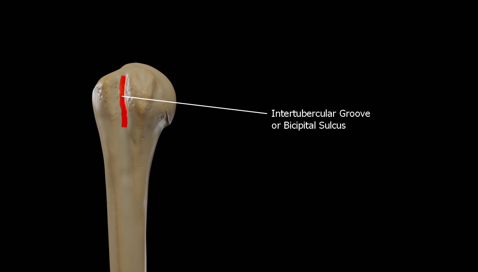
Intertubercular Groove
The bicipital groove (intertubercular groove, sulcus intertubercularis) is a deep groove on the humerus that separates the greater tubercle from the lesser tubercle. It allows for the long tendon of the biceps brachii muscle to pass. Structure The bicipital groove separates the greater tubercle from the lesser tubercle. It is usually around 8 cm long and 1 cm wide in adults. It lodges the long tendon of the biceps brachii muscle between the tendon of the pectoralis major muscle on the lateral lip and the tendon of the teres major muscle on the medial lip. It also transmits a branch of the anterior humeral circumflex artery to the shoulder joint. The insertion of the latissimus dorsi muscle is found along the floor of the bicipital groove. The teres major muscle inserts on the medial lip of the groove. It runs obliquely downward, and ends near the junction of the upper with the middle third of the bone. It is the lateral wall of the axilla. Function The bicipital groove al ... [...More Info...] [...Related Items...] OR: [Wikipedia] [Google] [Baidu] |
Coracobrachialis Muscle
The coracobrachialis muscle is the smallest of the three muscles that attach to the coracoid process of the scapula. (The other two muscles are pectoralis minor and the short head of the biceps brachii.) It is situated at the upper and medial part of the arm. Structure Coracobrachialis muscle arises from the apex of the coracoid process, in common with the short head of the biceps brachii, and from the intermuscular septum between the two muscles. It is inserted by means of a flat tendon into an impression at the middle of the medial surface and border of the body of the humerus (shaft of the humerus) between the origins of the triceps brachii and brachialis. Innervation Coracobrachialis muscle is perforated by and innervated by the musculocutaneous nerve, which arises from the anterior division of the upper trunk ( C5, C6) and middle trunk ( C7) of the brachial plexus. Development Variation Function The action of the coracobrachialis is to flex and adduct the arm at the ... [...More Info...] [...Related Items...] OR: [Wikipedia] [Google] [Baidu] |
Brachialis Muscle
The brachialis (brachialis anticus), also known as the Teichmann muscle, is a muscle in the upper arm that flexes the elbow. It lies deeper than the biceps brachii, and makes up part of the floor of the region known as the cubital fossa (elbow pit). The brachialis is the prime mover of elbow flexion generating about 50% more power than the biceps.Saladin, Kenneth S, Stephen J. Sullivan, and Christina A. Gan. Anatomy & Physiology: The Unity of Form and Function. 2015. Print. Structure The brachialis originates from the anterior surface of the distal half of the humerus, near the insertion of the deltoid muscle, which it embraces by two angular processes. Its origin extends below to within 2.5 cm of the margin of the articular surface of the humerus at the elbow joint. Its fibers converge to a thick tendon, which is inserted into the tuberosity of the ulna and the rough depression on the anterior surface of the coronoid process of the ulna. Blood supply The brachialis is suppl ... [...More Info...] [...Related Items...] OR: [Wikipedia] [Google] [Baidu] |
Fascial Compartments Of Arm
The fascial compartments of arm refers to the specific anatomical term of the compartments within the upper segment of the upper limb (the arm) of the body. The upper limb is divided into two segments, the arm and the forearm. Each of these segments is further divided into two compartments which are formed by deep fascia – tough connective tissue septa (walls). Each compartment encloses specific muscles and nerves. The compartments of the arm are the anterior compartment of the arm and the posterior compartment of the arm, divided by the lateral and the medial intermuscular septa. The compartments of the forearm are the anterior compartment of the forearm and posterior compartment of the forearm. Intermuscular septa The lateral intermuscular septum extends from the lower part of the crest of the greater tubercle of the humerus, along the lateral supracondylar ridge, to the lateral epicondyle; it is blended with the tendon of the deltoid muscle, gives attachment to the tricep ... [...More Info...] [...Related Items...] OR: [Wikipedia] [Google] [Baidu] |
Biceps Brachii Muscle06
The biceps or biceps brachii ( la, musculus biceps brachii, "two-headed muscle of the arm") is a large muscle that lies on the front of the upper arm between the shoulder and the elbow. Both heads of the muscle arise on the scapula and join to form a single muscle belly which is attached to the upper forearm. While the biceps crosses both the shoulder and elbow joints, its main function is at the elbow where it flexes the forearm and supinates the forearm. Both these movements are used when opening a bottle with a corkscrew: first biceps screws in the cork (supination), then it pulls the cork out (flexion). Structure The biceps is one of three muscles in the anterior compartment of the upper arm, along with the brachialis muscle and the coracobrachialis muscle, with which the biceps shares a nerve supply. The biceps muscle has two heads, the short head and the long head, distinguished according to their origin at the coracoid process and supraglenoid tubercle of the sca ... [...More Info...] [...Related Items...] OR: [Wikipedia] [Google] [Baidu] |
Flexion
Motion, the process of movement, is described using specific anatomical terms. Motion includes movement of organs, joints, limbs, and specific sections of the body. The terminology used describes this motion according to its direction relative to the anatomical position of the body parts involved. Anatomists and others use a unified set of terms to describe most of the movements, although other, more specialized terms are necessary for describing unique movements such as those of the hands, feet, and eyes. In general, motion is classified according to the anatomical plane it occurs in. ''Flexion'' and ''extension'' are examples of ''angular'' motions, in which two axes of a joint are brought closer together or moved further apart. ''Rotational'' motion may occur at other joints, for example the shoulder, and are described as ''internal'' or ''external''. Other terms, such as ''elevation'' and ''depression'', describe movement above or below the horizontal plane. Many anatomica ... [...More Info...] [...Related Items...] OR: [Wikipedia] [Google] [Baidu] |


