|
Baffle (medical)
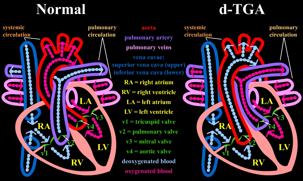
A baffle is a surgically created tunnel or wall within the heart or major blood vessels used to redirect the flow of blood. They are used in some types of heart abnormalities that a child is born with known as congenital heart defects. Baffles are usually constructed, at least in part, from a person's own heart tissue, while other methods of redirecting blood using artificial material are known by the more generic term 'conduits'. Baffle does not refer to surgical techniques that redirect blood outside the heart or blood vessels such as coronary artery bypass grafting. Baffles can be made between different structures depending on the heart condition that needs to be treated. The Mustard and Senning 'atrial switch' procedures use a baffle within the atria, redirecting blood from the superior and inferior vena cava to the left ventricle and blood from the pulmonary veins to the right ventricle, to treat transposition of the great arteries. The lateral tunnel form of the Fontan ... [...More Info...] [...Related Items...] OR: [Wikipedia] [Google] [Baidu] |
Heart
The heart is a muscular organ in most animals. This organ pumps blood through the blood vessels of the circulatory system. The pumped blood carries oxygen and nutrients to the body, while carrying metabolic waste such as carbon dioxide to the lungs. In humans, the heart is approximately the size of a closed fist and is located between the lungs, in the middle compartment of the chest. In humans, other mammals, and birds, the heart is divided into four chambers: upper left and right atria and lower left and right ventricles. Commonly the right atrium and ventricle are referred together as the right heart and their left counterparts as the left heart. Fish, in contrast, have two chambers, an atrium and a ventricle, while most reptiles have three chambers. In a healthy heart blood flows one way through the heart due to heart valves, which prevent backflow. The heart is enclosed in a protective sac, the pericardium, which also contains a small amount of fluid. The wall ... [...More Info...] [...Related Items...] OR: [Wikipedia] [Google] [Baidu] |
Great Vessels
Great vessels are the large vessels that bring blood to and from the heart. These are: *Superior vena cava *Inferior vena cava *Pulmonary arteries *Pulmonary veins *Aorta Transposition of the great vessels is a group of congenital heart defects A congenital heart defect (CHD), also known as a congenital heart anomaly and congenital heart disease, is a defect in the structure of the heart or great vessels that is present at birth. A congenital heart defect is classed as a cardiovascular ... involving an abnormal spatial arrangement of any of the great vessels. References Angiology {{Anatomy-stub ... [...More Info...] [...Related Items...] OR: [Wikipedia] [Google] [Baidu] |
Congenital Heart Defect
A congenital heart defect (CHD), also known as a congenital heart anomaly and congenital heart disease, is a defect in the structure of the heart or great vessels that is present at birth. A congenital heart defect is classed as a cardiovascular disease. Signs and symptoms depend on the specific type of defect. Symptoms can vary from none to life-threatening. When present, symptoms may include rapid breathing, bluish skin (cyanosis), poor weight gain, and feeling tired. CHD does not cause chest pain. Most congenital heart defects are not associated with other diseases. A complication of CHD is heart failure. The cause of a congenital heart defect is often unknown. Risk factors include certain infections during pregnancy such as rubella, use of certain medications or drugs such as alcohol or tobacco, parents being closely related, or poor nutritional status or obesity in the mother. Having a parent with a congenital heart defect is also a risk factor. A number of genetic conditio ... [...More Info...] [...Related Items...] OR: [Wikipedia] [Google] [Baidu] |
Coronary Artery Bypass Surgery
Coronary artery bypass surgery, also known as coronary artery bypass graft (CABG, pronounced "cabbage") is a surgical procedure to treat coronary artery disease (CAD), the buildup of plaques in the arteries of the heart. It can relieve chest pain caused by CAD, slow the progression of CAD, and increase life expectancy. It aims to bypass narrowings in heart arteries by using arteries or veins harvested from other parts of the body, thus restoring adequate blood supply to the previously ischemic (deprived of blood) heart. There are two main approaches. The first uses a cardiopulmonary bypass machine, a machine which takes over the functions of the heart and lungs during surgery by circulating blood and oxygen. With the heart in arrest, harvested arteries and veins are used to connect across problematic regions—a construction known as surgical anastomosis. In the second approach, called the off-pump coronary artery bypass graft (OPCABG), these anastomoses are constructed while t ... [...More Info...] [...Related Items...] OR: [Wikipedia] [Google] [Baidu] |
Mustard Procedure
The Mustard procedure was developed in 1963 by Dr. William Mustard at the Hospital for Sick Children in Toronto, Ontario, Canada. Dr. Mustard, with support from the Heart and Stroke Foundation of Canada, developed an alternative and simplified technique to the Senning procedure which was used to correct a congenital heart defect that produced “ blue babies”. The technique was adopted by other surgeons and became the standard operation for d-TGA. In his autobiography, South African cardiac surgeon Christiaan Barnard claims to have been the first to perform the operation, with Mustard only following 'several years later'. Background The defect is called transposition of the great vessels, or transposition of the great arteries (TGV or TGA). Until the late 1950s, and Senning's operation, the condition was commonly fatal. The defect causes blood from the lungs to flow back to the lungs and blood from the body to flow back to the body. This occurs because the aorta and the pul ... [...More Info...] [...Related Items...] OR: [Wikipedia] [Google] [Baidu] |
Senning Procedure
The Senning procedure is an atrial switch heart operation performed to treat transposition of the great arteries. It is named after its inventor, the Swedish cardiac surgeon Åke Senning (1915–2000), also known for implanting the first permanent cardiac pacemaker in 1958. Brief History This procedure, a form of atrial switch, was developed and first performed by Senning in 1957 as a treatment for d-TGA (dextro-Transposition of the great arteries) before improvements in cardiopulmonary bypass made more curative surgical techniques feasible.Atrial Switch Operation: Past, Present, and Future. Konstantinov IE, Alexi-Meskishvili VV, Williams WG et al.Ann Thorac Surg 2004;77:2250 – 8 In this congenital heart defect, the venous circulation drains into the right ventricle but from this chamber, blood is directed towards the systemic circulation through the aorta. This is also expressed by the term ventriculoarterial discordance, that is the ventricles are connected to the wrong great ... [...More Info...] [...Related Items...] OR: [Wikipedia] [Google] [Baidu] |
Atrium (heart)
The atrium ( la, ātrium, , entry hall) is one of two upper chambers in the heart that receives blood from the circulatory system. The blood in the atria is pumped into the heart ventricles through the atrioventricular valves. There are two atria in the human heart – the left atrium receives blood from the pulmonary circulation, and the right atrium receives blood from the venae cavae of the systemic circulation. During the cardiac cycle the atria receive blood while relaxed in diastole, then contract in systole to move blood to the ventricles. Each atrium is roughly cube-shaped except for an ear-shaped projection called an atrial appendage, sometimes known as an auricle. All animals with a closed circulatory system have at least one atrium. The atrium was formerly called the 'auricle'. That term is still used to describe this chamber in some other animals, such as the ''Mollusca''. They have thicker muscular walls than the atria do. Structure Humans have a four-chambered ... [...More Info...] [...Related Items...] OR: [Wikipedia] [Google] [Baidu] |
Venae Cavae
In anatomy, the venae cavae (; singular: vena cava ; ) are two large veins (great vessels) that return deoxygenated blood from the body into the heart. In humans they are the superior vena cava and the inferior vena cava, and both empty into the right atrium. They are located slightly off-center, toward the right side of the body. The right atrium receives deoxygenated blood through coronary sinus and two large veins called venae cavae. The inferior vena cava (or caudal vena cava in some animals) travels up alongside the abdominal aorta with blood from the lower part of the body. It is the largest vein in the human body. MadSci Network: Anatomy. Retrieved 19 September 2013. The superior vena cava (or cranial vena cava in animals) is above the heart, and ... [...More Info...] [...Related Items...] OR: [Wikipedia] [Google] [Baidu] |
Ventricle (heart)
A ventricle is one of two large chambers toward the bottom of the heart that collect and expel blood towards the peripheral beds within the body and lungs. The blood pumped by a ventricle is supplied by an atrium, an adjacent chamber in the upper heart that is smaller than a ventricle. Interventricular means between the ventricles (for example the interventricular septum), while intraventricular means within one ventricle (for example an intraventricular block). In a four-chambered heart, such as that in humans, there are two ventricles that operate in a double circulatory system: the right ventricle pumps blood into the pulmonary circulation to the lungs, and the left ventricle pumps blood into the systemic circulation through the aorta. Structure Ventricles have thicker walls than atria and generate higher blood pressures. The physiological load on the ventricles requiring pumping of blood throughout the body and lungs is much greater than the pressure generated by the atria ... [...More Info...] [...Related Items...] OR: [Wikipedia] [Google] [Baidu] |
Pulmonary Vein
The pulmonary veins are the veins that transfer oxygenated blood from the lungs to the heart. The largest pulmonary veins are the four ''main pulmonary veins'', two from each lung that drain into the left atrium of the heart. The pulmonary veins are part of the pulmonary circulation. Structure There are four main pulmonary veins, two from each lung – an inferior and a superior main vein, emerging from each hilum. The main pulmonary veins receive blood from three or four feeding veins in each lung, and drain into the left atrium. The peripheral feeding veins do not follow the bronchial tree. They run between the pulmonary segments from which they drain the blood. At the root of the lung, the right superior pulmonary vein lies in front of and a little below the pulmonary artery; the inferior is situated at the lowest part of the lung hilum. Behind the pulmonary artery is the bronchus. The right main pulmonary veins (contains oxygenated blood) pass behind the right atrium and ... [...More Info...] [...Related Items...] OR: [Wikipedia] [Google] [Baidu] |
Transposition Of The Great Vessels
Transposition of the great vessels (TGV) is a group of congenital heart defects involving an abnormal spatial arrangement of any of the great vessels: superior and/or inferior venae cavae, pulmonary artery, pulmonary veins, and aorta. Congenital heart diseases involving only the primary arteries (pulmonary artery and aorta) belong to a sub-group called transposition of the great arteries (TGA), which is considered the most common congenital heart lesion that presents in neonates. Types Transposed vessels can present with atriovenous, ventriculoarterial and/or arteriovenous discordance. The effects may range from a slight change in blood pressure to an interruption in circulation depending on the nature and degree of the misplacement, and on which specific vessels are involved. Although "transposed" literally means "swapped", many types of TGV involve vessels that are in abnormal positions, while not actually being swapped with each other. The terms TGV and TGA are most c ... [...More Info...] [...Related Items...] OR: [Wikipedia] [Google] [Baidu] |
Fontan Procedure
The Fontan procedure or Fontan–Kreutzer procedure is a palliative surgical procedure used in children with univentricular hearts. It involves diverting the venous blood from the inferior vena cava (IVC) and superior vena cava (SVC) to the pulmonary arteries without passing through the morphologic right ventricle; i.e., the systemic and pulmonary circulations are placed in series with the functional single ventricle. The procedure was initially performed in 1968 by Francis Fontan and Eugene Baudet from Bordeaux, France, published in 1971, simultaneously described in 1971 by Guillermo Kreutzer from Buenos Aires, Argentina, and finally published in 1973. Indications The Fontan Kreutzer procedure is used in pediatric patients who possess only a single functional ventricle, either due to lack of a heart valve (e.g. tricuspid or mitral atresia), an abnormality of the pumping ability of the heart (e.g. hypoplastic left heart syndrome or hypoplastic right heart syndrome), or a complex ... [...More Info...] [...Related Items...] OR: [Wikipedia] [Google] [Baidu] |


