|
Burst Suppression
Burst suppression is an electroencephalography (EEG) pattern that is characterized by periods of high-voltage electrical activity alternating with periods of no activity in the brain. The pattern is found in patients with inactivated brain states, such as from general anesthesia, coma, or hypothermia. This pattern can be physiological, as during early development, or pathological, as in diseases such as Ohtahara syndrome. History The burst suppression pattern was first observed by Derbyshire et al. while studying effects of anesthetics on feline cerebral cortices in 1936, where the researchers noticed mixed slow and fast electrical activity with decreasing amplitude as anesthesia deepened. In 1948, Swank and Watson coined the term "burst-suppression pattern" to describe the alternation of spikes and flatlines in electrical activity in deep anesthesia. It wasn't until after the early 1960s that the burst suppression pattern began being used in medical settings; it had been primaril ... [...More Info...] [...Related Items...] OR: [Wikipedia] [Google] [Baidu] |
Ion Transporter
In biology, a transporter is a transmembrane protein that moves ions (or other small molecules) across a biological membrane to accomplish many different biological functions including, cellular communication, maintaining homeostasis, energy production, etc. There are different types of transporters including, pumps, uniporters, antiporters, and symporters. Active transporters or ion pumps are transporters that convert energy from various sources—including adenosine triphosphate (ATP), sunlight, and other redox reactions—to potential energy by pumping an ion up its concentration gradient. This potential energy could then be used by secondary transporters, including ion carriers and ion channels, to drive vital cellular processes, such as ATP synthesis. This page is focused mainly on ion transporters acting as pumps, but transporters can also function to move molecules through facilitated diffusion. Facilitated diffusion does not require ATP and allows molecules, that are unable ... [...More Info...] [...Related Items...] OR: [Wikipedia] [Google] [Baidu] |
Maryam Shanechi
Maryam M. Shanechi is an Iran-born American neuroengineer. She studies ways of decoding the brain's activity to control brain-machine interfaces. She was honored as one of MIT Technology Review's Innovators under 35 in 2014 and one of the Science News 10 scientists to watch in 2019. She is Professor and Viterbi Early Career Chair in Electrical and Computer Engineering at the Viterbi School of Engineering, and a member of the Neuroscience Graduate Program at the University of Southern California. Early life and career Shanechi was born in Iran and moved to Canada with her family when she was 16. She received her bachelor's degree in engineering from the University of Toronto in 2004. She then moved to MIT, where she completed her master's degree in electrical engineering and computer science in 2006 and her PhD in 2011. She completed a postdoc at Harvard Medical School before moving to the University of California, Berkeley, in 2012. She held a faculty position at Cornell Univ ... [...More Info...] [...Related Items...] OR: [Wikipedia] [Google] [Baidu] |
Delta Wave
Delta waves are high amplitude neural oscillations with a frequency between 0.5 and 4 hertz. Delta waves, like other brain waves, can be recorded with electroencephalography (EEG) and are usually associated with the deep stage 3 of NREM sleep, also known as slow-wave sleep (SWS), and aid in characterizing the depth of sleep. Suppression of delta waves leads to inability of body rejuvenation, brain revitalization and poor sleep. Background and history "Delta waves" were first described in the 1930s by W. Grey Walter, who improved upon Hans Berger's electroencephalograph machine (EEG) to detect alpha and delta waves. Delta waves can be quantified using quantitative electroencephalography. Classification and features Delta waves, like all brain waves, can be detected by electroencephalography (EEG). Delta waves were originally defined as having a frequency between 1 and 4 Hz, although more recent classifications put the boundaries at between 0.5 and 2 Hz. They are the s ... [...More Info...] [...Related Items...] OR: [Wikipedia] [Google] [Baidu] |
Isothalamus
The isothalamus is a division used by some researchers in describing the thalamus. The isothalamus constitutes 90% or more of the thalamus, and despite the variety of functions it serves, follows a simple organizational scheme. The constituting neurons belong to two different neuronal genera. The first correspond to the ''thalamocortical neurons'' (or principal). They have a "tufted" (or radiate) morphology, as their dendritic arborisation is made up of straight dendritic distal branches starting from short and thick stems. The number of branches and the diameter of the arborisation are linked to the specific system of which they are a part of, and to the animal species. They have the rather rare property of having no initial axonal collaterals, which implies that one emitting thalamocortical neuron does not send information to its neighbor. They send long-range glutamatergic projections to the cerebral cortex where they end electively at the layer IV (or around) level. The other ... [...More Info...] [...Related Items...] OR: [Wikipedia] [Google] [Baidu] |
Potassium Channel
Potassium channels are the most widely distributed type of ion channel found in virtually all organisms. They form potassium-selective pores that span cell membranes. Potassium channels are found in most cell types and control a wide variety of cell functions. Function Potassium channels function to conduct potassium ions down their electrochemical gradient, doing so both rapidly (up to the diffusion rate of K+ ions in bulk water) and selectively (excluding, most notably, sodium despite the sub-angstrom difference in ionic radius). Biologically, these channels act to set or reset the resting potential in many cells. In excitable cells, such as neurons, the delayed counterflow of potassium ions shapes the action potential. By contributing to the regulation of the cardiac action potential duration in cardiac muscle, malfunction of potassium channels may cause life-threatening arrhythmias. Potassium channels may also be involved in maintaining vascular tone. They also regulate ce ... [...More Info...] [...Related Items...] OR: [Wikipedia] [Google] [Baidu] |
Adenosine Triphosphate
Adenosine triphosphate (ATP) is an organic compound that provides energy to drive many processes in living cells, such as muscle contraction, nerve impulse propagation, condensate dissolution, and chemical synthesis. Found in all known forms of life, ATP is often referred to as the "molecular unit of currency" of intracellular energy transfer. When consumed in metabolic processes, it converts either to adenosine diphosphate (ADP) or to adenosine monophosphate (AMP). Other processes regenerate ATP. The human body recycles its own body weight equivalent in ATP each day. It is also a precursor to DNA and RNA, and is used as a coenzyme. From the perspective of biochemistry, ATP is classified as a nucleoside triphosphate, which indicates that it consists of three components: a nitrogenous base (adenine), the sugar ribose, and the Polyphosphate, triphosphate. Structure ATP consists of an adenine attached by the 9-nitrogen atom to the 1′ carbon atom of a sugar (ribose), which i ... [...More Info...] [...Related Items...] OR: [Wikipedia] [Google] [Baidu] |
Isoflurane
Isoflurane, sold under the brand name Forane among others, is a general anesthetic. It can be used to start or maintain anesthesia; however, other medications are often used to start anesthesia rather than isoflurane, due to airway irritation with isoflurane. Isoflurane is given via inhalation. Side effects of isoflurane include a decreased ability to breathe (respiratory depression), low blood pressure, and an irregular heartbeat. Serious side effects can include malignant hyperthermia or high blood potassium. It should not be used in patients with a history of malignant hyperthermia in either themselves or their family members. It is unknown if its use during pregnancy is safe for the fetus, but use during a cesarean section appears to be safe. Isoflurane is a halogenated ether. Isoflurane was approved for medical use in the United States in 1979. It is on the World Health Organization's List of Essential Medicines. Medical uses Isoflurane is always administered in con ... [...More Info...] [...Related Items...] OR: [Wikipedia] [Google] [Baidu] |
Muscimol
Muscimol (also known as agarin or pantherine) is one of the principal psychoactive constituents of ''Amanita muscaria'' and related species of mushroom. Muscimol is a potent and selective orthosteric agonist for the GABAA receptors and displays sedative-hypnotic, depressant and hallucinogenic psychoactivity. This colorless or white solid is classified as an isoxazole. Muscimol went under clinical trial phase I for epilepsy, but the trial was discontinued. Biochemistry Muscimol is one of the psychoactive compounds responsible for the effects of ''Amanita muscaria'' intoxication. Ibotenic acid, a neurotoxic secondary metabolite of ''Amanita muscaria'', serves as a prodrug to muscimol when the mushroom is ingested or dried, converting to muscimol via decarboxylation. Muscimol is produced in the mushrooms ''Amanita muscaria'' (fly agaric) and ''Amanita pantherina'', along with muscarine (which is present in trace amounts and it is not active), muscazone, and ibotenic acid. ''A ... [...More Info...] [...Related Items...] OR: [Wikipedia] [Google] [Baidu] |
Agonist
An agonist is a chemical that activates a receptor to produce a biological response. Receptors are cellular proteins whose activation causes the cell to modify what it is currently doing. In contrast, an antagonist blocks the action of the agonist, while an inverse agonist causes an action opposite to that of the agonist. Etymology From the Greek αγωνιστής (agōnistēs), contestant; champion; rival < αγων (agōn), contest, combat; exertion, struggle < αγω (agō), I lead, lead towards, conduct; drive Types of agonists can be activated by either endogenous agonists (such as |
GABA Receptor
The GABA receptors are a class of receptors that respond to the neurotransmitter gamma-aminobutyric acid (GABA), the chief inhibitory compound in the mature vertebrate central nervous system. There are two classes of GABA receptors: GABAA and GABAB. GABAA receptors are ligand-gated ion channels (also known as ionotropic receptors); whereas GABAB receptors are G protein-coupled receptors, also called metabotropic receptors. Ligand-gated ion channels GABAA receptor It has long been recognized that the fast response of neurons to GABA that is stimulated by bicuculline and picrotoxin is due to direct activation of an anion channel. This channel was subsequently termed the GABAA receptor. Fast-responding GABA receptors are members of a family of Cys-loop ligand-gated ion channels. Members of this superfamily, which includes nicotinic acetylcholine receptors, GABAA receptors, glycine and 5-HT3 receptors, possess a characteristic loop formed by a disulfide bond between two cys ... [...More Info...] [...Related Items...] OR: [Wikipedia] [Google] [Baidu] |
Membrane Potential
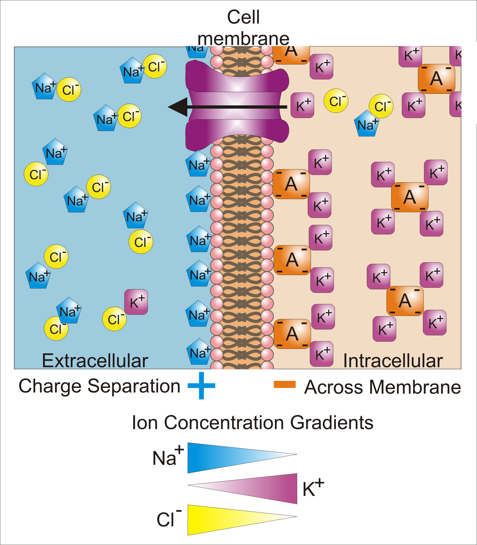
Membrane potential (also transmembrane potential or membrane voltage) is the difference in electric potential between the interior and the exterior of a biological cell. That is, there is a difference in the energy required for electric charges to move from the internal to exterior cellular environments and vice versa, as long as there is no acquisition of kinetic energy or the production of radiation. The concentration gradients of the charges directly determine this energy requirement. For the exterior of the cell, typical values of membrane potential, normally given in units of milli volts and denoted as mV, range from –80 mV to –40 mV. All animal cells are surrounded by a membrane composed of a lipid bilayer with proteins embedded in it. The membrane serves as both an insulator and a diffusion barrier to the movement of ions. Transmembrane proteins, also known as ion transporter or ion pump proteins, actively push ions across the membrane and establish concentration gradi ... [...More Info...] [...Related Items...] OR: [Wikipedia] [Google] [Baidu] |


