|
Bulbus Cordis
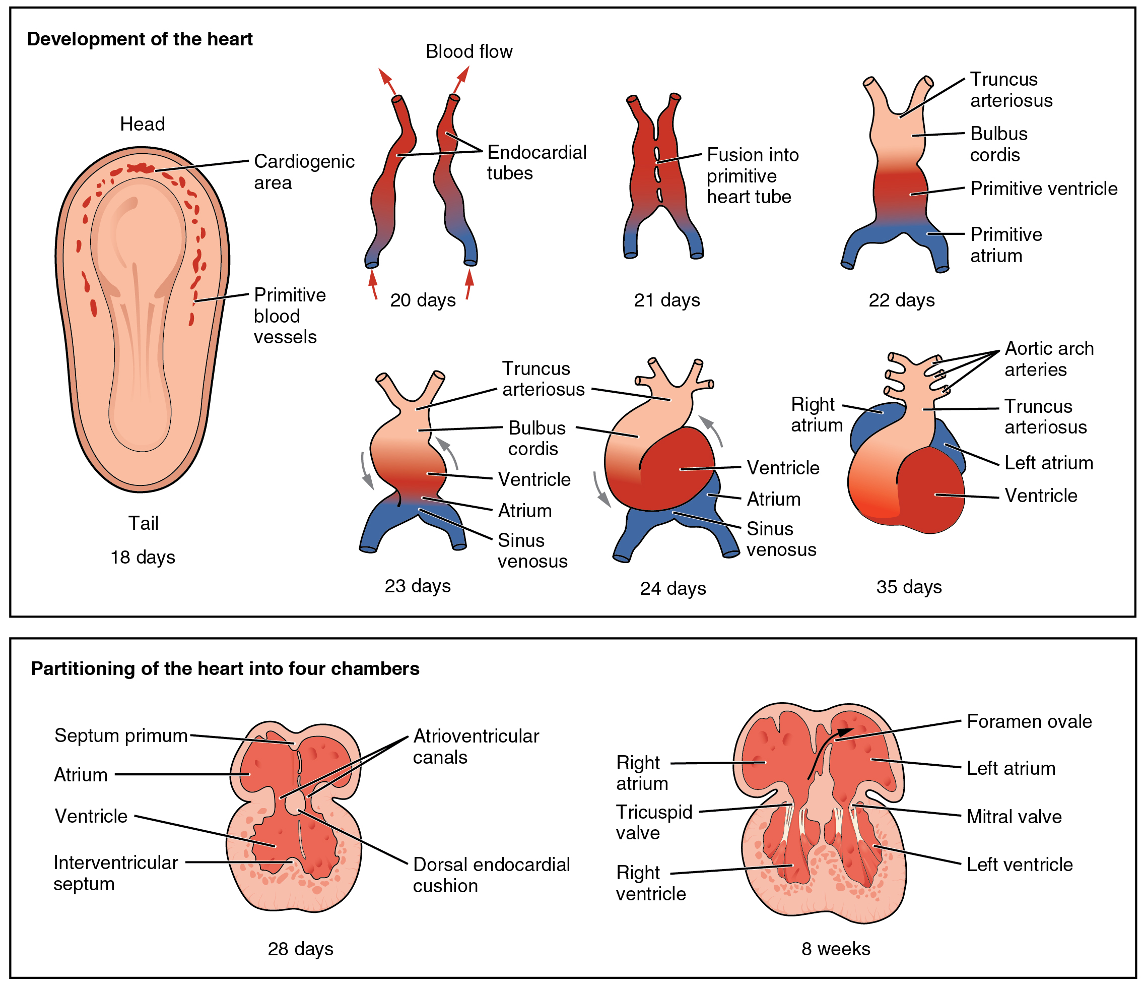
The bulbus cordis (the bulb of the heart) is a part of the developing heart that lies ventral to the primitive ventricle after the heart assumes its S-shaped form. The superior end of the bulbus cordis is also called the conotruncus. Structure In the early tubular heart, the bulbus cordis is the major outflow pathway. It receives blood from the primitive ventricle, and passes it to the truncus arteriosus. After heart looping, it is located slightly to the left of the ventricle. Development The early bulbus cordis is formed by the fifth week of development. The truncus arteriosus is derived from it later. The adjacent walls of the bulbus cordis and ventricle approximate, fuse, and finally disappear, and the bulbus cordis now communicates freely with the right ventricle, while the junction of the bulbus with the truncus arteriosus is brought directly ventral to and applied to the atrial canal. By the upgrowth of the ventricular septum the bulbus cordis is separated from the ... [...More Info...] [...Related Items...] OR: [Wikipedia] [Google] [Baidu] |
Atrium (heart)
The atrium ( la, ātrium, , entry hall) is one of two upper chambers in the heart that receives blood from the circulatory system. The blood in the atria is pumped into the heart ventricles through the atrioventricular valves. There are two atria in the human heart – the left atrium receives blood from the pulmonary circulation, and the right atrium receives blood from the venae cavae of the systemic circulation. During the cardiac cycle the atria receive blood while relaxed in diastole, then contract in systole to move blood to the ventricles. Each atrium is roughly cube-shaped except for an ear-shaped projection called an atrial appendage, sometimes known as an auricle. All animals with a closed circulatory system have at least one atrium. The atrium was formerly called the 'auricle'. That term is still used to describe this chamber in some other animals, such as the ''Mollusca''. They have thicker muscular walls than the atria do. Structure Humans have a four-c ... [...More Info...] [...Related Items...] OR: [Wikipedia] [Google] [Baidu] |
Heart Development
Heart development, also known as cardiogenesis, refers to the prenatal development of the heart. This begins with the formation of two endocardial tubes which merge to form the tubular heart, also called the primitive heart tube. The heart is the first functional organ in vertebrate embryos. The tubular heart quickly differentiates into the truncus arteriosus, bulbus cordis, primitive ventricle, primitive atrium, and the sinus venosus. The truncus arteriosus splits into the ascending aorta and the pulmonary trunk. The bulbus cordis forms part of the ventricles. The sinus venosus connects to the fetal circulation. The heart tube elongates on the right side, looping and becoming the first visual sign of left-right asymmetry of the body. Septa form within the atria and ventricles to separate the left and right sides of the heart. Early development The heart derives from embryonic mesodermal germ layer cells that differentiate after gastrulation into mesothelium, endothel ... [...More Info...] [...Related Items...] OR: [Wikipedia] [Google] [Baidu] |
Fish
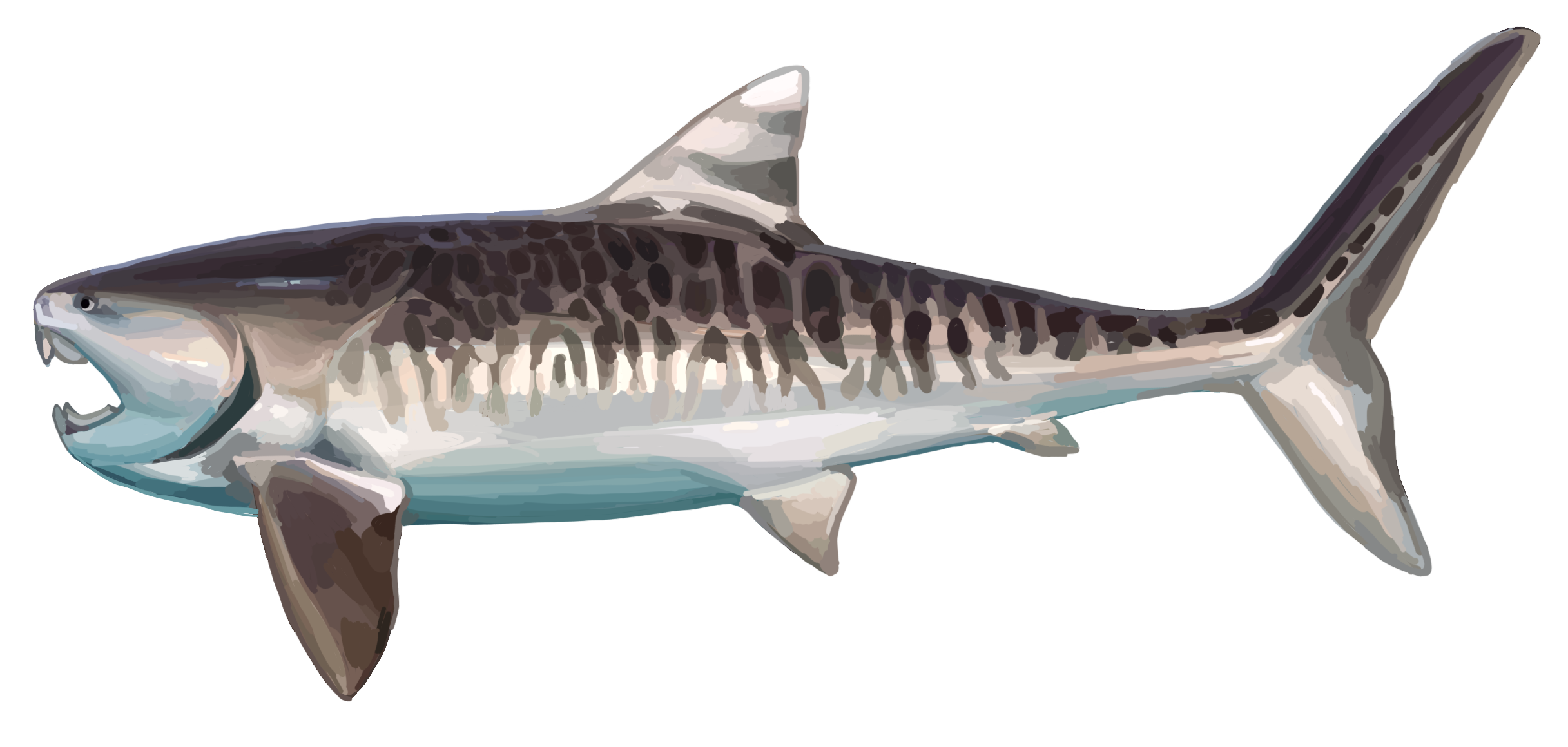
Fish are aquatic, craniate, gill-bearing animals that lack limbs with digits. Included in this definition are the living hagfish, lampreys, and cartilaginous and bony fish as well as various extinct related groups. Approximately 95% of living fish species are ray-finned fish, belonging to the class Actinopterygii, with around 99% of those being teleosts. The earliest organisms that can be classified as fish were soft-bodied chordates that first appeared during the Cambrian period. Although they lacked a true spine, they possessed notochords which allowed them to be more agile than their invertebrate counterparts. Fish would continue to evolve through the Paleozoic era, diversifying into a wide variety of forms. Many fish of the Paleozoic developed external armor that protected them from predators. The first fish with jaws appeared in the Silurian period, after which many (such as sharks) became formidable marine predators rather than just the prey of arthrop ... [...More Info...] [...Related Items...] OR: [Wikipedia] [Google] [Baidu] |
Frog
A frog is any member of a diverse and largely carnivorous group of short-bodied, tailless amphibians composing the order Anura (ανοὐρά, literally ''without tail'' in Ancient Greek). The oldest fossil "proto-frog" '' Triadobatrachus'' is known from the Early Triassic of Madagascar, but molecular clock dating suggests their split from other amphibians may extend further back to the Permian, 265 million years ago. Frogs are widely distributed, ranging from the tropics to subarctic regions, but the greatest concentration of species diversity is in tropical rainforest. Frogs account for around 88% of extant amphibian species. They are also one of the five most diverse vertebrate orders. Warty frog species tend to be called toads, but the distinction between frogs and toads is informal, not from taxonomy or evolutionary history. An adult frog has a stout body, protruding eyes, anteriorly-attached tongue, limbs folded underneath, and no tail (the tail of tailed frogs ... [...More Info...] [...Related Items...] OR: [Wikipedia] [Google] [Baidu] |
Ventricle (heart)
A ventricle is one of two large chambers toward the bottom of the heart that collect and expel blood towards the peripheral beds within the body and lungs. The blood pumped by a ventricle is supplied by an atrium (heart), atrium, an adjacent chamber in the upper heart that is smaller than a ventricle. Interventricular means between the ventricles (for example the interventricular septum), while intraventricular means within one ventricle (for example an intraventricular block). In a four-chambered heart, such as that in humans, there are two ventricles that operate in a double circulatory system: the right ventricle pumps blood into the pulmonary circulation to the lungs, and the left ventricle pumps blood into the systemic circulation through the aorta. Structure Ventricles have thicker walls than atria and generate higher blood pressures. The physiological load on the ventricles requiring pumping of blood throughout the body and lungs is much greater than the pressure generated ... [...More Info...] [...Related Items...] OR: [Wikipedia] [Google] [Baidu] |
Infundibulum (heart)
The infundibulum (also known as ''conus arteriosus'') is a conical pouch formed from the upper and left angle of the right ventricle in the chordate heart, from which the pulmonary trunk arises. It develops from the bulbus cordis. Typically, the infundibulum refers to the corresponding internal structure, whereas the conus arteriosus refers to the external structure. Defects in infundibulum development can result in a heart condition known as tetralogy of Fallot. A tendinous band extends upward from the right atrioventricular fibrous ring and connects the posterior surface of the infundibulum to the aorta. The infundibulum is the entrance from the right ventricle into the pulmonary artery and pulmonary trunk. The wall of the infundibulum is smooth. Additional images File:Gray490.png, Front view of human Humans (''Homo sapiens'') are the most abundant and widespread species of primate, characterized by bipedalism and exceptional cognitive skills due to a large and ... [...More Info...] [...Related Items...] OR: [Wikipedia] [Google] [Baidu] |
Ventricular Septum
The interventricular septum (IVS, or ventricular septum, or during development septum inferius) is the stout wall separating the ventricles, the lower chambers of the heart, from one another. The ventricular septum is directed obliquely backward to the right and curved with the convexity toward the right ventricle; its margins correspond with the anterior and posterior interventricular sulci. The lower part of the septum, which is the major part, is thick and muscular, and its much smaller upper part is thin and membraneous. During each cardiac cycle the interventricular septum contracts by shortening longitudinally and becoming thicker. Structure The interventricular septum is the stout wall separating the ventricles, the lower chambers of the heart, from one another. The ventricular septum is directed obliquely backward to the right and curved with the convexity toward the right ventricle; its margins correspond with the anterior and posterior longitudinal sulci. The gr ... [...More Info...] [...Related Items...] OR: [Wikipedia] [Google] [Baidu] |
Atrial Canal
The proper development of the atrioventricular canal into its prospective components (The heart septum and associated valves) to create a clear division between the four compartments of the heart and ensure proper blood movement through the heart, are essential for proper heart function. When this process does not happen correctly, a child will develop atrioventricular canal defect which occurs in 2 out of every 10,000 births. It also has a correlation with Down syndrome because 20% of children with Down syndrome have atrioventricular canal disease as well. This is a very serious condition and surgery is necessary within the first six months of life for a child.Brown, David MD "Atrioventricular Canal Defect" Children's Hospital Boston 20120 http://www.childrenshospital.org/az/Site521/mainpageS521P0.html Half of the children who are untreated with this condition die during their first year due to heart failure or pneumonia. Atrioventricular canal defect is a combination of abnormal ... [...More Info...] [...Related Items...] OR: [Wikipedia] [Google] [Baidu] |
Truncus Arteriosus (embryology)
The truncus arteriosus is a structure that is present during embryonic development. It is an arterial trunk that originates from both ventricles of the heart that later divides into the aorta and the pulmonary trunk. Structure The truncus arteriosus and bulbus cordis are divided by the aorticopulmonary septum. The truncus arteriosus gives rise to the ascending aorta and the pulmonary trunk. The caudal end of the bulbus cordis gives rise to the smooth parts (outflow tract) of the left and right ventricles (aortic vestibule & conus arteriosus respectively). The cranial end of the bulbus cordis (also known as the conus cordis) gives rise to the aorta and pulmonary trunk with the truncus arteriosus. This makes its appearance in three portions. # Two distal ridge-like thickenings project into the lumen of the tube: the truncal and bulbar ridges. These increase in size, and ultimately meet and fuse to form a septum (aorticopulmonary septum), which takes a spiral course toward the ... [...More Info...] [...Related Items...] OR: [Wikipedia] [Google] [Baidu] |
Embryonic Development
An embryo is an initial stage of development of a multicellular organism. In organisms that reproduce sexually, embryonic development is the part of the life cycle that begins just after fertilization of the female egg cell by the male sperm cell. The resulting fusion of these two cells produces a single-celled zygote that undergoes many cell divisions that produce cells known as blastomeres. The blastomeres are arranged as a solid ball that when reaching a certain size, called a morula, takes in fluid to create a cavity called a blastocoel. The structure is then termed a blastula, or a blastocyst in mammals. The mammalian blastocyst hatches before implantating into the endometrial lining of the womb. Once implanted the embryo will continue its development through the next stages of gastrulation, neurulation, and organogenesis. Gastrulation is the formation of the three germ layers that will form all of the different parts of the body. Neurulation forms the ... [...More Info...] [...Related Items...] OR: [Wikipedia] [Google] [Baidu] |
Truncus Arteriosus
The truncus arteriosus is a structure that is present during embryonic development. It is an arterial trunk that originates from both ventricles of the heart that later divides into the aorta and the pulmonary trunk. Structure The truncus arteriosus and bulbus cordis are divided by the aorticopulmonary septum. The truncus arteriosus gives rise to the ascending aorta and the pulmonary trunk. The caudal end of the bulbus cordis gives rise to the smooth parts (outflow tract) of the left and right ventricles (aortic vestibule & conus arteriosus respectively). The cranial end of the bulbus cordis (also known as the conus cordis) gives rise to the aorta and pulmonary trunk with the truncus arteriosus. This makes its appearance in three portions. # Two distal ridge-like thickenings project into the lumen of the tube: the truncal and bulbar ridges. These increase in size, and ultimately meet and fuse to form a septum ( aorticopulmonary septum), which takes a spiral course toward the p ... [...More Info...] [...Related Items...] OR: [Wikipedia] [Google] [Baidu] |


_Ranomafana.jpg)


