|
Buccinator
The buccinator () is a thin quadrilateral muscle occupying the interval between the maxilla and the mandible at the side of the face. It forms the anterior part of the cheek or the lateral wall of the oral cavity.Illustrated Anatomy of the Head and Neck, Fehrenbach and Herring, Elsevier, 2012, page 91 Structure It arises from the outer surfaces of the alveolar processes of the maxilla and mandible, corresponding to the three pairs of molar teeth and in the mandible, it is attached upon the buccinator crest posterior to the third molar; and behind, from the anterior border of the pterygomandibular raphe which separates it from the constrictor pharyngis superior. The fibers converge toward the angle of the mouth, where the central fibers intersect each other, those from below being continuous with the upper segment of the orbicularis oris, and those from above with the lower segment; the upper and lower fibers are continued forward into the corresponding lip without decussation. ... [...More Info...] [...Related Items...] OR: [Wikipedia] [Google] [Baidu] |
Buccinator Crest
The buccinator crest (Latin ''crista buccinatoria'') is a bony crest of the human mandible, that passes from the base of the coronoid process to the area of the third molar.https://medical-dictionary.thefreedictionary.com/buccinator+crest The free dictionary's medical dictionary defenition of buccinator crest The alveolar border of the buccinator muscle The buccinator () is a thin quadrilateral muscle occupying the interval between the maxilla and the mandible at the side of the face. It forms the anterior part of the cheek or the lateral wall of the oral cavity.Illustrated Anatomy of the Head ... attaches upon it. References Bones of the head and neck {{musculoskeletal-stub ... [...More Info...] [...Related Items...] OR: [Wikipedia] [Google] [Baidu] |
Buccal Artery
The buccal artery (buccinator artery) is a small artery in the head. It branches off the second part of the maxillary artery and supplies the cheek and buccinator muscle. Course It runs obliquely forward, between the pterygoideus internus and the insertion of the temporalis, to the outer surface of the buccinator, to which it is distributed, anastomosing with branches of the facial artery and with the infraorbital. From the infraorbital area, it descends bilaterally in the superficial face along the lateral margin of the nose, then running anti-parallel to the facial artery The facial artery (external maxillary artery in older texts) is a branch of the external carotid artery that supplies structures of the superficial face. Structure The facial artery arises in the carotid triangle from the external carotid arte ... across the lateral oral region. Additional images file:Gray508.png, The arteries of the face and scalp. References External links * () {{Authority ... [...More Info...] [...Related Items...] OR: [Wikipedia] [Google] [Baidu] |
Pterygomandibular Raphe
The pterygomandibular raphe (pterygomandibular ligament) is a ligamentous band of the buccopharyngeal fascia. It is attached superiorly to the pterygoid hamulus of the medial pterygoid plate, and inferiorly to the posterior end of the mylohyoid line of the mandible. It connects the buccinator muscle in front to the superior pharyngeal constrictor muscle behind. Structure The pterygomandibular raphe is a ligament that forms from the buccopharyngeal fascia. It is a paired structure, with one on each side of the mouth. Superiorly, it is attached to the pterygoid hamulus of the medial pterygoid plate of the sphenoid bone. Inferiorly, it is attached to the posterior end of the mylohyoid line of the mandible. * Its ''medial surface'' is covered by the mucous membrane of the mouth. * Its ''lateral surface'' is separated from the ramus of the mandible by a quantity of adipose tissue. * Its ''posterior border'' gives attachment to the superior pharyngeal constrictor muscle. * Its ''an ... [...More Info...] [...Related Items...] OR: [Wikipedia] [Google] [Baidu] |
Orbicularis Oris
In human anatomy, the orbicularis oris muscle is a complex of muscles in the lips that encircles the mouth. It is a sphincter, or circular muscle, but it is actually composed of four independent quadrants that interlace and give only an appearance of circularity.Saladin, "Anatomy & Physiology: The Unity of Form and Function". 5th edition. McGraw Hill. Page 330 It is also one of the muscles used in the playing of all brass instruments and some woodwind instruments. This muscle closes the mouth and puckers the lips when it contracts. Structure The orbicularis oris is not a simple sphincter muscle like the orbicularis oculi; it consists of numerous strata of muscular fibers surrounding the orifice of the mouth, but having different direction. It consists partly of fibers derived from the other facial muscles which are inserted into the lips, and partly of fibers proper to the lips. Of the former, a considerable number are derived from the buccinator and form the deeper stratum of th ... [...More Info...] [...Related Items...] OR: [Wikipedia] [Google] [Baidu] |
Buccal Branch Of The Facial Nerve
The buccal branches of the facial nerve (infraorbital branches), are of larger size than the rest of the branches, pass horizontally forward to be distributed below the orbit and around the mouth. Branches The ''superficial branches'' run beneath the skin and above the superficial muscles of the face, which they supply: some are distributed to the procerus, joining at the medial angle of the orbit with the infratrochlear and nasociliary branches of the ophthalmic. The ''deep branches'' pass beneath the zygomaticus and the quadratus labii superioris, supplying them and forming an infraorbital plexus with the infraorbital branch of the maxillary nerve. These branches also supply the small muscles of the nose. The ''lower deep branches'' supply the buccinator and orbicularis oris, and join with filaments of the buccinator branch of the mandibular nerve. Muscles of facial expression The facial nerve innervates the muscles of facial expression. The buccal branch supplies these mu ... [...More Info...] [...Related Items...] OR: [Wikipedia] [Google] [Baidu] |
Bucina
A buccina ( lat, buccina) or bucina ( lat, būcina, link=no), anglicized buccin or bucine, is a brass instrument that was used in the ancient Roman army, similar to the cornu. An ''aeneator'' who blew a buccina was called a "buccinator" or "bucinator" ( lat, buccinātor, būcinātor, link=no). Design It was originally designed as a tube made of either bronze or shells. However, as time went on more materials started to be used. It measured in length, of narrow cylindrical bore, and played by means of a cup-shaped mouthpiece. The tube is bent round upon itself from the mouthpiece to the bell in the shape of a broad C and is strengthened by means of a bar across the curve, which the performer grasps while playing to steady the instrument; the bell curves over his head or shoulder. Usage The buccina was used for the announcement of night watches, to summon soldiers by means of the special signal known as ''classicum'', and to give orders. Frontinus relates that a Roman genera ... [...More Info...] [...Related Items...] OR: [Wikipedia] [Google] [Baidu] |
Stenson's Duct
The parotid duct, or Stensen duct, is a salivary duct. It is the route that saliva takes from the major salivary gland, the parotid gland, into the mouth. Structure The parotid duct is formed when several interlobular ducts, the largest ducts inside the parotid gland, join. It emerges from the parotid gland. It runs forward along the lateral side of the masseter muscle for around 7 cm. In this course, the duct is surrounded by the buccal fat pad. It takes a steep turn at the border of the masseter and passes through the buccinator muscle, opening into the vestibule of the mouth, the region of the mouth between the cheek and the gums, at the parotid papilla, which lies across the second Maxillary (upper) molar tooth. The buccinator acts as a valve that prevents air forcing into the duct, which would cause pneumoparotitis. Running along with the duct superiorly is the transverse facial artery and upper buccal nerve; running along with the duct inferiorly is the lower buccal nerve ... [...More Info...] [...Related Items...] OR: [Wikipedia] [Google] [Baidu] |
Human Mandible
In anatomy, the mandible, lower jaw or jawbone is the largest, strongest and lowest bone in the human facial skeleton. It forms the lower jaw and holds the lower tooth, teeth in place. The mandible sits beneath the maxilla. It is the only movable bone of the skull (discounting the ossicles of the middle ear). It is connected to the temporal bones by the temporomandibular joints. The bone is formed prenatal development, in the fetus from a fusion of the left and right mandibular prominences, and the point where these sides join, the mandibular symphysis, is still visible as a faint ridge in the midline. Like other symphyses in the body, this is a midline articulation where the bones are joined by fibrocartilage, but this articulation fuses together in early childhood.Illustrated Anatomy of the Head and Neck, Fehrenbach and Herring, Elsevier, 2012, p. 59 The word "mandible" derives from the Latin word ''mandibula'', "jawbone" (literally "one used for chewing"), from ''wikt:mandere ... [...More Info...] [...Related Items...] OR: [Wikipedia] [Google] [Baidu] |
Parotid
The parotid gland is a major salivary gland in many animals. In humans, the two parotid glands are present on either side of the mouth and in front of both ears. They are the largest of the salivary glands. Each parotid is wrapped around the mandibular ramus, and secretes serous saliva through the parotid duct into the mouth, to facilitate mastication and swallowing and to begin the digestion of starches. There are also two other types of salivary glands; they are submandibular and sublingual glands. Sometimes accessory parotid glands are found close to the main parotid glands. Etymology The word ''parotid'' literally means "beside the ear". From Greek παρωτίς (stem παρωτιδ-) : (gland) behind the ear < παρά - pará : in front, and οὖς - ous (stem ὠτ-, ōt-) : ear. Structure The parotid glands are a pair of mainly |
Alveolar Processes
The alveolar process () or alveolar bone is the thickened ridge of bone that contains the tooth sockets on the jaw bones (in humans, the maxilla and the mandible). The structures are covered by gums as part of the oral cavity. The synonymous terms ''alveolar ridge'' and ''alveolar margin'' are also sometimes used more specifically to refer to the ridges on the inside of the mouth which can be felt with the tongue, either on roof of the mouth between the upper teeth and the hard palate or on the bottom of the mouth behind the lower teeth. Terminology The term ''alveolar'' () ('hollow') refers to the cavities of the tooth sockets, known as dental alveoli. The alveolar process is also called the ''alveolar bone'' or ''alveolar ridge''. The curved portion is referred to as the alveolar arch. The alveolar bone proper, also called bundle bone, directly surrounds the teeth. The term alveolar crest describes the extreme rim of the bone nearest to the Crown (tooth), crowns of the Human ... [...More Info...] [...Related Items...] OR: [Wikipedia] [Google] [Baidu] |
Breastfeeding
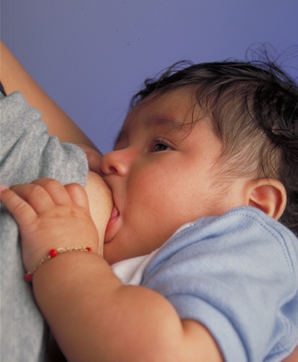
Breastfeeding, or nursing, is the process by which human breast milk is fed to a child. Breast milk may be from the breast, or may be expressed by hand or pumped and fed to the infant. The World Health Organization (WHO) recommends that breastfeeding begin within the first hour of a baby's life and continue as often and as much as the baby wants. Health organizations, including the WHO, recommend breastfeeding exclusively for six months. This means that no other foods or drinks, other than vitamin D, are typically given. WHO recommends exclusive breastfeeding for the first 6 months of life, followed by continued breastfeeding with appropriate complementary foods for up to 2 years and beyond. Of the 135 million babies born every year, only 42% are breastfed within the first hour of life, only 38% of mothers practice exclusive breastfeeding during the first six months, and 58% of mothers continue breastfeeding up to the age of two years and beyond. Breastfeeding has a numb ... [...More Info...] [...Related Items...] OR: [Wikipedia] [Google] [Baidu] |
Ancient Greek
Ancient Greek includes the forms of the Greek language used in ancient Greece and the ancient world from around 1500 BC to 300 BC. It is often roughly divided into the following periods: Mycenaean Greek (), Dark Ages (), the Archaic period (), and the Classical period (). Ancient Greek was the language of Homer and of fifth-century Athenian historians, playwrights, and philosophers. It has contributed many words to English vocabulary and has been a standard subject of study in educational institutions of the Western world since the Renaissance. This article primarily contains information about the Epic and Classical periods of the language. From the Hellenistic period (), Ancient Greek was followed by Koine Greek, which is regarded as a separate historical stage, although its earliest form closely resembles Attic Greek and its latest form approaches Medieval Greek. There were several regional dialects of Ancient Greek, of which Attic Greek developed into Koine. Dia ... [...More Info...] [...Related Items...] OR: [Wikipedia] [Google] [Baidu] |


.jpg)
