|
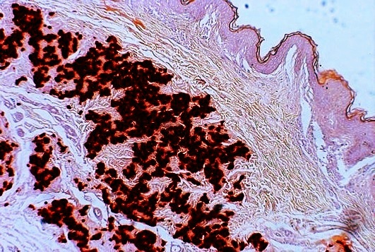
Bruch's Membrane
Bruch's membrane is the innermost layer of the choroid of the eye. It is also called the ''vitreous lamina'' or ''Membrane vitriae'', because of its glassy microscopic appearance. It is 2–4 μm thick. Layers Bruch's membrane consists of five layers (from inside to outside): #the basement membrane of the retinal pigment epithelium #the inner collagenous zone #a central band of elastic fibers #the outer collagenous zone #the basement membrane of the choriocapillaris The retinal pigment epithelium transports metabolic waste from the photoreceptors across Bruch's membrane to the choroid. Embryology Bruch's membrane is present by midterm in fetal development as an elastic sheet. Pathology Bruch's membrane thickens with age, slowing the transport of metabolites. This may lead to the formation of drusen in age-related macular degeneration. There is also a buildup of deposits (Basal Linear Deposits or BLinD and Basal Lamellar Deposits BLamD) on and within the membrane, primarily co ... [...More Info...] [...Related Items...] OR: [Wikipedia] [Google] [Baidu] |
Visual System
The visual system comprises the sensory organ (the eye) and parts of the central nervous system (the retina containing photoreceptor cells, the optic nerve, the optic tract and the visual cortex) which gives organisms the sense of sight (the ability to perception, detect and process visible light) as well as enabling the formation of several non-image photo response functions. It detects and interprets information from the optical spectrum perceptible to that species to "build a representation" of the surrounding environment. The visual system carries out a number of complex tasks, including the reception of light and the formation of monocular neural representations, colour vision, the neural mechanisms underlying stereopsis and assessment of distances to and between objects, the identification of a particular object of interest, motion perception, the analysis and integration of visual information, pattern recognition, accurate motor coordination under visual guidance, and mor ... [...More Info...] [...Related Items...] OR: [Wikipedia] [Google] [Baidu] |
Choroid
The choroid, also known as the choroidea or choroid coat, is a part of the uvea, the vascular layer of the eye, and contains connective tissues, and lies between the retina and the sclera. The human choroid is thickest at the far extreme rear of the eye (at 0.2 mm), while in the outlying areas it narrows to 0.1 mm. The choroid provides oxygen and nourishment to the outer layers of the retina. Along with the ciliary body and iris, the choroid forms the uveal tract. The structure of the choroid is generally divided into four layers (classified in order of furthest away from the retina to closest): *Haller's layer - outermost layer of the choroid consisting of larger diameter blood vessels; *Sattler's layer - layer of medium diameter blood vessels; * Choriocapillaris - layer of capillaries; and *Bruch's membrane (synonyms: Lamina basalis, Complexus basalis, Lamina vitra) - innermost layer of the choroid. Blood supply There are two circulations of the eye: the retin ... [...More Info...] [...Related Items...] OR: [Wikipedia] [Google] [Baidu] |
Basement Membrane
The basement membrane is a thin, pliable sheet-like type of extracellular matrix that provides cell and tissue support and acts as a platform for complex signalling. The basement membrane sits between Epithelium, epithelial tissues including mesothelium and endothelium, and the underlying connective tissue. Structure As seen with the electron microscope, the basement membrane is composed of two layers, the basal lamina and the reticular lamina. The underlying connective tissue attaches to the basal lamina with collagen VII anchoring fibrils and fibrillin microfibrils. The basal lamina layer can further be subdivided into two layers based on their visual appearance in electron microscopy. The lighter-colored layer closer to the epithelium is called the lamina lucida, while the denser-colored layer closer to the connective tissue is called the lamina densa. The Electron microscope, electron-dense lamina densa layer is about 30–70 nanometers thick and consists of an underlying ... [...More Info...] [...Related Items...] OR: [Wikipedia] [Google] [Baidu] |
Retinal Pigment Epithelium
The pigmented layer of retina or retinal pigment epithelium (RPE) is the pigmented cell layer just outside the neurosensory retina that nourishes retinal visual cells, and is firmly attached to the underlying choroid and overlying retinal visual cells. History The RPE was known in the 18th and 19th centuries as the pigmentum nigrum, referring to the observation that the RPE is dark (black in many animals, brown in humans); and as the tapetum nigrum, referring to the observation that in animals with a tapetum lucidum, in the region of the tapetum lucidum the RPE is not pigmented. Anatomy The RPE is composed of a single layer of hexagonal cells that are densely packed with pigment granules. When viewed from the outer surface, these cells are smooth and hexagonal in shape. When seen in section, each cell consists of an outer non-pigmented part containing a large oval nucleus and an inner pigmented portion which extends as a series of straight thread-like processes between the rods, ... [...More Info...] [...Related Items...] OR: [Wikipedia] [Google] [Baidu] |
Photoreceptor Cell
A photoreceptor cell is a specialized type of neuroepithelial cell found in the retina that is capable of visual phototransduction. The great biological importance of photoreceptors is that they convert light (visible electromagnetic radiation) into signals that can stimulate biological processes. To be more specific, photoreceptor proteins in the cell absorb photons, triggering a change in the cell's membrane potential. There are currently three known types of photoreceptor cells in mammalian eyes: rods, cones, and intrinsically photosensitive retinal ganglion cells. The two classic photoreceptor cells are rods and cones, each contributing information used by the visual system to form an image of the environment, sight. Rods primarily mediate scotopic vision (dim conditions) whereas cones primarily mediate to photopic vision (bright conditions), but the processes in each that supports phototransduction is similar. A third class of mammalian photoreceptor cell was discovered ... [...More Info...] [...Related Items...] OR: [Wikipedia] [Google] [Baidu] |
Drusen
Drusen, from the German word for ''node'' or ''geode'' (singular, "Druse"), are tiny yellow or white accumulations of extracellular material that build up between Bruch's membrane and the retinal pigment epithelium of the eye. The presence of a few small ("hard") drusen is normal with advancing age, and most people over 40 have some hard drusen. However, the presence of larger and more numerous drusen in the macula is a common early sign of age-related macular degeneration (AMD). Classification Drusen are associated with aging and macular degeneration are distinct from another clinical entity, optic disc drusen, which is present on the optic nerve head. Both age-related drusen and optic disc drusen can be observed by ophthalmoscopy. Optical coherence tomography scans of the orbits or head, calcification at the head of the optic nerve without change in size of globe strongly suggests drusen in a middle-age or elderly patient. Whether drusen promote AMD or are symptomatic of an un ... [...More Info...] [...Related Items...] OR: [Wikipedia] [Google] [Baidu] |
Age-related Macular Degeneration
Macular degeneration, also known as age-related macular degeneration (AMD or ARMD), is a medical condition which may result in blurred or no vision in the center of the visual field. Early on there are often no symptoms. Over time, however, some people experience a gradual worsening of vision that may affect one or both eyes. While it does not result in complete blindness, loss of central vision can make it hard to recognize faces, drive, read, or perform other activities of daily life. Visual hallucinations may also occur. Macular degeneration typically occurs in older people. Genetic factors and smoking also play a role. It is due to damage to the macula of the retina. Diagnosis is by a complete eye exam. The severity is divided into early, intermediate, and late types. The late type is additionally divided into "dry" and "wet" forms with the dry form making up 90% of cases. The difference between the two forms is the change of macula. Those with dry form AMD have drusen, ce ... [...More Info...] [...Related Items...] OR: [Wikipedia] [Google] [Baidu] |
Macular Degeneration
Macular degeneration, also known as age-related macular degeneration (AMD or ARMD), is a medical condition which may result in blurred or no vision in the center of the visual field. Early on there are often no symptoms. Over time, however, some people experience a gradual worsening of vision that may affect one or both eyes. While it does not result in complete blindness, loss of central vision can make it hard to recognize faces, drive, read, or perform other activities of daily life. Visual hallucinations may also occur. Macular degeneration typically occurs in older people. Genetic factors and smoking also play a role. It is due to damage to the macula of the retina. Diagnosis is by a complete eye exam. The severity is divided into early, intermediate, and late types. The late type is additionally divided into "dry" and "wet" forms with the dry form making up 90% of cases. The difference between the two forms is the change of macula. Those with dry form AMD have drusen, ce ... [...More Info...] [...Related Items...] OR: [Wikipedia] [Google] [Baidu] |
Pseudoxanthoma Elasticum
Pseudoxanthoma elasticum (PXE) is a genetic disease that causes mineralization of elastic fibers in some tissues. The most common problems arise in the skin and eyes, and later in blood vessels in the form of premature atherosclerosis. PXE is caused by autosomal recessive mutations in the '' ABCC6'' gene on the short arm of chromosome 16 (16p13.1). Signs and symptoms Usually, pseudoxanthoma elasticum affects the skin first, often in childhood or early adolescence. Small, yellowish papular lesions form and cutaneous laxity mainly affect the neck, axillae (armpits), groin, and flexural creases (the inside parts of the elbows and knees). Skin may become lax and redundant. Many individuals have "oblique mental creases" (horizontal grooves of the chin) PXE first affects the retina through a dimpling of the Bruch membrane (a thin membrane separating the blood vessel-rich layer from the pigmented layer of the retina), that is only visible during ophthalmologic examinations. This is ca ... [...More Info...] [...Related Items...] OR: [Wikipedia] [Google] [Baidu] |
Myopia
Near-sightedness, also known as myopia and short-sightedness, is an eye disease where light focuses in front of, instead of on, the retina. As a result, distant objects appear blurry while close objects appear normal. Other symptoms may include headaches and eye strain. Severe near-sightedness is associated with an increased risk of retinal detachment, cataracts, and glaucoma. The underlying mechanism involves the length of the eyeball growing too long or less commonly the lens being too strong. It is a type of refractive error. Diagnosis is by eye examination. Tentative evidence indicates that the risk of near-sightedness can be decreased by having young children spend more time outside. This decrease in risk may be related to natural light exposure. Near-sightedness can be corrected with eyeglasses, contact lenses, or a refractive surgery. Eyeglasses are the easiest and safest method of correction. Contact lenses can provide a wider field of vision, but are associated with ... [...More Info...] [...Related Items...] OR: [Wikipedia] [Google] [Baidu] |
Alport's Syndrome
Alport syndrome is a genetic disorder affecting around 1 in 5,000-10,000 children, characterized by glomerulonephritis, end-stage kidney disease, and hearing loss. Alport syndrome can also affect the eyes, though the changes do not usually affect vision, except when changes to the lens occur in later life. Blood in urine is universal. Proteinuria is a feature as kidney disease progresses. The disorder was first identified in a British family by the physician Cecil A. Alport in 1927. Alport syndrome once also had the label hereditary nephritis, but this is misleading as there are many other causes of hereditary kidney disease and 'nephritis'. Alport syndrome is caused by an inherited defect in type IV collagen—a structural material that is needed for the normal function of different parts of the body. Since type IV collagen is found in the ears, eyes, and kidneys, this explains why Alport syndrome affects different seemingly unrelated parts of the body (ears, eyes, kidneys, etc. ... [...More Info...] [...Related Items...] OR: [Wikipedia] [Google] [Baidu] |
Angioid Streaks
Angioid streaks, also called Knapp streaks or Knapp striae, are small breaks in Bruch's membrane, an elastic tissue containing membrane of the retina that may become calcified and crack. Up to 50% of angioid streak cases are idiopathic. It may occur secondary to blunt trauma, or it may be associated with many systemic diseases. The condition is usually asymptomatic, but decrease in vision may occur due to choroidal neovascularization. Clinical features Angioid streaks are often associated with pseudoxanthoma elasticum, but have been found to occur in conjunction with other disorders, including Paget's disease, sickle cell disease and Ehlers–Danlos syndrome. These streaks can have a negative impact on vision due to choroidal neovascularization or choroidal rupture. Also, vision can be impaired if the streaks progress to the fovea and damage the retinal pigment epithelium. Signs Retinal fundus examination may reveal grey or dark red spoke like lesions around optic disk and radi ... [...More Info...] [...Related Items...] OR: [Wikipedia] [Google] [Baidu] |


