|
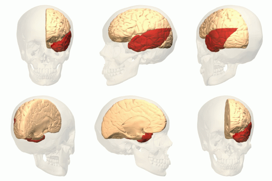
Brodmann Area 37
Brodmann area 37, or BA37, is part of the temporal cortex in the human brain. It contains the fusiform gyrus which in turn contains the fusiform face area, an area important for the recognition of faces. This area is also known as occipitotemporal area 37 (H). It is a subdivision of the cytoarchitecturally defined temporal region of cerebral cortex. It is located primarily in the caudal portions of the fusiform gyrus and inferior temporal gyrus on the mediobasal and lateral surfaces at the caudal extreme of the temporal lobe. Cytoarchitecturally, it is bounded caudally by the peristriate Brodmann area 19, rostrally by the inferior temporal area 20 and middle temporal area 21, and dorsally on the lateral aspect of the hemisphere by the angular area 39 (H) (Brodmann-1909). See also * Fusiform face area * Brodmann area A Brodmann area is a region of the cerebral cortex, in the human or other primate brain, defined by its cytoarchitecture, or histological structure and org ... [...More Info...] [...Related Items...] OR: [Wikipedia] [Google] [Baidu] |
Temporal Lobe
The temporal lobe is one of the four Lobes of the brain, major lobes of the cerebral cortex in the brain of mammals. The temporal lobe is located beneath the lateral fissure on both cerebral hemispheres of the mammalian brain. The temporal lobe is involved in processing sensory input into derived meanings for the appropriate retention of visual memory, language comprehension, and emotion association. ''Temporal'' refers to the head's Temple (anatomy), temples. Structure The Temple (anatomy)#Etymology, temporal Lobe (anatomy), lobe consists of structures that are vital for declarative or long-term memory. Declarative memory, Declarative (denotative) or Explicit memory, explicit memory is conscious memory divided into semantic memory (facts) and episodic memory (events). Medial temporal lobe structures that are critical for long-term memory include the hippocampus, along with the surrounding Hippocampal formation, hippocampal region consisting of the Perirhinal cortex, perirhinal, ... [...More Info...] [...Related Items...] OR: [Wikipedia] [Google] [Baidu] |
Cerebral Cortex
The cerebral cortex, also known as the cerebral mantle, is the outer layer of neural tissue of the cerebrum of the brain in humans and other mammals. The cerebral cortex mostly consists of the six-layered neocortex, with just 10% consisting of allocortex. It is separated into two cortices, by the longitudinal fissure that divides the cerebrum into the left and right cerebral hemispheres. The two hemispheres are joined beneath the cortex by the corpus callosum. The cerebral cortex is the largest site of neural integration in the central nervous system. It plays a key role in attention, perception, awareness, thought, memory, language, and consciousness. The cerebral cortex is part of the brain responsible for cognition. In most mammals, apart from small mammals that have small brains, the cerebral cortex is folded, providing a greater surface area in the confined volume of the cranium. Apart from minimising brain and cranial volume, cortical folding is crucial for the brain ... [...More Info...] [...Related Items...] OR: [Wikipedia] [Google] [Baidu] |
Human Brain
The human brain is the central organ of the human nervous system, and with the spinal cord makes up the central nervous system. The brain consists of the cerebrum, the brainstem and the cerebellum. It controls most of the activities of the body, processing, integrating, and coordinating the information it receives from the sense organs, and making decisions as to the instructions sent to the rest of the body. The brain is contained in, and protected by, the skull bones of the head. The cerebrum, the largest part of the human brain, consists of two cerebral hemispheres. Each hemisphere has an inner core composed of white matter, and an outer surface – the cerebral cortex – composed of grey matter. The cortex has an outer layer, the neocortex, and an inner allocortex. The neocortex is made up of six neuronal layers, while the allocortex has three or four. Each hemisphere is conventionally divided into four lobes – the frontal, temporal, parietal, and occipital lo ... [...More Info...] [...Related Items...] OR: [Wikipedia] [Google] [Baidu] |
Fusiform Gyrus
The fusiform gyrus, also known as the ''lateral occipitotemporal gyrus'','' ''is part of the temporal lobe and occipital lobe in Brodmann area 37. The fusiform gyrus is located between the lingual gyrus and parahippocampal gyrus above, and the inferior temporal gyrus below. Though the functionality of the fusiform gyrus is not fully understood, it has been linked with various neural pathways related to recognition. Additionally, it has been linked to various neurological phenomena such as synesthesia, dyslexia, and prosopagnosia. Anatomy Anatomically, the fusiform gyrus is the largest macro-anatomical structure within the ventral temporal cortex, which mainly includes structures involved in high-level vision. The term fusiform gyrus (lit. "spindle-shaped convolution") refers to the fact that the shape of the gyrus is wider at its centre than at its ends. This term is based on the description of the gyrus by Emil Huschke in 1854. (see also section on history). The fusiform gyrus ... [...More Info...] [...Related Items...] OR: [Wikipedia] [Google] [Baidu] |
Fusiform Face Area
The fusiform face area (FFA, meaning spindle-shaped face area) is a part of the human visual system (while also activated in people blind from birth) that is specialized for facial recognition. It is located in the inferior temporal cortex (IT), in the fusiform gyrus (Brodmann area 37). Structure The FFA is located in the ventral stream on the ventral surface of the temporal lobe on the lateral side of the fusiform gyrus. It is lateral to the parahippocampal place area. It displays some lateralization, usually being larger in the right hemisphere. The FFA was discovered and continues to be investigated in humans using positron emission tomography (PET) and functional magnetic resonance imaging (fMRI) studies. Usually, a participant views images of faces, objects, places, bodies, scrambled faces, scrambled objects, scrambled places, and scrambled bodies. This is called a functional localizer. Comparing the neural response between faces and scrambled faces will reveal areas ... [...More Info...] [...Related Items...] OR: [Wikipedia] [Google] [Baidu] |
Cytoarchitecture
Cytoarchitecture (Greek '' κύτος''= "cell" + '' ἀρχιτεκτονική''= "architecture"), also known as cytoarchitectonics, is the study of the cellular composition of the central nervous system's tissues under the microscope. Cytoarchitectonics is one of the ways to parse the brain, by obtaining sections of the brain using a microtome and staining them with chemical agents which reveal where different neurons are located. The study of the parcellation of ''nerve fibers'' (primarily axons) into layers forms the subject of myeloarchitectonics ( History of the cerebral cytoarchitecture Defining cerebral cytoarchitecture began with the advent of —the science of slicing a ...[...More Info...] [...Related Items...] OR: [Wikipedia] [Google] [Baidu] |
Inferior Temporal Gyrus
The inferior temporal gyrus is one of three gyri of the temporal lobe and is located below the middle temporal gyrus, connected behind with the inferior occipital gyrus; it also extends around the infero-lateral border on to the inferior surface of the temporal lobe, where it is limited by the inferior Sulcus (neuroanatomy), sulcus. This region is one of the higher levels of the ventral stream of visual processing, associated with the representation of objects, places, faces, and colors. It may also be involved in face perception, and in the recognition of numbers and words. The inferior temporal gyrus is the anterior region of the temporal lobe located underneath the central temporal sulcus. The primary function of the occipital temporal gyrus – otherwise referenced as IT cortex – is associated with visual stimuli processing, namely visual object recognition, and has been suggested by recent experimental results as the final location of the ventral cortical visual system. The ... [...More Info...] [...Related Items...] OR: [Wikipedia] [Google] [Baidu] |
Brodmann Area 19
Brodmann area 19, or BA 19, is part of the occipital lobe cortex in the human brain. Along with area 18, it comprises the extrastriate (or peristriate) cortex. In humans with normal sight, extrastriate cortex is a visual association area, with feature-extracting, shape recognition, attentional, and multimodal integrating functions. This area is also known as peristriate area 19, and it refers to a subdivision of the cytoarchitecturally defined occipital region of cerebral cortex. In the human it is located in parts of the lingual gyrus, the cuneus, the lateral occipital gyrus (H) and the superior occipital gyrus (H) of the occipital lobe where it is bounded approximately by the parieto-occipital sulcus. It is bounded on one side by the parastriate area 18, which it surrounds. It is bounded rostrally by the angular area 39 (H) and the occipitotemporal area 37 (H) (Brodmann-1909). In animals Brodmann area 19-1909 is a subdivision of the cerebral cortex of the guenon defined on t ... [...More Info...] [...Related Items...] OR: [Wikipedia] [Google] [Baidu] |
Inferior Temporal Area 20
Brodmann area 20, or BA20, is part of the temporal cortex in the human brain. The region encompasses most of the ventral temporal cortex, a region believed to play a part in high-level visual processing and recognition memory. This area is also known as inferior temporal area 20, and it refers to a subdivision of the cytoarchitecturally defined temporal region of cerebral cortex. In the human it corresponds approximately to the inferior temporal gyrus. Cytoarchitecturally it is bounded medially by the ectorhinal area 36 (H), laterally by the middle temporal area 21, rostrally by the temporopolar area 38 (H) and caudally by the occipitotemporal area 37 (H) (Brodmann-1909). Guenon Brodmann area 20 is a subdivision of the cerebral cortex of the guenon defined on the basis of cytoarchitecture. It is cytoarchitecturally homologous to the inferior temporal area 20 of the human (Brodmann-1909). Distinctive features (Brodmann-1905): area 20 is similar to area 19 of Brodmann-1909 in th ... [...More Info...] [...Related Items...] OR: [Wikipedia] [Google] [Baidu] |
Middle Temporal Area 21
Brodmann area 21, or BA21, is part of the temporal cortex in the human brain. The region encompasses most of the lateral temporal cortex and is also known as middle temporal area 21. In the human it corresponds approximately to the middle temporal gyrus. Function BA 21 is a higher-order visual association cortex with medium myelin content. As a lateral temporal region, it is likely to play a part in auditory processing and language. Language function is left lateralized in most individuals. Location BA21 is a subdivision of the cytoarchitecturally defined temporal region of the cerebral cortex. BA21 is superior to BA20 and inferior to BA40 and BA41. It is bounded rostrally by the temporopolar area 38 (H), ventrally by the inferior temporal area 20, caudally by the occipitotemporal area 37 (H), and dorsally by the superior temporal area 22 (Brodmann-1909). Guenon Brodmann area 21 is a subdivision of the cerebral cortex of the guenon defined on the basis of cytoarchitecture ... [...More Info...] [...Related Items...] OR: [Wikipedia] [Google] [Baidu] |
Angular Area 39
Brodmann area 39, or BA39, is part of the parietal cortex in the human brain. BA39 encompasses the angular gyrus, lying near to the junction of temporal, occipital and parietal lobes. This area is also known as angular area 39 (H). It corresponds to the angular gyrus surrounding the caudal tip of the superior temporal sulcus. It is bounded dorsally approximately by the intraparietal sulcus. In terms of its cytoarchitecture, it is bounded rostrally by the supramarginal area 40 (H), dorsally and caudally by the peristriate area 19, and ventrally by the occipitotemporal area 37 (H) (Brodmann-1909). Function Area 39 was regarded by Alexander Luria as a part of the parietal-temporal-occipital area, which includes Brodmann area 40, Brodmann area 19, and Brodmann area 37. Damage to the left Brodmann area 39 may result in dyslexia or in semantic aphasia. Albert Einstein had less neurones (relative to glial cells) in this (left sided) area than normal. The right Brodmann area 39 a ... [...More Info...] [...Related Items...] OR: [Wikipedia] [Google] [Baidu] |


