|
Brachial Plexus
The brachial plexus is a network () of nerves formed by the anterior rami of the lower four cervical nerves and first thoracic nerve ( C5, C6, C7, C8, and T1). This plexus extends from the spinal cord, through the cervicoaxillary canal in the neck, over the first rib, and into the armpit, it supplies afferent and efferent nerve fibers the to chest, shoulder, arm, forearm, and hand. Structure The brachial plexus is divided into five ''roots'', three ''trunks'', six ''divisions'' (three anterior and three posterior), three ''cords'', and five ''branches''. There are five "terminal" branches and numerous other "pre-terminal" or "collateral" branches, such as the subscapular nerve, the thoracodorsal nerve, and the long thoracic nerve, that leave the plexus at various points along its length. A common structure used to identify part of the brachial plexus in cadaver dissections is the M or W shape made by the musculocutaneous nerve, lateral cord, median nerve, medial cord, and ... [...More Info...] [...Related Items...] OR: [Wikipedia] [Google] [Baidu] |
Cadaver
A cadaver or corpse is a dead human body that is used by medical students, physicians and other scientists to study anatomy, identify disease sites, determine causes of death, and provide tissue to repair a defect in a living human being. Students in medical school study and dissect cadavers as a part of their education. Others who study cadavers include archaeologists and arts students. The term ''cadaver'' is used in courts of law (and, to a lesser extent, also by media outlets such as newspapers) to refer to a dead body, as well as by recovery teams searching for bodies in natural disasters. The word comes from the Latin word ''cadere'' ("to fall"). Related terms include ''cadaverous'' (resembling a cadaver) and ''cadaveric spasm'' (a muscle spasm causing a dead body to twitch or jerk). A cadaver graft (also called “postmortem graft”) is the grafting of tissue from a dead body onto a living human to repair a defect or disfigurement. Cadavers can be observed for their sta ... [...More Info...] [...Related Items...] OR: [Wikipedia] [Google] [Baidu] |
Spinal Nerve
A spinal nerve is a mixed nerve, which carries motor, sensory, and autonomic signals between the spinal cord and the body. In the human body there are 31 pairs of spinal nerves, one on each side of the vertebral column. These are grouped into the corresponding cervical, thoracic, lumbar, sacral and coccygeal regions of the spine. There are eight pairs of cervical nerves, twelve pairs of thoracic nerves, five pairs of lumbar nerves, five pairs of sacral nerves, and one pair of coccygeal nerves. The spinal nerves are part of the peripheral nervous system. Structure Each spinal nerve is a mixed nerve, formed from the combination of nerve fibers from its dorsal and ventral roots. The dorsal root is the afferent sensory root and carries sensory information to the brain. The ventral root is the efferent motor root and carries motor information from the brain. The spinal nerve emerges from the spinal column through an opening (intervertebral foramen) between adjacent vertebrae. ... [...More Info...] [...Related Items...] OR: [Wikipedia] [Google] [Baidu] |
Cutaneous Innervation
Cutaneous innervation refers to the area of the skin which is supplied by a specific cutaneous nerve. Dermatomes are similar; however, a dermatome only specifies the area served by a spinal nerve. In some cases, the dermatome is less specific (when a spinal nerve is the source for more than one cutaneous nerve), and in other cases it is more specific (when a cutaneous nerve is derived from multiple spinal nerves.) Modern texts are in agreement about which areas of the skin are served by which nerves, but there are minor variations in some of the details. The borders designated by the diagrams in the 1918 edition of Gray's Anatomy are similar, but not identical, to those generally accepted today. Importance of the peripheral nervous system The peripheral nervous system (PNS) is divided into the somatic nervous system, the autonomic nervous system, and the enteric nervous system. However, it is the somatic nervous system, responsible for body movement and the reception of exter ... [...More Info...] [...Related Items...] OR: [Wikipedia] [Google] [Baidu] |
Radial Nerve
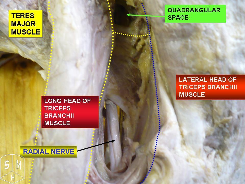
The radial nerve is a nerve in the human body that supplies the posterior portion of the upper limb. It innervates the medial and lateral heads of the triceps brachii muscle of the arm, as well as all 12 muscles in the posterior osteofascial compartment of the forearm and the associated joints and overlying skin. It originates from the brachial plexus, carrying fibers from the ventral roots of spinal nerves C5, C6, C7, C8 & T1. The radial nerve and its branches provide motor innervation to the dorsal arm muscles (the triceps brachii and the anconeus) and the extrinsic extensors of the wrists and hands; it also provides cutaneous sensory innervation to most of the back of the hand, except for the back of the little finger and adjacent half of the ring finger (which are innervated by the ulnar nerve). The radial nerve divides into a deep branch, which becomes the posterior interosseous nerve, and a superficial branch, which goes on to innervate the dorsum (back) of the hand. Th ... [...More Info...] [...Related Items...] OR: [Wikipedia] [Google] [Baidu] |
Axillary Nerve
The axillary nerve or the circumflex nerve is a nerve of the human body, that originates from the brachial plexus (upper trunk, posterior division, posterior cord) at the level of the axilla (armpit) and carries nerve fibers from C5 and C6. The axillary nerve travels through the quadrangular space with the posterior circumflex humeral artery and vein to innervate the deltoid and teres minor. Structure The nerve lies at first behind the axillary artery, and in front of the subscapularis, and passes downward to the lower border of that muscle. It then winds from anterior to posterior around the neck of the humerus, in company with the posterior humeral circumflex artery, through the quadrangular space (bounded above by the teres minor, below by the teres major, medially by the long head of the triceps brachii, and laterally by the surgical neck of the humerus), and divides into an anterior, a posterior, and a collateral branch to the long head of the triceps brachii branch. * The ... [...More Info...] [...Related Items...] OR: [Wikipedia] [Google] [Baidu] |
Medial Cord
The medial cord is the part of the brachial plexus formed by of the anterior division of the lower trunk (C8-T1). Its name comes from it being medial to the axillary artery as it passes through the axilla. The other cords of the brachial plexus are the posterior cord and lateral cord. The medial cord gives rise to the following nerves from proximal to distal: *medial pectoral nerve (C8-T1) *medial brachial cutaneous nerve (T1) *medial antebrachial cutaneous nerve (C8-T1) *medial head of median nerve (C8-T1) [other part of median nerve comes from lateral cord The lateral cord is the part of the brachial plexus formed by the anterior divisions of the upper (C5-C6) and middle trunks (C7). Its name comes from it being lateral to the axillary artery as it passes through the axilla. The other cords of the br ...] *ulnar nerve (C8-T1, occasionally C7) Additional images File:PLEXUS BRACHIALIS.jpg, Brachial plexus References Nerves of the upper limb {{Neuroanatomy-stub ... [...More Info...] [...Related Items...] OR: [Wikipedia] [Google] [Baidu] |
Lateral Cord
The lateral cord is the part of the brachial plexus formed by the anterior divisions of the upper (C5-C6) and middle trunks (C7). Its name comes from it being lateral to the axillary artery as it passes through the axilla. The other cords of the brachial plexus are the posterior cord and medial cord. The lateral cord gives rise to the following nerves from proximal to distal: *lateral pectoral nerve (C5-C7) *musculocutaneous nerve (C5-C7) *lateral head of median nerve (C5-C7) [other part of median nerve comes from medial cord The medial cord is the part of the brachial plexus formed by of the anterior division of the lower trunk (C8-T1). Its name comes from it being medial to the axillary artery as it passes through the axilla. The other cords of the brachial plexus ar ...] Additional images File:Slide10EEEE.JPG, Lateral cord File:Slide1cord.JPG, Brachial plexus.Deep dissection. Nerves of the upper limb {{neuroanatomy-stub ... [...More Info...] [...Related Items...] OR: [Wikipedia] [Google] [Baidu] |
Posterior Cord
The posterior cord is a part of the brachial plexus The brachial plexus is a network () of nerves formed by the anterior rami of the lower four cervical nerves and first thoracic nerve ( C5, C6, C7, C8, and T1). This plexus extends from the spinal cord, through the cervicoaxillary canal in th .... It consists of contributions from all of the roots of the brachial plexus. The posterior cord gives rise to the following nerves: Additional images File:PLEXUS BRACHIALIS.jpg, Brachial plexus File:Slide12OOO.JPG, Posterior cord File:Slide1SSS.JPG, Posterior cord File:Slide1cord.JPG, Brachial plexus.Deep dissection. File:Slide1ecc.JPG, Brachial plexus.Deep dissection.Anterolateral view References MBBS resources http://mbbsbasic.googlepages.com/ External links * - "Axilla, dissection, anterior view" Nerves of the upper limb {{neuroscience-stub ... [...More Info...] [...Related Items...] OR: [Wikipedia] [Google] [Baidu] |
Axillary Artery
In human anatomy, the axillary artery is a large blood vessel that conveys oxygenated blood to the lateral aspect of the thorax, the axilla (armpit) and the upper limb. Its origin is at the lateral margin of the first rib, before which it is called the subclavian artery. After passing the lower margin of teres major it becomes the brachial artery. Structure The axillary artery is often referred to as having three parts, with these divisions based on its location relative to the Pectoralis minor muscle, which is superficial to the artery. * First part – the part of the artery superior to the pectoralis minor * Second part – the part of the artery posterior to the pectoralis minor * Third part – the part of the artery inferior to the pectoralis minor. Relations The axillary artery is accompanied by the axillary vein, which lies medial to the artery, along its length. In the axilla, the axillary artery is surrounded by the brachial plexus. The second part of the axi ... [...More Info...] [...Related Items...] OR: [Wikipedia] [Google] [Baidu] |
Cervical Spinal Nerve 6
The cervical spinal nerve 6 (C6) is a spinal nerve of the cervical segment. It originates from the spinal column from above the cervical vertebra 6 (C6). The C6 nerve root shares a common branch from C5, and has a role in innervating many muscles of the rotator cuff and distal arm, including: *Subclavius *Supraspinatus *Infraspinatus * Biceps Brachii * Brachialis * Deltoid *Teres Minor *Brachioradialis *Serratus Anterior *Subscapularis *Pectoralis Major *Coracobrachialis *Teres Major * Supinator *Extensor Carpi Radialis Brevis * Extensor Carpi Radialis Longus * Latissimus Dorsi Damage to the C6 motor neuron, by way of impingement, ischemia, trauma, or degeneration of nerve tissue, can cause denervation Denervation is any loss of nerve supply regardless of the cause. If the nerves lost to denervation are part of the neuronal communication to a specific function in the body then altered or a loss of physiological functioning can occur. Denervati ... of one or more of the asso ... [...More Info...] [...Related Items...] OR: [Wikipedia] [Google] [Baidu] |
Upper Trunk
The upper (superior) trunk is part of the brachial plexus. It is formed by joining of the ventral rami of the fifth (C5) and sixth (C6) cervical nerves. The upper trunk divides into an anterior and posterior division. The branches of the upper trunk from proximal to distal are: * subclavian nerve (C5-C6) *suprascapular nerve (C5-C6) *anterior division of upper trunk (C5-C6, forms part of lateral cord) *posterior division of upper trunk (C5-C6, forms part of posterior cord) The axillary, radial, musculocutaneous and median nerve The median nerve is a nerve in humans and other animals in the upper limb. It is one of the five main nerves originating from the brachial plexus. The median nerve originates from the lateral and medial cords of the brachial plexus, and has contr ...s all contain axons derived from the upper trunk. Additional images File:Slide1cord.JPG, Brachial plexus.Deep dissection. File:Slide1ecc.JPG, Brachial plexus.Deep dissection.Anterolateral view {{Authori ... [...More Info...] [...Related Items...] OR: [Wikipedia] [Google] [Baidu] |
Serratus Anterior Muscle
The serratus anterior is a muscle that originates on the surface of the 1st to 8th ribs at the side of the chest and inserts along the entire anterior length of the medial border of the scapula. The serratus anterior acts to pull the scapula forward around the thorax. The muscle is named from Latin: ''serrare'' = to saw, referring to the shape, ''anterior'' = on the front side of the body. Structure Serratus anterior normally originates by nine or ten muscle slips – branches from either the first to ninth ribs or the first to eighth ribs. Because two slips usually arise from the second rib, the number of slips is greater than the number of ribs from which they originate. The muscle is inserted along the medial border of the scapula between the superior and inferior angles along with being inserted along the thoracic vertebrae. The muscle is divided into three named parts depending on their points of insertions: #the serratus anterior superior is inserted near the superior a ... [...More Info...] [...Related Items...] OR: [Wikipedia] [Google] [Baidu] |


